Advances in Animal and Veterinary Sciences
Research Article
Advances in Animal and Veterinary Sciences 1 (3S): 9 – 11Special Issue–3 (Epidemiology and Animal Disease Investigations)
Prevalence and Haemato–biochemical Studies on Naturally Occurring Gout in Chhattisgarh
Nitin Singh*, Ratan Chandra Ghosh, Amit Singh
-
Department of Veterinary Pathology, College of Veterinary Science and Animal Husbandry, Anjora, Durg Indira Gandhi Krishi Vishwa Vidyalaya, Raipur, Chattisgarh, India
*Corresponding author:drnitinsinghvet@gmail.com
ARTICLE CITATION:
Singh N, Ghosh RC, Singh A (2013). Prevalence and haemato; biochemical studies on naturally occurring gout in chhattisgarh. Adv. Anim. Vet. Sci. 1 (3S): 9 – 11.
Received: 2013–12–09, Revised: 2013–12–16, Accepted: 2013–12–17
The electronic version of this article is the complete one and can be found online at
(
http://nexusacademicpublishers.com/table_contents_detail/4/154/html
)
which permits unrestricted use, distribution, and reproduction in any medium, provided the original work is properly cited
ABSTRACT
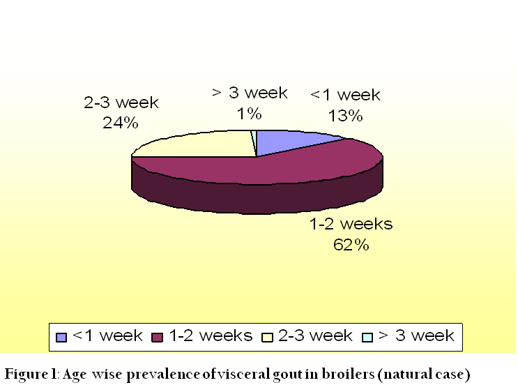
The present investigation was undertaken to study the prevalence of visceral gout and haemato–biochemical alterations in the gout affected broilers. The overall prevalence of visceral gout from different farms was found to be 21.47%. Out of total cases of visceral gout recorded (332 birds), 74.09% were found during winter season i.e. from November to February as compared to summer (21.38 %) i.e. from March to June, followed by monsoon season (4.51%) i.e. from July to October. . Of the susceptible age (first three weeks), the highest autopsy incidences (62.34%) was recorded during the second week of age, followed by 24.09% in third week and 12.65% in first week of age, while incidence of 0.9% was observed in birds of more than three weeks of age.. Haematological results showed increase in total erythrocyte count (TEC), total leukocyte count (TLC) and haemoglobin (Hb) concentration. There was significant (P≤0.05) increase in the level of uric acid in gout positive birds. Significant (P≤0.05) increase in serum amino transferase, alkaline phosphatases and albumin were also found in natural case of gout positive birds.
INTRODUCTION
Gout is a metabolic disorder that results in hyperuricemia and deposition of monosodium urate crystals in various parts of the body. The gout is characterized by retention and build–up of urates in tissues. It usually occurs in two separate syndromes: visceral gout and articular gout. Phalenet al; (1990) defined visceral gout as the accumulation ofuric acid tophi on serosal surfaces of the pericardium, liver capsule, airsacs, and within the kidney but may be found in any tissue. These crystals stimulate phagocytosis by neutrophils and initiate the inflammatory cascade. Outbreaks are seen in young chicks in the first week of life (baby chick nephropathy) or in flocks suffering kidney damage, or reduced water intake. The kidney damage can arise from infection with certain strains of Infectious bronchitis virus, exposure to some mycotoxins or inadequate water intake. The study was considered with the objective to study the seasonal prevalence of gout and to further evaluate the pathogenesis by studying Haemato–biochemical findings.
Methodology
The prevalence of gout was studied on the basis of live and dead birds collected from different poultry farms of different age groups in Durg district, Chattisgarh, India with respect to different seasons in broilers for a period of 12 months. Feed samples were collected from gout affected farms and analyzed for its crude protein content. Nitrogen present in the samples of feed was estimated by Micro–Kjeldahl’s method of AOAC (1995).
Assay of Hematological Parameters
The blood samples were collected from jugular vein/ heart using EDTA as an anticoagulant @ 1mg/ml of blood. The parameters like i) Total erythrocyte Count (TEC) (million/µl), (ii) Total leukocyte Count (TLC) (thousand/µl), (iii) Packed cell volume (PCV) (percent), (iv) Haemoglobin (Hb) (gram percent), (v) Differential leukocyte count (DLC) (percent) were estimated. TEC and TLC were done as per the method of Nambiar (1960) using diluting fluid recommended by Natt and Herick (1954). Hb was estimated by Acid haematin method using Sahli’s instrument. PCV and DLC were determined as per the method described by Jain (1986).
Assay of Biochemical Parameters
For biochemical parameters serum was collected as per standard procedure and biochemical parameters were carried out by using a semi autoanalyser using standard kit (Bayer Diagnostic India Ltd). The parameters includes (i) Total serum protein (g/dl) (ii) Serum albumin (g/dl) (iii) Serum globulin (g/dl), (iv) Serum alkaline phosphatase (ALP) (U/L) (v) Serum alanine amino transferase (ALT) (U/L) (vi) Serum aspartate amino transferase (AST) (U/L) (vii) Serum uric acid (mg/dl) (viii) Serum creatinine (mg/dl).
RESULTS AND DISCUSSION
Clinically the affected birds revealed the prevalence of visceral gout which was found to be highly influenced by atmospheric temperature. A total of 332 suspected cases of gout were collected for duration of 12 months. The study revealed that 74.09% of the total (332 birds) suspected cases of visceral gout were recorded during colder months i.e. from November to February as compared to hotter months (21.38%) i.e. from March to June, followed by monsoon (4.51%) i.e. from July to October during the year. In the present study, it was observed that the prevalence of visceral gout was highest in the month of January (43.64%), followed by the month of December (38.60%) and May (23.52%), whereas the lowest prevalence was observed in the month of August (3.26%). Age wise susceptibility due to visceral gout among broiler chicks was found to be maximum below three weeks of age. Age wise prevalence was recorded to be higher (62.34%) in birds below 2 weeks of age (Out of 332 positive case) followed by (24.09%) in birds between 2 to 3 weeks of age and (12.65%) was recorded in birds below 1 week of age. Only 0.90% of gout was observed in birds above 3 weeks of age (Figure 1). Similarly, Shrivastava (2001) and Karasawa et al. (1991) also reported higher mortality (68.28%) due to visceral gout during colder months of December to March. There is also excess formation and decreased dissolution of uric acid at colder temperature (Sayed, 2001). High protein diet is one of the major etiological agents in the production of gout and in the present survey the highest amount of crude protein found was 26% whereas the lowest amount was 12.25% and the average amount being 20.59%. Thus it showed that higher protein level in the diet is not only the major etiological agent in production of gout but it result in with combination of other factors like infectious bronchitis virus, diets containing higher amount of protein, cryptosporidiosis, Vitamin A deficiency, water deprivation (Schmidt, R. E. et al; 2003, Trampel et al; 2000, Hocking and Bernard, 1997).
Haematological Parameters
Significantly (P≤0.05) higher levels of TEC (4.12±1.380 million/µl), TLC (34.27±2.28 thousand/µl), Hb (12.8±2.46%) and PCV (38.23±3.06%) were observed in suspected gout affected birds as compared to normal birds in which TEC, TLC, Hb and PCV was found to be (3.25±1.58 million/µl) (26.35±2.34 thousand/µl) (8.8±1.08%) (29.36±0.97%), respectively, whereas the results of DLC showed significantly (P≤0.05) higher lymphocyte (61.86±2.71%) count in suspected gout affected birds as compared to normal birds (56.23±3.46%). There was no significant (P≤0.05) difference observed in heterophils, eosinophils, monocytes and basophils counts. Our findings were in agreement with Christopher (1977), who also observed increase in the lymphocyte counts in birds with gout. Koutsoset al. (2001) found no significant differences in any of the hematological parameters, whereas Rahamatulla and Mohiyuddeen (1973) found only increase in number of monocytes.
Higher values of TEC, TLC, Hb and PCV in gout affected birds may be due to adverse climatic conditions. The birds huddle together or scatter due to low and high temperature respectively leading to low water intake. This results in progressive dehydration and simultaneously haemo–concentration (Schmidt, R. E. et al; 2003), Julian, R. 1982). Higher lymphocyte count might be due to metabolic acidosis leading to uremia, which may have stimulatory effect on bone marrow leading to leukocytosis.
Biochemical Parameters
Significantly (P≤0.05) higher level of uric acid was observed in birds suspected to be positive for gout (86.13±12.10 mg/dl) as compared to the level of uric acid in normal birds (5.33±0.274 mg/dl). No significant alterations were observed in the level of creatinine in the suspected gout affected birds. There was significant (P≤0.05) differences observed in the activity of AST (374.83±24.99 U/L), ALT (16.71±0.4628 U/L) and ALP (3966.6±147.0 U/L) in serum sample of birds suspected to be gout affected as compare to AST (232.56±1.156 U/L), ALT (8.83±0.30U/L) and ALP (766.8±7.0 U/L) of normal birds (Table 1). The activity of AST is high in acute and chronic liver injury (Tennant, 1997). Alanine amino transferase (ALT) is employed as a marker of hepato–cellular damage and in general ALT is considered a more sensitive indicator of liver cell injury than AST (Oser, 1976). Increase level of AST and ALT in the blood indicates cellular damage (Ramazzotto and Carlin, 1978). Elevated plasma alkaline phosphatase might be due to acute hepato–cellular damage (Zimmerman and Henby, 1969) and enhance activity indicates renal damage.
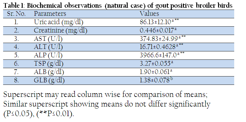
There was no significant (P≤0.05) alteration observed in the level of total serum protein, however serum albumin level was found to be significantly (P≤0.05) higher (1.90±0.061 g/dl), whereas the globulin values (1.38±0.078 g/dl) were lowered significantly (P≤0.05) in birds suspected to be gout affected as compared to normal birds (2.51±0.058 g/dl). This finding was in concurrence with Chandra et al. (1985) who reported that dehydration could be considered as an important factor in the precipitation of gout. This may be due to non–availability of adequate water to flush out the urinary system leading to its clogging. The ammonia and carbon monoxide if accumulated in sheds may affect the normal ventilation and ultimately reduce water intake and subsequently cause visceral gout in young chicks (Phatak, 2001). As AST is chiefly a mitochondrial enzyme. Mitochondrion plays an important role in maintaining hepatocyte integrity and function, which may be hampered due to excessive physiologic stress (Hassanein, 2004). The elevated levels of TEC, PCV and Hb, which were found in the natural cases of gout, are in accordance with our findings of albuminemia. Globulin part of protein was found to be increased only because of dehydration and emaciation of the birds. Immunoglobulin production might have been enhanced, as lymphocytosis occurrences are common in the natural cases of gout.
CONCLUSIONS
Gout is a metabolic disorder that results in hyperuricemia and deposition of monosodium urate crystals in various parts of the body. The study showed that highest suspected cases of visceral gout were recorded during colder months. Age wise prevalence in broiler chicks was recorded to be higher in birds below 2 weeks of age. The results also concluded that significant higher levels of TEC, TLC, Hb and PCV were observed in suspected gout affected birds as compared to normal birds. The uric acid estimation show significant higher in birds suspected to be positive for gout. There was significant (P≤0.05) differences in the activity of AST, ALT and ALP in serum sample of birds suspected to be gout affected as compare to normal birds, while no significant difference in creatinine level is found in affected birds.
REFERENCES
AOAC (1995). Official Methods of Analysis. 16th edition.
Chandra, M., Singh B., Gupta, P. P., Ahuja, S. P. and Singh N. (1985). Clinicopathological, Haematological and Biochemical studies in some outbreaks of nephritis in poultry. Avian Dis.29(3): 590–600.
http://dx.doi.org/10.2307/1590650
PMid:4074233
Christopher, K. J. (1977). Some aspects of gout in poultry. Irish Veterinary Journal. 31: 180–185. (c.f.Vet.Bull.48: 4463).
Hassanein, T. (2004). Mitochondrial dysfunction in liver disease and organ transplantation. Mitochondrion. 4: 609–620.
http://dx.doi.org/10.1016/j.mito.2004.07.015
PMid:16120418
Hocking, D.M. and Bernard, R., 1997. Effect of dietary crude protein content on the prevalence of articular gout in different line of broiler males. Br. Poul. Sci., 38: S21–S23.
http://dx.doi.org/10.1080/00071669708417969
PMid:9158897
Jain, N. C. (1986). Haematological techniques – In Schalms. Veterinary Haematology (4th Ed) Lea and Febinger, Philadelphia. pp. 20–86.
Julian R. Water deprivation as a cause of renal disease of chickens. Avian Pathol 1982;11:615–7.
http://dx.doi.org/10.1080/03079458208436137
PMid:18770228
Karasawa, Y., Tsubota, R., Sakiyama, C., Meeda, M., Yamaguchi, M. and Maruyama, T. (1991). Incidence of gout in broiler chickens during the early life in mid– summer and late autumn. Jap. Poult. Sci. 28 (5): 278–283 {c.f. Winspris Cab Abstr.}.
http://dx.doi.org/10.2141/jpsa.28.278
Koutsos, E. A., Smith, J., Woods, L. W. and Klasing, K. (2001). Adult Cockatiels (Nymphicushollandicus) metabolically adapt to high protein. J. Nutr. 131: 2014–2020.
PMid:11435523
Nambiar, K. T. K. (1960). Studies on haematology of the domestic fowl. M.V.Sc. thesis submitted to the University of Madras.
Natt, M. P. and Herick, C. A. (1954). A new blood diluent for counting erythrocytes and leucocytes of chickens. Poult. Sci.31: 735–738.
http://dx.doi.org/10.3382/ps.0310735
Oser, B. L. (1976). Hawk's physiological chemistry. McGraw – Hill Book Company, New York. London.
Phalen, D.N., Ambrus S., Graham D.L. The avian urinary system: form, function, diseases. In:Association of Avian Veterinarians Annual Conference Proceedings. Boca Raton (FL): Association of Avian Veterinarians; 1990. p. 44–57.
Phatak, R. K. (2001). Management of gout in tropical countries. Conference Proceedings. South Asian Regional Poultry Conference and Exhibition, 24–26 September 2001, Pune, India.
Rahamathulla, P. M. and Mohiyuddeen, S. (1973). Studies on gouty nephritis in poultry. Indian Vet. J. 50: 841–844.
Ramazzotto, L. J. and Carlin, R. (1978). Effects of DMSO on SGOT during hypothermia in adrenalectomized rats. Life science, 22: 329–336.
http://dx.doi.org/10.1016/0024-3205(78)90140-6
Sayed, A. (2001). Gout in poultry. Kemin Direct. 2(3): 10–12.
Schmidt, R. E., Reavill, D. R., Phalen, D.N. Urinary system. In: Pathology of pet and aviarybirds. Ames (IA): Iowa State Press; 2003. p. 95–107.
http://dx.doi.org/10.1002/9780470376836.ch5
PMCid:PMC1180594
Shrivastava, N. (2001). Etiopathological studies on visceral gout in broiler chicks, M.V.Sc. thesis submitted to Gujarat Agricultural University,S. K. Nagar.
Trampel, D.W., Pepper, T.M. and Blagburn,B.L., 2000. Urinary tract cryptosporidiosis in commercial laying hens. Avian Dis., 44: 479–484.
http://dx.doi.org/10.2307/1592566
PMid:10879932
Zimmerman, H. J. and Henby, J. B. (1969). Serum enzyme determination as an aid to diagnosis. In Clinical diagnosis by laboratory methods. W. B. Saunders. Philadelphia.


