Advances in Animal and Veterinary Sciences
Research Article
Advances in Animal and Veterinary Sciences 1 (5): 134 – 139A retrospective Study of Canine Tumors in Grenada, West Indies
Muhammed Iqbal Bhaiyat1, Alfred Chikweto1, Keshaw Prasad Tiwari1, Claude DeAllie1, Rajveer Singh Pawaiya3, Allison Inga1, Cecilia Hegamin–Younger2, Ravindra Nath Sharma 1*
- Pathobiology Academic Program, School of Veterinary Medicine, St George’s University, Grenada
- Department of Public Health and Preventive Medicine, School of Medicine, St George’s University, Grenada
- Principal Scientist, Division of Animal Health, Central Institute for Research on Goats (ICAR), Makhdoom, P.O. Farah – 281122, Mathura (U.P.) India
*Corresponding author:rsharma@sgu.edu
ARTICLE CITATION:
MI Bhaiyat, A Chikweto, KP Tiwari, C DeAllie, RVS Pawaiya, A Inga, C Hegamin–Younger, and RN Sharma (2013). A retrospective study of canine tumors in Grenada, West Indies. Adv. Anim. Vet. Sci. 1 (5): 134 – 139.
Received: 2013–08–27, Revised: 2013–09–03, Accepted: 2013–09–03
The electronic version of this article is the complete one and can be found online at
(
http://nexusacademicpublishers.com/table_contents_detail/4/87/html
)
which permits unrestricted use, distribution, and reproduction in any medium, provided the original work is properly cited
ABSTRACT
A retrospective study on 462 cases of tumor conditions in dogs was carried out in Pathology laboratory, School of Veterinary Medicine, St George’s University, Grenada, West Indies for a period of ten years, from 2001 to 2010. Out of these 462 tumors, 242 and 220 tumors were recorded from males and females of varying age groups respectively. Among all these tumor conditions, the occurrence of haemangiosarcoma was found to be highest (13%) followed by mammary gland tumors (10.8%), transmissible venereal tumors (7.6%), cutaneous hemangioma (7.4%), cutaneous histiocytoma (6.3%), lymphoma and papilloma (5.0% each), mast cell tumor (4.5%), lipoma and squamous cell carcinoma (3.5% each), melanocytoma (3.2%), fibrosarcoma, hemangiopericytoma and melanoma (2.6% each), fibroma (2.2%) and others including basal cell tumor and chondrosarcoma (1.5% each), seminoma (1.3%), plasmacytoma (1.1%), perianal gland adenoma (0.9%) and adenocarcinoma (0.6%), hepatocellular carcinoma (0.6%), pancreatic adenocarcinoma (0.6%), pheochromocytoma (0.4%), sertoli cell tumor (0.2%), etc. Age–wise, the highest tumor occurence (28.1%) was in the >8–12 year age group, followed by 25.8% in >5–8 year, 19.7% in >12 year, 14.1% in >3–5 year and 10.2% in >1–3 year age groups. Mixed breed dogs showed highest tumor incidence (62.6%) followed by German shepherd (6.5%), Labrador Retriever (5.6%), Rottweiler (4.3%), Doberman Pinscher (3.2%), Pompek (3.0%) and Pitbull (2.2%).
INTRODUCTION
Cancer is one of the biggest threats to humans and animals, claiming 7.6 millions of human lives in 2008 and 13.2 million expected cancer deaths by 2030 (American Cancer Society, 2011), in spite of several interdisciplinary approaches that have contributed significantly to the progress in cancer diagnosis and treatment. It is the leading cause of mortality in pet animals (Bonnett et al., 2005; Merlo et al., 2008) and second most in humans (1 in every 4 deaths in United States) (Siegel et al., 2013). The frequency of cancer occurrence in dogs is twice that in humans (Rungsipipat, 2003).
There are about 70 million pet dogs in USA of which about 10% are diagnosed with naturally occurring tumors (Paoloni and Khanna, 2007). No data are available on the population of dogs in Grenada or West Indies. Lack of reliable pet tumor registries have made it difficult to ascertain the increasing or decreasing prevalence of cancer in dogs. However, neoplasms have gained much importance in pet animals owing to the increased awareness among the people towards animal sufferings. As a result of improvements in health and welfare animals are living longer, leading to increased diagnosis of cancer in dogs (Dobson, 2013). In a recent mortality investigation in the UK, cancer accounted for 27% of all deaths in purebred dogs (Adams et al., 2010). Studies from various animal tumor registries in the world have reported dog tumor incidence rate from 99.3 to 282 to 1126 cases per 100,000 per annum, with the highest tumor type occurrence of mammary tumors, skin tumors or transmissible venereal tumor (MacVean et al., 1978, Merlo et al., 2008, Vascellari et al., 2009). Merlo et al 2008 reported mammary tumors being 70% of all tumor cases followed by non–Hodgkin's lymphoma, whereas Vascellari et al 2009 from an animal tumor registry in Italy reported skin tumors being 40.8% followed by mammary tumors being 38.8% of a total 2509 dog tumor cases. In another study on 403 cases of dog neoplasms, transmissible venereal tumors (42.9%) were the highest followed by 33.5% mammary tumors (Khimta et al., 2010). The present study documents the occurrence of canine tumors in Grenada, West Indies.
MATERIALS AND METHODS
Tumor tissue specimens
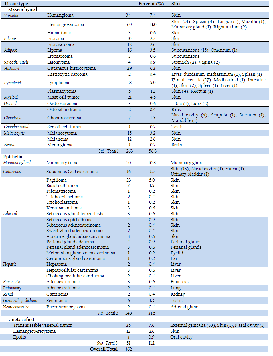
The data on canine tumors diagnosed from 2001 to 2010 in the Department of Pathobiology Academic Program of School of Veterinary Medicine, Saint George’s University, Grenada, West Indies, inclusive of clinical history were taken from the archived postmortem and biopsy reports and analyzed for the study. Confirmed histopathological diagnosis was done in missing cases after processing the archival formalin–fixed canine tumor tissues. The details of tumor types, their origin and primary sites are given in Table 1.
Histopathological examination
The tissue samples were processed routinely through graded alcohol and xylene in automatic tissue processor (Leica, LP 1080 Model) to obtain paraffin–embedded tissue blocks. The blocks were cut using semi motorized microtome (Leica, RM 2045 Model) to obtain 4 um thick sections. The sections were stained manually by hematoxylin and eosin staining method and examined under the microscope. The diagnosis of various tumor conditions was made based on the characteristic histopathological features.
Statistical Analysis
History and clinical data of each case with regard to age, sex and breed of the dogs were collected and analyzed statistically using student’s t test.
RESULTS
A total of 462 cases of tumor conditions were diagnosed during the period from 2001 to 2010 in the dogs which comprised 242 (52.4%) males and 220 (47.6%) females of different age groups. The various tumor conditions diagnosed are listed in Table–1. The occurrence of hemangiosarcoma was the highest at 13.0%. followed by mammary gland tumors (10.8%), transmissible venereal tumors (7.6%), cutaneous hemangioma (7.4%), cutaneous histiocytoma (6.3%), lymphoma and papilloma (5.0% each), mast cell tumor (4.5%), lipoma and squamous cell carcinoma (3.5% each), melanocytoma (3.2%), fibrosarcoma, hemangiopericytoma & melanoma (2.6% each), fibroma (2.2%) and others including basal cell tumor and chondrosarcoma (1.5% each), seminoma (1.3%), plasmacytoma (1.1%), perianal gland adenoma (0.9%) and adenocarcinoma (0.6%), hepatocellular carcinoma (0.6%), pancreatic adenocarcinoma (0.6%), pheochromocytoma (0.4%), sertoli cell tumor (0.2%). A list of the tumors and their primary sites are presented in Table 1.
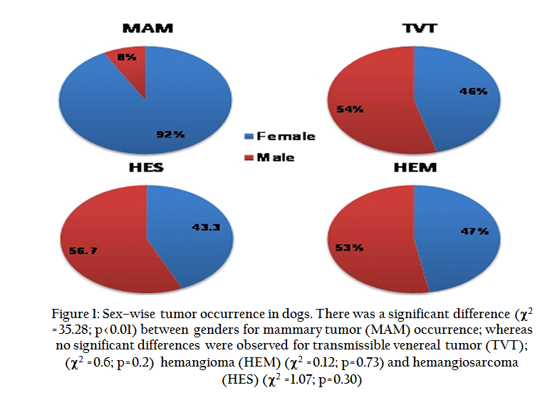
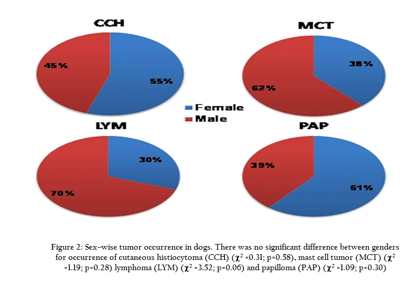
Sex–wise analysis revealed that although more tumors occurred in males (52.4%) than females (47.6%), there was no significant difference between gender (χ2 =1.05, p=0.31). Among tumor types, the occurrence of mammary tumor was significantly high (χ2 =35.28, p<0.01). in females than males (Figure 1), whereas, for other tumors no significant difference was observed between gender (Figure 1 and 2).
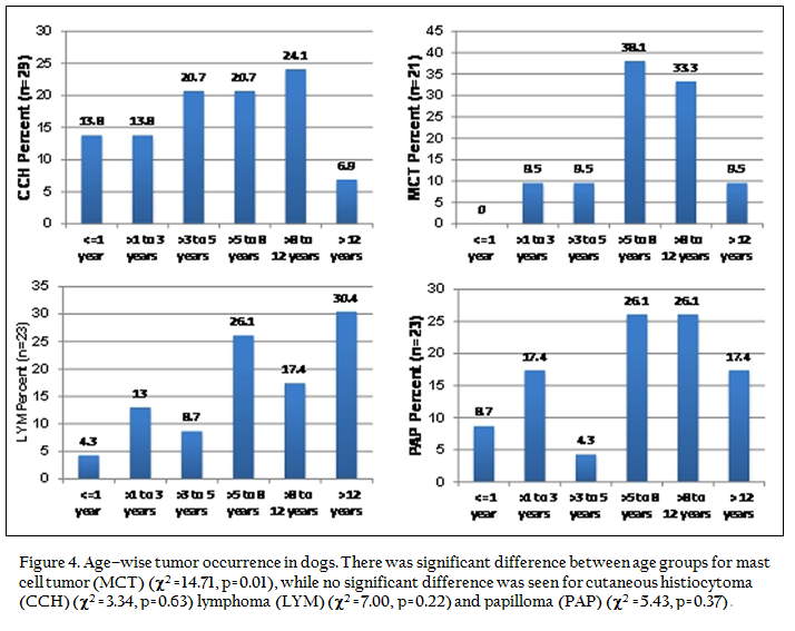
Age–wise, the highest occurrence of tumors (28.1%) was seen in the >8–12 years age group, followed by 25.8% in >5–8 years, 19.7% in >12 years, 14.1% in >3–5 years and 10.2% in >1–3 years age groups. Notably, three major tumor types, namely cutaneous histiocytoma, papilloma and lymphoma also involved animals younger than 1 year of age (Figure 3 and 4), whereas other tumors occurred in animals above 1 year of age. Transmissible venereal tumors specifically affected the age group of >1–3 years and over 12 years (Figure 3), whereas lymphoma appeared mostly between 5–8 years and after 12 years of age (Figure 4). Interestingly, although not significant ((χ2 =3.34, p=.62), there is a trend that cutaneous histiocytoma started occurring early on in the age and continued to increase up to 12 years of age, declining thereafter (Figure 4). Other tumors including papilloma, hemangioma, hemagiosarcoma, mast cell tumor and mammary tumor occurred at peak between 5 and 12 years of age (Figure 3 and 4)

Breed–wise analysis showed that mixed breed dogs (62.6%) were the most affected ones, followed by German Shepherd Dog (GSD) (6.5%), Labrador Retriever (5.6%), Rottweiler (4.3%), Doberman Pinscher (3.2%), Pompek (3.0%) and Pitbull (2.2%). The order of affected breeds remained similar for most of the tumors except for papilloma, mammary and mast cell tumors (Figure 5). Mammary tumors ranked highest in mixed breed (69.8%) followed by Doberman Pinscher (13.9%) and GSD (11.6%). Mast cell tumors affected mostly mixed breed (75%) followed by Labrador Retriever (16.7%) and GSD (8.3%), whereas papilloma occurred mostly in mixed breed (76.2%) followed by Pompek (14.3%) and GSD and Labrador Retriever (4.8% each)

DISCUSSION
The analysis of 462 canine tumor cases from different organs diagnosed in Grenada during the period from 2001 through 2010 revealed hemangiosarcoma (13%) as the most frequent neoplasm followed by mammary tumors (10.8%), transmissible venereal tumor (7.6%), cutaneous hemangioma (7.4%), cutaneous histiocytoma (6.3%) and others. Similarily, a previous study on tumors affecting the skin of dogs in Grenada revealed hemangiosarcoma as the most frequent neoplasm (Chikweto et al., 2009). In previous studies from other countries of the world, the distribution of tumor occurrence varied from region to region, with mostly either mammary tumors or skin tumors or transmissible venereal tumor being recorded (MacVean et al., 1978, Merlo et al., 2008, Vascellari et al., 2009). In their study in Italy, Merlo et al., 2008 reported mammary tumors being 70% of all neoplastic cases followed by non–Hodgkin's lymphoma, whereas Vascellari et al 2009 reported 40.8% skin tumors followed by 38.8% mammary tumors – out of a total – of 2509 dog tumor cases. In a study from India on 403 cases of dog neoplasms, transmissible venereal tumors (42.9%) were the highest followed by mammary tumors (33.5%) (Khimta et al., 2010). Mukaratirwa et al 2005 in Zimbabwe found 73.7% cutaneous neoplasms out of 540 canine tumor biopsies, with the prevalence of epithelial, mesenchymal, lymphohistiocytic and melanocytic tumors being 39.4%, 44.4%, 7.4% and 8.7%, respectively. Pakhrin et al. (2007) diagnosed 25.34% cutaneous tumors from 2,952 canine biopsy specimens, comprising epithelial and melanocytic tumors (56.9%), mesenchymal tumors (38.9%), and hematopoietic tumors (4.1%) in Korea. Although we did not analyze organ–wise locations, the combination of tumors that mostly originated from skin totaled 58.8% followed by mammary tumors (10.8%) and transmissible venereal tumors (7.6%), consistent with the findings of Vascellari et al. (2009). In dogs, it is now generally accepted that solar radiation is a contributing factor in the development of several skin neoplasms such as hemangioma, hemangiosarcoma and squamous cell carcinoma (Nikula et al., 1992; Ward et al., 1994). It is likely that geographical location may also be a contributing factor to the predominance of tumor type, as Grenada is located nearer to the equator receiving more direct sun exposure and thus, higher skin tumor occurrence in dogs (Nikula et al., 1992; Ward et al., 1994).
In the present study, male dogs (52.4%) were affected more than females (47.6%), in contrast to the finding of other studies where females were affected significantly more than males (Merlo et al 2008), who also recorded a high incidence of mammary tumors. However, studies recording a higher incidence of mammary tumors are likely to have more females affected than males as mammary tumors occur mostly in females. In our study, mammary tumors affected more female (92%) than male dogs (8.0%), with the percentage of male dogs being on the higher side in comparison to previous reports where a mere 1 to 2% male dogs suffered from mammary tumors (Mulligan 1975, Priester 1979). Lymphoma also showed a gender predisposition with 70% occurrence in males and 30% in females. No significant sex predisposition was noticed for transmissible venereal tumor. This observation was in congruence with that of Calvert et al 1982 but in disagreement with Khimta et al 2010 who found significantly more TVTs in females (67.6%) than males (32.4%).
Generally, the occurrence of all tumors increased with age, with the highest tumor prevalence (28.1%) seen in dogs of the >8–12 years age group followed by >5–8 years (25.8%), >12 years (19.7%), >3–5 years (14.1%) and >1–3 years (10.2%) age groups. These observations were consistent with those of previous studies (Pakhrin et al 2007; Merlo et al 2008; Vascellari et al 2009; Khimta et al 2010). Pakhrin et al 2007 recorded a mean age of 8.3 years for the ten most frequent tumors, with a range of 2 months to 19 years., whereas, Khimta et al 2010 observed highest tumor cases in the age group of 8–10 years (23.08%) followed by 4–6 years (20.35%), 2–4 years (18.1%), 6–8 years (16.4%), 10–12 years (9.9%), ≥12 years (6.9%) and <2 years (5.2%). Although the prevalence of all tumors increased with age in the present study, the tumors generally appeared after 1 year of age except for cutaneous histiocytoma, lymphoma and papilloma, which started appearing in dogs less than 1 year of age. Interestingly, 13.8% of all cutaneous histiocytomas, 8.7% of all papillomas and 4.3% of all lymphomas occurred at <1 year age. These observations corroborate the previous findings in canine pediatric oncology where, of 9522 neoplasms, 89% were cutaneous histiocytomas and 2% were papillomas in juvenile dogs up to the age of 12 months (Schmidt et al 2010). Another interesting finding was the occurrence of transmissible venereal tumor which was highest in dogs >12 years of age (31.4%) followed by the 1–3 year age group (25.7%), >5–8 year (17.1%), >3–5 year (14.3% and >8–12 year (11.5%) age groups. In contrast, Khimta et al 2010 observed a low (4.6%) prevalence of transmissible venereal tumor in dogs ≥12 years of age; however, their findings of a high prevalence of TVT (28.3%) in the 2–4 years age group almost concurs with our findings of a higher prevalence (25.7%) of TVT cases in the >1–3 year age group. Our study population comprised mostly intact mixed breed dogs which are often allowed to wander at will, and thus, the sexually mature dogs above 1 year of age were significantly affected by TVT ((χ2 =11, p<.0001) which is usually transmitted through coitus.
The age–wise distribution of mammary tumors showed the highest frequency in dogs of the >8–12 year age group (30%) followed by >3–5 year and >12 year (22% each), >5–8 year (20%) and >1–3 (0%) year age groups. These results are comparable to the findings of previous studies (Moe, 2001; Reddy et al 2009; Khimta et al 2010). Mammary tumors are thought to be age dependent, as bitches less than two years of age encounter it rarely, but there is a sharp increase in the incidence after six years of age with a peak incidence at 8–12 years of age (Brodey et al 1983; Rungsipipat et al 2003). Moe (2001) observed a mean age of 8.8 years for histologically diagnosed mammary tumors in all breeds of dogs in Norway. In a study on 128 canine mammary tumors in India (Reddy et al 2009), the most affected age group was 8–10 years (35.94%), followed by 6–8 years (30.47%), 10–12 years (17.97%), ≤ 6 years (12.50%) and >12 years (3.13%). Almost similar trends were observed by Khimta et al 2010 with the most affected age group being 8–10 years (33.33%), followed by 6–8 years (18.52%), 4–6 years (15.55%), 10–12 years (13.33%), ≥12 years and 2–4 years (8.88% each) and <2 years (1.48%). Moulton (1999) observed that mammary gland tumors occurred rarely in female dogs younger than 2 years of age and that the incidence increased after the 5th year of age with its peak at the age of 10 yrs. This is consistent with our findings.
Mixed breed dogs were affected most (62.6%), followed by GSD (6.5%), Labrador Retriever (5.6%), Rottweiler (4.3%), Doberman Pinscher (3.2%), Pompek (3.0%) and Pitbull (2.2%). Mixed breeds comprising local stray dogs or mongrels remained the highest affected for all tumor types and the order of occurrence for the other breeds remained more or less similar. However, for mammary tumors, the second highest affected breed was the Doberman Pinscher (13.95%) followed by GSD (11.63%); for mast cell tumors the second highest affected breed was Labrador Retriever (16.67%) followed by GSD (8.33%); and for papilloma the second highest affected breed was Pompek (14.29%) followed by GSD and Labrador Retriever (4.76% each). Earlier reports on breed predisposition of mammary tumors and other tumors differ from the present observations which could be attributed to the fact that different geographical areas have different patterns of breed distribution (Schneider 1970, Rosen and Oberman 1993, Moe 2001, Reddy et al 2009, Khimta et al 2010). Overall tumor prevalence in India was highest in the Spitz (34.49%), followed by Mongrels (28.54%), GSD (16.13%), Doberman Pinscher (7.94%), Labrador Retriever (3.97%) and Great Dane (2.98%) in a study on 403 canine neoplasm (Khimta et al 2010). However, Reddy et al (2009) in India observed the highest number of mammary tumors in GSD (35.0%) followed by Spitz (24.22%), Mongrels (19.53%), Pomeranian (10.94%), Labrador Retriever (6.25%), Boxer (3.91%), Doberman Pinscher (4.69%), Cocker Spaniel (3.13%), Bhutia (1.56%) and Great Dane (0.78%). Moe (2001) from Norway observed the highest relative risk ratio of mammary tumors in Boxers, Cocker Spaniels, English Springer Spaniels and Dachshunds. The finding of highest tumor occurrence in mixed breed dogs in the present study evidently reflects on the pattern of breed distribution in Grenada.
In conclusion, the present study revealed a high incidence (20.4%) of hemangioma and hemangiosarcoma, followed by mammary gland tumor (10.8%) and transmissible venereal tumor (7.6%). Mixed breed dogs were mostly affected (62.2%), followed by GSD (6.5%) and Labrador Retriever (5.6%). Tumor occurrence was highest in dogs of the >8–12 year age group (28.1%), followed by >5–8 year (25.8%) and >12 year (19.7%) age groups. These data provide valuable epidemiological information on spontaneous tumors occurring in the Grenadian dog population.
ACKNOWLEDGEMENT
Authors are grateful to the Dean, SVM, Provost and Chanceller of the Saint George’s University, Grenada for providing necessary facilities to carry out the study.
CONFLICT OF INTEREST
Authors do not have any conflict of interest.
REFERENCES
Adams VJ, Evans KM, Sampson J and Wood JLN (2010). Methods and mortality results of a health survey of purebred dogs in the UK. J. Small Anim. Pract. 51: 512–524.
http://dx.doi.org/10.1111/j.1748-5827.2010.00974.x
PMid:21029096
American Cancer Society (2011). Global Cancer Facts & Figures, 2nd Edition. Atlanta: American Cancer Society.
Bonnett B, NA Egenvall, Hedhammar A and Olson P (2005). Mortality in over 350000 insured Swedish dogs from 1995−2000: I. Breed, gender, age and causes pecific rates. Acta Vet. Scand. 46: 105−120.
http://dx.doi.org/10.1186/1751-0147-46-105
PMid:16261924 PMCid:PMC1624819
Brodey RS, Goldschmidt MA and JR Roszel (1983). Canine mammary gland neoplasm. J. Cancer Res. Treat. 50: 11–25.
Calvert CA Leifer CE and MacEwen EG (1982). Vincristine for treatment of transmissible venereal tumor in the dog. J. Am. Vet. Med. Assoc. 181: 163–164.
PMid:6749780
Chikweto A, Bhaiyat MI, Sharma R, DeAllie C and McNeil PE (2009). Angiomatous lesions of the skin of dogs in Grenada. J. Commonw. Vet. Assoc. 25: 29–31.
Dobson JM (2013). Breed–predispositions to cancer in pedigree dogs. ISRN Vet. Sci. vol. 2013, Article ID 941275, 23 pages, 2013. doi:10.1155/2013/941275. Available online at: http://www.hindawi.com/isrn/veterinary.science/2013/941275/#B170. Accessed 03 September, 2013.
http://dx.doi.org/10.1155/2013/941275
Khimta S, Maiti SK, Kumar N and Sharma AK (2010). Occurrence of neoplasms in canine – a retrospective study. Indian J. Anim. Sci. 80: 7–11.
MacVean DW, Monlux AW, Anderson PS, Silberg SL and Roszel JF (1978). Frequency of canine and feline tumors in a defined population. Vet. Path. 15: 700–715.
http://dx.doi.org/10.1177/030098587801500602
PMid:220774
Merlo DF, Rossi L, Pellegrino C, Ceppi M, Cardellino U, Capurro C, Ratto A, Sambucco PL, Sestito V, Tanara G and Bocchini V (2008). Cancer Incidence in Pet Dogs: Findings of the Animal Tumor Registry of Genoa, Italy. J. Vet. Intern. Med. 22: 976 – 984.
http://dx.doi.org/10.1111/j.1939-1676.2008.0133.x
PMid:18564221
Moe L (2001). Population–based incidence of mammary tumors in some dog breeds. J. Reprod. Fertil.(Suppl) 57: 439–443.
Mukaratirwa S, Chipunza J, Chitanga S, Chimonyo M and Bhebhe E (2005). Canine cutaneous neoplasms: prevalence and influence of age, sex and site on the presence and potential malignancy of cutaneous neoplasms in dogs from Zimbabwe. J. S. Afr. Vet. Assoc. 76: 59–62.
http://dx.doi.org/10.4102/jsava.v76i2.398
PMid:16108522
Mulligan RM (1975). Mammary cancer in the dog: a study of 120 cases. Am. J. Vet. Res. 36: 1391–1396.
PMid:169716
Nikula KJ, Benjamin SA, Angleton GM, Saunders WJ and Lee AC (1992). Ultraviolet radiation, solar dermatosis, and cutaneous neoplasia in beagle dogs. Radiat. Res. 129: 11–18.
http://dx.doi.org/10.2307/3577898
PMid:1728052
Pakhrin B, Kang MS, Bae IH, Park MS, Jee H, You MH, Kim JH, Yoon BI, Choi YK and Kim DY (2007). Retrospective study of canine cutaneous tumors in Korea. J. Vet. Sci. 8: 229–236.
http://dx.doi.org/10.4142/jvs.2007.8.3.229
PMid:17679768 PMCid:PMC2868128
Paoloni MC and Khanna C (2007). Comparative Oncology Today. Vet. Clin. North Am. Small Anim. Pract. 37: 1023–1032.
http://dx.doi.org/10.1016/j.cvsm.2007.08.003
PMid:17950880 PMCid:PMC2174910
Priester WA (1979). Occurrence of mammary neoplasms in bitches in relation to breed, age, tumor type, geographic area from which reported. J. Am. Anim. Pract. 20: 1–10.
http://dx.doi.org/10.1111/j.1748-5827.1979.tb07014.x
Reddy GBM, Kumar P, Ramkumar, Pawaiya RVS and Ravindran R (2009). Histopathological classification and incidence of canine mammary tumors. Indian J. Vet. Pathol. 33: 152–155.
Rosen PP and Oberman HA (1993). Tumors of mammary gland. In: Atlas of Tumor Pathology. 3rd series. Fascicle 7, (Ed.) J. Rosai and LH Sobin, Armed Forces Institute of Pathology, Washington, D.C. USA
Rungsipipat A, Sunyasootcharee B, Ousawaphlangchai L, Sailasuta A, Thanawongnuwech R and Teankum K (2003). Neoplasms of dogs in Bangkok. Thailand J. Vet. Med. 33: 59–66.
Schmidt JM, North SM, Freeman KP and Ramiro–Iba-ez F (2010). Canine paediatric oncology: retrospective assessment of 9522 tumors in dogs up to 12 months (1993–2008). Vet. Comp. Oncol. 8: 283–292.
http://dx.doi.org/10.1111/j.1476-5829.2010.00226.x
PMid:21062410
Schneider A (1970). Comparison of age, sex and incidence rates in humans and canine breast cancer. Cancer 26: 419–426.
http://dx.doi.org/10.1002/1097-0142(197008)26:2<419::AID-CNCR2820260225>3.0.CO;2-U
Siegel R, Naishadham D and Jemal A (2013). Cancer statistics, 2013. CA: A Cancer J. Clin. 63: 11–30.
http://dx.doi.org/10.3322/caac.21166
PMid:23335087
Vascellari M, Baioni E, Ru G, Carminato A and Mutinelli F (2009). Animal tumor registry of two provinces in northern Italy: incidence of spontaneous tumors in dogs and cats. BMC Vet. Res. 5: 39–47.
http://dx.doi.org/10.1186/1746-6148-5-39
PMid:19825169 PMCid:PMC2763881
Ward H, Fox LE, Calderwood–Mays MB, Hammer AS and Couto CG (1994). Cutaneous hemangiosarcoma in 25 dogs: a retrospective study. J. Vet. Int. Med. 8: 345–348, 1994.
http://dx.doi.org/10.1111/j.1939-1676.1994.tb03248.x
PMid:7837111


