Advances in Animal and Veterinary Sciences
Short Communication
Advances in Animal and Veterinary Sciences. 1 (3S): 21 – 24Special Issue–3 (Epidemiology and Animal Disease Investigations)
Outbreaks of Inclusion Body Hepatitis (IBH) in Chickens; Pathological Studies and Isolation of Fowl Adenovirus
Vipan Kumar1, Rajesh Kumar2*, Rajesh Chandra3, Prakash Bhatt4, Kuldeep Dhama5
- Department of Veterinary Microbiology, Khalsa College of Veterinary and Animal Sciences, Amritsar, Punjab
- Department of Veterinary Microbiology, College of Veterinary and Animal Sciences, G.B. Pant University of Agriculture & Technology, Pantnagar-263145
- Uttarakhand; College of Veterinary & Animal Sciences, Central Agricultural University, Selseih, Aizawal
- Veterinary Clinics, College of Veterinary and Animal Sciences, G.B. Pant University of Agriculture & Technology, Pantnagar, Uttarakhand
- Division of Pathology, Indian Veterinary Research Institute, Izatnagar (UP), India
*Corresponding author:rajeshvet@rediffmail.com
ARTICLE CITATION:
Kumar V, Kumar R, Chandra R, Bhatt P and Dhama K (2013). Outbreaks of inclusion body hepatitis (IBH) in chickens; pathological studies and isolation of fowl adenovirus. Adv. Anim. Vet. Sci. 1 (3S): 21 – 24.
Received: 2013–01–30, Revised: 2013–02–27, Accepted: 2013–02–28
The electronic version of this article is the complete one and can be found online at
(
http://nexusacademicpublishers.com/table_contents_detail/4/197/html
)
which permits unrestricted use, distribution, and reproduction in any medium, provided the original work is properly cited
ABSTRACT
Fowl adenoviruses (FAdV) are known to cause many diseases in birds including poultry and wild birds including inclusion body hepatitis (IBH). The IBH is endemic in many states of India. The disease outbreaks of IBH in poultry farms have been investigated through clinical and histopathological examination of affected birds, isolation via chicken embryo liver cell (CEL) and detection of virus using immunofluorescent test. Clinically, the birds showed depression and greenish diarrhea. Necropsy showed jaundice, enlargement of kidneys, greyish white area of necrosis on liver and atrophy of bursa of Fabricius. Histopathology revealed large intracellular inclusion bodies in hepatocyte, degeneration and congestion of sinusoids and degeneration of real tubules. Virus infected CEL cells revealed intranuclear fluorescence in indirect fluorescent antibody test (IFAT) using FAdV specific antisera. Present study demonstrated the involvement of fowl adenovirus in inclusion hepatitis outbreaks and adds to the epidemiological data available in India.
Fowl adenoviruses are known to cause many diseases in poultry birds like Inclusion body hepatitis (IBH), hydropericardium syndrome (HPS), and respiratory infections in chickens and quail bronchitis. Among these conditions, Inclusion body hepatitis has a worldwide distribution, and there are indications that its incidence is increasing in many poultry industries (Mase, 2012; Kataria et al., 2013). All eleven serotypes of fowl adenovirus (FAdV) have been isolated from natural infections of poultry birds (Kim et al., 2008; Choi et al., 2012; Asthana et al., 2013) and wild birds like wild black kites (Kumar et al., 2010) and pigeons (Schrenzel et al. 2005; Catroxo et al., 2011). The disease causes high morbidity among broiler birds leading to production losses although average mortality is low (5–10%) but 30% mortality has been reported from Australia (McFerran and Smyth, 2000). Recently, some of FAdVs have been implicated in immunosuppression (Hussain et al., 2012). The disease was first reported from USA as a necrotizing hepatitis in 7–weeks–old chickens (Helmboldt and Frazier, 1963). Since then, the disease has been reported from all continents. In India, many outbreaks of this disease have been investigated by earlier workers (Garewal et al., 1981; Sandhu et al., 1994; Deshmukh et al., 2000; Kumar et al., 2003a; Kataria et al., 2013).
In domestic poultry, the disease usually affects birds at the age of 2 to 6 weeks (Asthana et al., 2013). The onset of disease occurs in hot and humid weather (Shah et al., 2011; Shahzad et al., 2011). The disease, inclusion body hepatitis is characterized by sudden onset of mortality, severe anemia and necrotic hepatitis with basophilic or eosinophillic intranuclear inclusion bodies in hepatocytes (Kim et al., 2008; Shahzad et al., 2011; Dar et al., 2012). Present study deals with investigation of outbreaks of Inclusion body hepatitis, histopathology of naturally infected birds and isolation of causative agent and its identification by IFAT.
Disease outbreaks of Inclusion body hepatitis (IBH) were suspected in broiler poultry birds in two private poultry farms of Bilaspur region (28.80N/ 79.00E) of Rampur district, Uttar Pradesh and Kashipur (29.220N/ 78.950E) of Uttarakhand on the basis of clinical signs and post mortem examination carried out in dead and sacrificed birds. The flock size of Bilaspur farm was 2200 and mortality rate recorded was 10%. In Kashipur farm, flock size was 4000 and mortality was 3%. Morbidity in both farms reached 80% and average age of affected birds was between 6–8 weeks. Clinical samples of liver, kidneys and bursa of Fabricius were collected from dead and necropsied birds for histopathological and virus isolation studies. The liver samples were collected in 50% glycerol saline for virus isolation. Tissue samples of liver, kidneys and bursa of Fabricius were collected in 10% formal saline for histopathological examination.
The tissues (liver, kidneys and bursa of Fabricius) were processed for histopathological examination as per standard protocol, fixed in 10% formal saline, washed in running tap water overnight and then dehydrated for one hour in different concentrations of ethanol, 50%, 60%, 70%, 80%, 90% and absolute alcohol for dehydration of tissues in same order. Then the tissues were cleared in xylene and embedded in paraffin wax. Sections of 4–5 µ thickness were cut and stained with Haemotoxylin and Eosin (H & E) staining procedure as described by Kumar et al. (2003a).
Twenty percent (w/v) liver homogenates of both isolates were prepared in phosphate buffered saline (PBS) from the clinical samples collected from both the outbreaks. Virus isolation was done on 14 day–old chicken embryo liver (CEL) cell culture (Kumar and Chandra, 2009). A total of six passages were done for isolation of the virus and the virus was identified by observing characteristic cytopathic effects in CEL cell cultures.
The presence of fowl adenovirus (FAdV) was detected by indirect immunofluorescent antibody technique (Kumar et al., 2003b). Briefly, CEL cells were grown on cover slips and infected with both the virus isolates obtained from the two disease outbreaks. After 48 hours of infection cover slips were flooded with chilled acetone for 30 min for fixing of cells of monolayers. Then the cells were rehydrated with PBS (pH 7.2). Single washing of infected cells was done with wash buffer (Triton X–100 0.01% v/v in PBS). After 5 min. of incubation, the detergent was removed and blocking buffer (10% foetal calf serum in wash buffer) was added to block unreactive sites on monolayers. Then, after three washings with wash buffer, FAdV specific antiserum was added and incubated for 30 min at 37˚C again after three washings with PBS, rabbit anti–chicken FITC conjugate (Sigma) was added to detect the binding of anti adenoviral antibodies with adenoviral antigens present in infected cell cultures. The coverslips were washed thrice as mentioned above with wash buffer, air–dried and mounted in glycerol. The stained coverslips were examined under U.V. light in fluorescent microscope. The un–infected CEL cell culture monolayer was kept as negative control and processed in similar way.
In the present disease investigation, naturally affected birds did not show any clinical signs at initial stages, except greenish watery diarrhea. However at terminal stages birds were dull depressed and reluctant to move and showed typical changes in posture. Kumar et al. (2009) also reported varying degree of dullness, depression and diarrhea leading to prostration and death. Grgic et al. (2006) also had similar observations. Absence of clinical signs and sudden mortality may be attributed to relatively short course of disease and acute damage to vital organs like liver and kidney. In this investigation, the mortality rate was about 3–10%. Earlier workers have also reported low mortality rates in adult broiler birds in IBH infections (McFerran and Smyth, 2000; Kumar et al., 2003).
The predominant post mortem lesions included enlarged livers with friable texture and pin point or white necrotic foci (Figure 1). In some cases, petechial and ecchymotic haemorrhages were also present. Kidneys were congested and contained haemorrhagic patches on the surface (Figure 2). Haemorrhages were also evident on thigh muscles. Lungs were congested and odematous. Previously many investigators have noticed these findings (Sandhu et al., 1994; Dar et al., 2012). In some cases, livers were also covered with a fibrinous mass of exudates as reported by other workers (Sandhu et al., 1994; Nayak et al., 1990; Mitra et al., 1996).
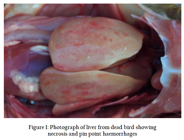
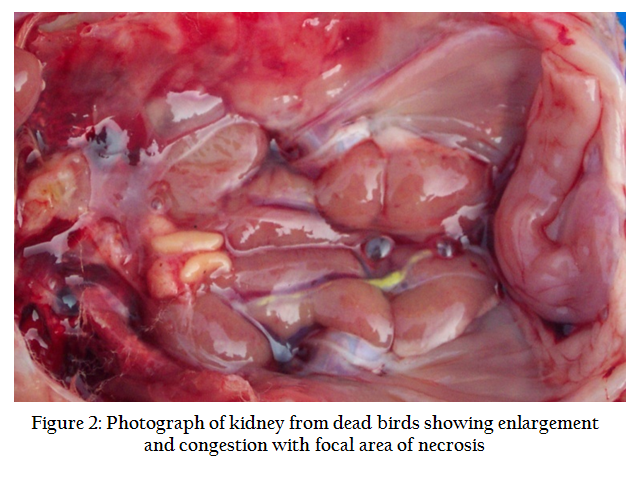
Figure 2: Photograph of kidney from dead birds showing enlargement and congestion with focal area of necrosis
The histopathological examination of liver revealed necrosis of hepatocytes, vacuolar degeneration and infiltration of mono nuclear cells. Many hepatocytes revealed large basophilic intranuclear inclusion bodies, which were round and compact and occupied almost entire nucleus. Large ‘bird eye’ basophilic intranuclear inclusion bodies and sinusoidal congestion were also observed in this study (Figure 3).
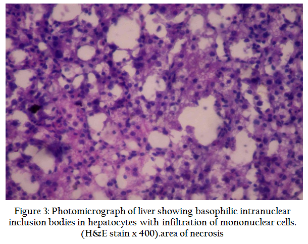
Figure 3: Photomicrograph of liver showing basophilic intranuclear inclusion bodies in hepatocytes with infiltration of mononuclear cells. (H&E stain x 400).area of necrosis
Theses findings are in accordance with those observed by earlier workers (Kaur et al., 2003; Kim et al., 2008). Kidneys showed congestion and haemorrhages, degeneration of renal epithelium and infiltration of mononuclear cells. Large basophilic intranuclear inclusions were occasionally observed (Figure 4). These findings were also reported by many earlier workers (Singh et al., 1996; Kaur et al., 2003). Bursa of Fabricius also had varying degree of degenerative changes and reduction in number of follicles (Figure 5). These findings are in agreement with earlier findings of Nayak et al., 1990 and Sandhu et al., 1994. However, Inclusion bodies were not demonstrated in the bursal tissues. The lesions in different organs were suggestive of acute extensive damage in the liver, kidneys and bursa of Fabricius. Increased vascular permeability may be attributed to deposition of serofibrinous exudates in the parenchyma of liver and kidney.
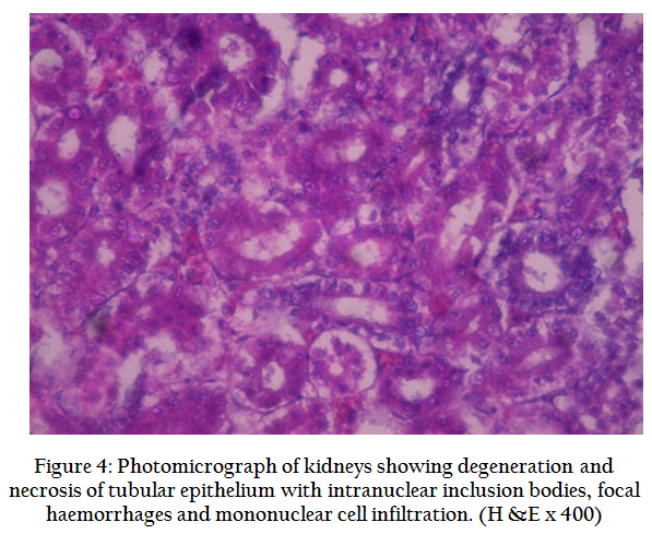
Figure 4: Photomicrograph of kidneys showing degeneration and necrosis of tubular epithelium with intranuclear inclusion bodies, focal haemorrhages and mononuclear cell infiltration. (H &E x 400)
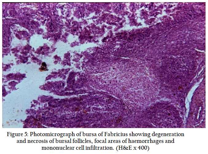
Figure 5: Photomicrograph of bursa of Fabricius showing degeneration and necrosis of bursal follicles, focal areas of haemorrhages and mononuclear cell infiltration. (H&E x 400)
On CEL cell cultures, the cytopathic effects (CPE) were characterized by rounding and degeneration of cells from first passage where microplaques were evident after 72 hours. By second passage, the CPE appeared within 24 hours. After 72 hours, almost 80% of the cell layer detached from its surface. Successive passaging showed more distinct and stable CPEs. Chicken embryo liver cell culture and chicken kidney cell culture are reported to support the growth of adenoviruses (Kumar and Chandra, 2004; Nakamura et al., 2002; Kaur et al., 2003). The cytopathic effects during isolation of the virus in the present study are in accordance with those reported by earlier workers (Kaur et al., 2003; Kumar et al., 2009). Micro and macro plaque formation is suggestive of cytolytic properties of virus.
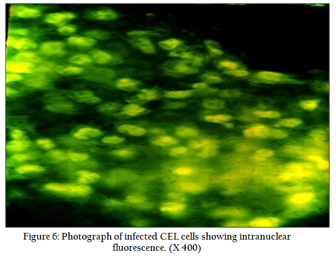
For confirmation of adenoviruses, different diagnostic tests are used including molecular tools, and among these, immunofluorescence detection of viral antigens during virus isolation has high diagnostic value because of the high degree of accuracy of detection of group specific antigens by using specific antibodies, and is in use since many years (Leland and Ginocchio, 2007). In the present study, the presence of viral antigens in CEL cell culture was demonstrated by indirect immunofluorescence antibody technique during the virus isolation in these cell cultures. Intense intranuclear immunofluorescence was observed in the virus infected CEL cell cultures at 7th passage for both the virus isolates, which confirmed the virus to be FAdV (Figure 6).
In conclusion, FAdVs are involved in IBH outbreaks. Degeneration of bursal follicles observed indicated that IBH virus may be involved in suppression of humoral immunity. Findings of present study add to epidemiological data of disease in the country and virus isolates obtained can be used for further molecular characterization, phylogenetic analysis, immunological and virological studies.
COMPETING INTEREST STATEMENT
The authors declare there is no competing interest associated with this study.
REFERENCES
Asthana M, Chandra R and Kumar R (2013). Hydropericardium syndrome: current state and future developments. Archiv. Virol. 158 (5):921–933
http://dx.doi.org/10.1007/s00705-012-1570-x
PMid:23242777
Catroxo MHB, Martins AMCRPF, Petrella S, Curi NA and Melo NA (2011) .Research of viral agents in free living pigeon feces (Columba livia) in São Paulo, SP, Brazil for transmission electron microscopy. Int. J. Morphol., 29(2):628–635,
http://dx.doi.org/10.4067/S0717-95022011000200055
Choi KS, Kye JY, Kim WJ, Jeon EK, Lee KY, Park and Sung HW (2012). Epidemiological investigation of outbreak of adenovirus infection in commercial chickens in Korea. Poultry Sci. 90(10): 2502–2506.
http://dx.doi.org/10.3382/ps.2012-02296
PMid:22991534
Dar A, Gomis S, Shirley I, Mutwiri G, Brownlie R, Potter A, Gerdts V and Tikoo SK (2012). Pathotypic and molecular characterization of a fowl adenovirus associated with inclusion body hepatitis in Saskatchewan chickens. Avian Dis. 56(1):73–81.
http://dx.doi.org/10.1637/9764-041911-Reg.1
PMid:22545531
Grgic H, Philippe C, Ojkic D and Nagy E (2006). Study of vertical transmission of fowl adenoviruses. Canadian J. Vet. Res. 70:230–233.
PMid:16850947 PMCid:PMC1477927
Hussain I, Mahmood MS, Arshadi MI, Masood A, Mahmood F and Rafique A (2012). Immune system dysfunction in broiler chickens experimentally inoculated with fowl adenovirus serotype–4 associated with inclusion body hepatitis hydropericardium syndrome. Turk. J. Vet. Anim. Sci.; 36(3): 223–230
Kataria JM, Dhama K, Nagarajan S, Chaktaborty S, Kaushal A and Deb R (2013). Fowl adenoviruses causing hydropericardium syndrome in poultry. Adv. Anim. Vet. Sci., 1(4S): 5–13.
Kaur G, Maiti NK and Oberoi MS (2003). Biological and immunological characterization of fowl adenovirus–4 isolates from inclusion body hepatitis–hydropericardium syndrome. Indian J. Anim. Sci. 73(1): 51–52.
Kim JN, Byun SH, Kim MJ, Kim JJ, Sung HW and Mo IP (2008). Outbreak of hydropericardium syndrome and molecular characterization of Korean fowl adenoviral isolates. Avian Dis. 52(3):526–30.
http://dx.doi.org/10.1637/8178-112207-Case
PMid:18939647
Kumar R and Chandra R (2009). Isolation and characterization of fowl adenovirus associated with hydropericardium syndrome. Indian Vet. J. 86(10): 1084–1086.
Kumar R, Kumar V, Asthana M, Shukla SK and Chandra R (2010). Isolation and identification of Fowl adenovirus from Wild Black Kites (Milvus migrans). J. Wild Dis. 46(1): 272–276.
http://dx.doi.org/10.7589/0090-3558-46.1.272
PMid:20090043
Kumar R, Shukla SK, Chandra R and Agarwal DK (2003a). Outbreaks of inclusion body hepatitis in domestic fowl. Indian J. Anim. Sci. 73(5): 477–480.
Kumar R, Chandra R and Shukla SK (2003b). Isolation of etiological agent of hydropericardium syndrome in chicken embryo liver cell culture and its serological characterization. Indian J. Exptl Biol. 41:821–826.
PMid:15248478
Kumar R and Chandra, R. (2004). Studies on structural and immunogenic polypeptides of hydropericardium syndrome virus by SDS-PAGE and western blotting. Comparative Immunology Microbiology & Infectious Diseases, 27 (3):155-161.
http://dx.doi.org/10.1016/j.cimid.2003.08.003
PMid:15001310
Leland DS and Ginocchio C (2007). Role of cell culture for virus detection in the age of technology. Clinic. Microbial. Rev. 20(1): 49–78.
http://dx.doi.org/10.1128/CMR.00002-06
PMid:17223623 PMCid:PMC1797634
Mase M, Nakamura K and Minami F (2012). Fowl adenoviruses isolated from chickens with inclusion body hepatitis in Japan, 2009–2010. J. Vet. Med. Sci. 74(8): 1087–1089
http://dx.doi.org/10.1292/jvms.11-0443
PMid:22516693
McFerran JB and Smyth JA (2000). Avian adenoviruses. Rev. Sci. Tech. Off. Int. Epiz. 19(2): 589–60.
Nakamura K, Tanaka H, Mase M, Imada T and Yamada M (2002). Pancreatic necrosis and ventricular erosion in adenovirus associated hydropericardium syndrome of broilers. Vet. Pathol. 39 (3):403–406.
http://dx.doi.org/10.1354/vp.39-3-403
PMid:12014508
Nayak HC, Chakraborty T, Chakraborty S and Chakraborty A (1990). An outbreak of inclusion body hepatitis in broiler chickens in West Bengal. Indian Vet. J. 76: 7–9.
Sandhu BS, Singh H and Singh B (1994). Prevalence and pathology of inclusion body hepatitis in chickens in Punjab. Indian Vet. J. 71: 438–442.
Scherenzel M, Oaks JL, Rotstein D, Maalouf G, Snook E, Sandfort C and Rideout B (2005). Characterization of a new species of adenovirus in Falcons, J. Clinicl. Microbiol. 43: 3402–3413.
http://dx.doi.org/10.1128/JCM.43.7.3402-3413.2005
PMid:16000466 PMCid:PMC1169131
Shah MS, Ashraf A, Khan MI, Rahman M, Habib M, Babapoor S, Ghaffar A, Malik IR, Khannum SA and Qureshi JA (2011). Molecular characterization of fowl adenoviruses associated with hydro–pericardium syndrome in broilers. African J. Microbiol. Res. 5(30):5407–5416.
Shahzad AK, Hussain T, Parkash O and Fazlanis A (2011). Pathogenicity of recent field isolate of Avian Adenovirus Serotype–IV of hydropericardium syndrome (Angara disease). African J. Microbiol. Res. 6(20): 4332–4335.
Singh A, Oberoi MS, Jand SK and Singh B (1996). Epidemiology of inclusion body hepatitis in poultry in northern India from 1990 to 1994. Rev. Sci. Tech. Off. Internatl. Epiz. 15(1): 1053–1060.


