Advances in Animal and Veterinary Sciences
Case Report
Advances in Animal and Veterinary Sciences 1 (2S): 26 – 28Special Issue–2 (Clinical Veterinary Practice– Trends)
Therapeutic Management of Chronic Generalized Demodicosis in a Pug
Neeraj Arora2*, Sukhdeep Vohra1, Satyavir Singh1, Sandeep Potliya2, Anshul Lather3, Akhil Gupta3, Devan Arora4, Davinder Singh4
- Veterinary Parasitology , College of Veterinary Sciences, Lala Lajpat Rai University of Veterinary and Animal Sciences, Hisar – 125004, Haryana
- Veterinary Surgery and Radiology, College of Veterinary Sciences, Lala Lajpat Rai University of Veterinary and Animal Sciences, Hisar – 125004, Haryana
- Veterinary Microbiology, College of Veterinary Sciences, Lala Lajpat Rai University of Veterinary and Animal Sciences, Hisar – 125004, Haryana
- Veterinary Public Health and Epidemiology, College of Veterinary Sciences, Lala Lajpat Rai University of Veterinary and Animal Sciences, Hisar – 125004, Haryana
*Corresponding author:neeru40@gmail.com
ARTICLE CITATION:
Arora N, Vohra S, Singh S, Potliya S, Lather A, Gupta A,Arora D and Davinder Singh D (2013). Therapeutic management of chronic generalized demodicosis in a pug. Adv. Anim. Vet. Sci. 1 (2S): 26 – 28.
Received: 2013–11–07, Revised: 2013–12–30, Accepted: 2013–12–30
The electronic version of this article is the complete one and can be found online at
(
http://nexusacademicpublishers.com/table_contents_detail/4/175/html
)
which permits unrestricted use, distribution, and reproduction in any medium, provided the original work is properly cited
ABSTRACT
A two year old female pug was presented to Teaching Veterinary Clinical Complex, LLRUVAS, Hisar with the history of itching, alopecia, crust formation, haemorrhage and thickening of the skin on face, neck, trunk and abdomen since last two months. The condition was laboratory diagnosed as chronic demodicosis and treated with amitraz and ivermectin along with supportive therapy. The female pug responded well to the treatment and recovered completely on 28th day after the start of the treatment.
Animal skin is exposed to attack by many kinds of parasites and each species has a particular effect on the skin; thatcan be mild or severe. In this regard, most of the ectoparasitic infestations produce irritation and sensitization of the skin. The reaction of the skin to these ectoparasites living in or on the skin results in inflammation, edema and an attempt to localize the foreign body, toxin or excretory products of the parasite. These reactions are often allergic and cause itching and burning sensation (Scott et al., 2001). Canine demodicosis, also called demodectic mange or follicular mange or red mange, is a common skin disease encountered in veterinary practice. Though the mite is a normal inhabitant of the hair follicles of all canines, clinical signs of demodecosis are common because of excessive proliferation of mite within the hair follicles (Scott et al. 2001). Canine demodicosis is most commonly caused by Demodex> canis; however, other species, such as, Demodex injai (a large bodied mite) and Demodex cornei (a short bodied mite), may also be involved (Tater and Patterson, 2008). Various drugs have been used against the Demodex mites with mixed results (Mueller, 2004). However, in view of increase in amitraz–resistant generalized demodicosis cases (Živičnjak, 2005) the current communication shows the therapeutic management of a generalized demodicosis in a pug with a combination therapy of acaricides and macrocyclic lactones along with adjunctive treatment.
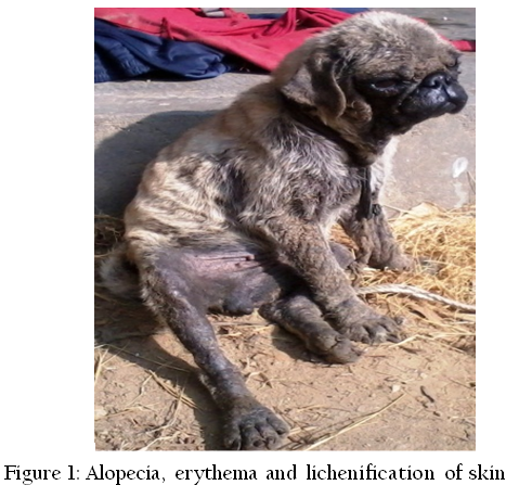
A two year old female pug weighing 8 Kg was presented to Teaching Veterinary Clinical Complex, Lala Lajpat Rai University of Veterinary and Animal Sciences, Hisar, Haryana with history of inappetance, hair shedding, foul smell from body and pruritis since 2 months. On clinical examination of the dog alopecia, crust formation, erythema and lichenification of skin was observed in the region of face, neck, trunk and limbs (Figure 1).
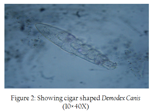
Deep skin scrapings from the skin of affected areas taken with proper sample collection technique were examined microscopically. The skin scrapings didn’t show any evidence of mite infestation, however, faecal sample examination showed the presence of elongate, cigar shaped mite with body divisible into head, thorax bearing four pairs of short and stumpy legs and abdomen bearing transverse striations. The morphology confirmed it to be Demodex canis (Soulsby, 1982) (Figure 2). Complete blood count, liver function test and mineral profile were also performed (Hb, TLCand DLC were performed as per standard procedures while iron, copper, ALT and AST were estimated by biochemical autoanalyser using Erba kits.) Skin scrapings /cm2 area were also collected before start of treatment and on subsequent days post–treatment (Singh and Chhabra, 1992) and the mite count/cm2 area was also correlated with improvement of lesions.
Amitraz (Diponil®) 12.5% was diluted @ 4 ml (500 mg)/L of water (at 500 ppm or 0.05% concentration) and carefully worked into the skin with a sponge after every week for twenty–one days. Before dipping, bathing with benzoyl peroxide shampoo was done for soothing of skin and removal of crusts and debris. Ivermectin (Tab. Neomec®) was given @ of 600µg/ kg body weight at weekly interval for three weeks. Oral cephalexin tablets (250mg) were also given daily for 15 days to check any secondary bacterial infections. Fatty acids supplement and liver tonic was also given along with adequate nutrition during the treatment period. New hair growth on affected skin started after 7th day of start of treatment (Figure 3) and complete uneventful recovery occurred on 28th day after start of the treatment (Figure 4).
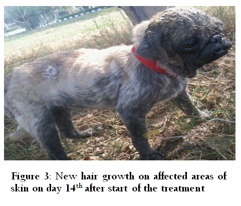

Figure 4: Complete recovery occurs on day 28th; after start of the treatment arrange the figures accordingly
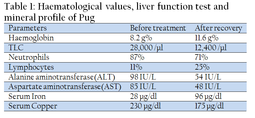
In present communication Demodex canis was not found in direct skin scrapping on day 0, which might be due to wrinkling as well as thickening and extensive involvement of skin of the animal. Pug licks the skin due to severe pruritis, as a result Demodex canis was found in faecal examination. So it is suggested that faecal examination of the animals suspected for demodicosis or with chronic form of mange must be done. Results of complete blood count, liver function test and mineral profile showed anaemia, leukocytosis due to neutrophilia, increase in values of aspartate aminotransferase and alanine aminotransferase, decrease in serum iron and increase in serum copper (Table 1). Similar findings were also reported by Jyotsna et al. (2005) reported decrease in serum iron and cobalt level and increase in serum copper but no change in serum zinc level in demodectic mange in dogs.
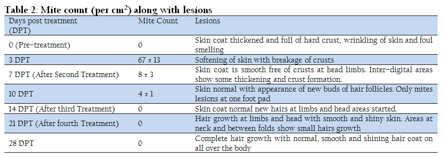
The detail of skin scraping along with mite count and improvement in skin lesions is given in Table 2. It was observed in the present study that although no mite was found in skin scraping on day 0 due to thickening, wrinkling and extensive involvement of skin, the active mites (67/cm2) along with eggs were found on day 3 when the softening of skin and breakage of thick crust started. From the improvement of lesions on skin and reduction of mites in scrapings, the animal started appearing healthy after second treatment. At this point most of animal owners or the veterinarian stops the acaricide treatment of the animal but some mites at the extremities i.e. inter–digital area still remains alive and proliferate with the appearance of favourable conditions (specially post–rainy season with the start of winters). After third treatment the animal was parasitologically free of any adult mite or its developmental stages and skin growth at all the body parts also started. To rule out any mite present on body fourth treatment was also given on 21th day. Complete hair growth along with smooth and shiny skin was observed on 28th day.
So it is suggested to field veterinarians that although the animal appears normal clinically after second treatment, atleast three to four dips in amitraz is recommended by parasitologist for complete recovery of animal and to prevent the occurrence of demdecosis.
ACKNOWLEDGEMENT
The authors are highly thankful to internee students for taking interest in care and management of this pug.
CONFLICT OF INTEREST
No conflict of interest to declare.
REFERENCES
Gunaseelan L, Bhavya S, SenthilKumar K and Balachandran C (2011). Influencing factors for mange mite infestation of dogs in Chennai city. Tamilnadu J. Vet. & Anim. Sci. 7: 247–249.
Jyotsna P, Maiti SK, Sanyal PK and Tiwari SP (2005). Haematobiochemical and mineral profiles in generalized canine demodicosis. Int. Pol. 6 No. 2 pp. 331–334.
Mueller RS (2004). Treatment protocols for demodicosis: an evidence–based review. Vet. Derm. 15: 75–98.
http://dx.doi.org/10.1111/j.1365-3164.2004.00344.x
PMid:15030556
Nayak DC, Dey PC, Parida GS and Biswal S (2000). Therapeutic evaluation of amitraz, deltamethrin and ivermectin in experimental canine demodicosis. Ind. Vet. J. 77: 883–886.
Paradis M and Laperriere E(1992). Efficacy of daily ivermectin treatment in a dog with amitraz–resistant, generalized demodicosis. Vet. Derm. 3:85–88.
http://dx.doi.org/10.1111/j.1365-3164.1992.tb00150.x
Scott DW, Miller WH and Griffin CE (2001). Parasitic skin diseases. Muller and Kirk's Small Animal Dermatology. 6th ed., W. B. Saunders, Philadelphia. pp. 423–516.
http://dx.doi.org/10.1016/B978-0-7216-7618-0.50010-9
Singh H, Jyoti, Haque M, Singh NK and Rath SS (2011). Prevalence of canine parasitic infections in and around Ludhiana, Punjab. J. Vet. Parasitol. 25: 179–180.
Singh S and Chhabra MB (1992). Comparative efficacy of ivermectin and fenvalerate against sarcoptic mange in pigs. Ind. Vet. J. 69:1037–1040.
Soulsby EJL (1982). Helminths, Arthropods and Protozoa of Demesticated Animals. 7th ed. ELBS, Bailliers Tindall and Cassel, London. PMCid:PMC370254
Tater K C and Patterson AC (2008). Canine and feline demodicosis. Vet. Med. pp: 444–461.
Živičnjak T (2005). A retrospective evaluation of efficiency in therapy for generalized canine demodicosis. Vet. Arh. 75(4):303–310.


