Advances in Animal and Veterinary Sciences
Case Report
Advances in Animal and Veterinary Sciences 1 (6): 202 – 204First Report of Moraxella bovis Infection in Indian Cattle
Lakshmanan Jeyabal1*, Debdatta Ray1, Sundaram Sureshkannan1, Kumaresan Nagarajan1, Sivam Visnuvinayagam1, Srikanth Ghosh1, Partha Sarathi Banerjee1, Shanmugam Chandra Sekar1, Madiajagan Bagath1, Krishanan Padmanath1, Kuppusamy Rajarajan1, Pentela Ravikumar2
- Indian Veterinary Research Institute (IVRI), Izatnagar, Uttar Pradesh India
- Department of Pharmacology, NTR–College of Veterinary Science, Sri Venkateswara Veterinary University, Gannavaram, Andhra Pradesh. India
*Corresponding author:drjeylaksh@gmail.com
ARTICLE CITATION:
Jeyabal L, Debdatta Ray D, Sureshkannan S, Nagarajan K, Visnuvinayagam S, Ghosh S, Banerjee PS, Sekar SC, Bagath M, Padmanath K, Rajarajan K and Ravikumar P(2013). First report of morexella bovis infection in Indian cattle. Adv. Anim. Vet. Sci. 1 (6): 202 – 204.
Received: 2013–09–19, Revised: 2013–10–19, Accepted: 2013–10–20
The electronic version of this article is the complete one and can be found online at
(
http://nexusacademicpublishers.com/table_contents_detail/4/126/html
)
which permits unrestricted use, distribution, and reproduction in any medium, provided the original work is properly cited
ABSTRACT
Infectious bovine keratoconjunctivitis (IBK) is a highly contagious, painful ocular disease that affects cattle of all ages and that occurs worldwide. This article focuses on the first report of a dairy herd IBK outbreak at Bareilly, Uttar Pradesh–State, India, during rainy season. The organism was isolated from seven crossbred male (HFx) calf of 6–7 month old from ocular discharge. Organism was identified on the basis of colonial morphology and biochemical characteristics. Examination revealed mild to severe swelling surrounding affected eyes, and profuse lacrimation. Lesions typically affected both the eyes, and involved the eyelid skin, conjunctiva and corneal opacity. Effective treatment of IBK is very important, as in untreated cases the corneal opacity may lead to corneal ulceration and blindness inturn it finally leads to production loss of animals. Drugs may be delivered to the eye in several ways: subconjunctival injection, topical application and systemic administration. Topical instillation of silver nitrate (1%) and zinc sulphate (0.4%) eye drops along with OTC parenterally, twice daily for 7–15 days to all the infected animals, which also exhibited corneal opacity were found to be more effective and led to cure within fortnight.
Infectious bovine keratoconjunctivitis (IBK), commonly known as pink eye disease is an economically important disease of cattle and may infect up to 80 % of herd within 3 weeks. The bacterium adheres to the cells via its fimbriae and pili proteins, and produces β–haemolysin toxins which lyse the corneal epithelial cells (Billson et al., 2000). Apart from the etiologic agent Moraxella bovis, many factors including exposure to UV light, accumulation of dust and trauma at ocular region etc., predisposes the infection. Pinkeye is a highly contagious and infectious disease. The ability of M. bovis to cause the disease is influenced by host (the cattle) and environmental factors. (Bedford, 1992; Ram Kasimanickam and Steve Parish, 2011). The face fly Musca autumnalis is the important species in transmission of M. bovis. Moreover, the ocular and nasal discharges of infected animals can carry the pathogens, hence direct transmission from animal–to–animal contact, contaminated equipment and animal handlers can also transmit the disease. The disease is characterized by increased ocular secretion, conjunctival hyperemia, edema, corneal opacity and ulceration of the infected eyes. M. bovis possesses virulence factors that allow it to colonize the eye and result in infection. However, current treatment and prevention measures are unrewarding and often do not circumvent the economic losses. (Kopecky et al., 1986).
A small dairy herd comprises totally of twenty cross breed cows, six female calves and seven male calves and all the animals except seven male calves were maintained in a closed system with face to face arrangement situated near Bareilly, U.P. state, India. The male calves of 6–7 months old kept separately in a calf pen situated 0.25 kilometer away from the dairy herd were diagnosed for Infectious Kerato Conjunctivitis (IBK) based on clinical symptoms and laboratory investigations in the month of August (rainy season).
All were showing symptoms like ocular discharge, corneal opacity, conjunctivitis and drastic reduction in feed intake. Sterile ocular swabs were obtained from all the infected animals and the samples were plated immediately on different agar plate’s viz. 10% sheep blood agar, Nutrient agar and MacConkey agar. The inoculated plates were incubated at 37°C for 48 hours under aerobic conditions. The causative organism was identified on the basis of cultural, morphological and biochemical characteristics (Collee et al 1989). The organism was isolated by streak plate method in Blood agar from twelve crossbred cattle of 7–8 months old (Figure 1). Further the laboratory results were correlated with clinical evidence such as blepharospasm, epiphora, photophobia, chemosis, corneal edema, corneal ulceration and blindness.
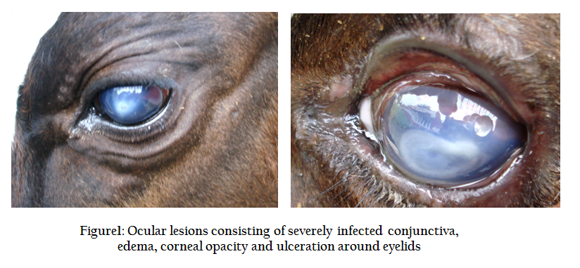
Figure 1: Ocular lesions consisting of severely infected conjunctiva, edema, corneal opacity and ulceration around eyelids
Characteristic hemolytic colonies were observed on blood agar but no colonies were developed on MacConkey agar plate. The pattern of hemolysis was very peculiar 1–2 mm diameter with corrosion of the agar at the edges of colony. Further, some of the colonies were found to be surface spreading (Figure 2). The organism was gram negative diplococci resembling tumbles and was non motile, catalase and oxidase positive. Gelatin agar was liquefied by the organism within in 24hrs of stab culture and was able to autoagglutinate normal saline in sugar tubes.
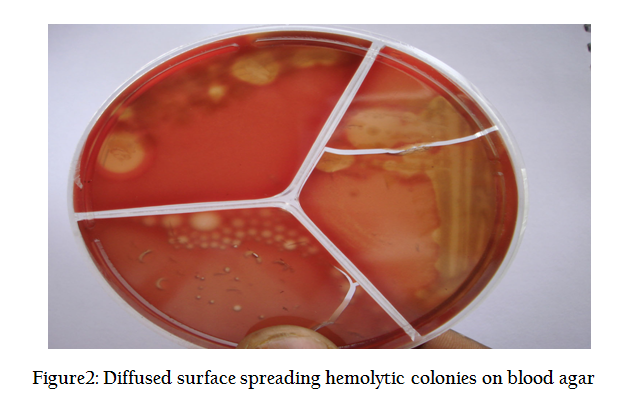
Bacteriological examination revealed the production of virulent factors such as hemolysin and proteolytic enzyme production which could have caused opacity or cloudiness of the affected eye (Pedersen et al., 1972; Frank Sandra et al., 1981). However, fimbriae also help in colonization of the organism in cornea along with capsule, the main virulence factor of Moraxella bovis (Hoien-Dalen et al., 1990). Spreading nature of the hemolysis may be due to the presence of fimbriae which is also responsible for the auto agglutination of normal saline.
The sensitivity of the isolate against various antibiotics was determined by Kirby– Bauer disc diffusion method. The antibacterial drug sensitive profile revealed that the organisms were more sensitive to Oxytetracycline (OTC) followed by the other antibiotics such as penicillin, gentamicin, neomycin, and kanamycin and found resistant to streptomycin.
Eye drop solution consisting of 1% Silver nitrate and 0.4% of zinc sulphate in sterile water was prepared and applied twice daily for 7–15 days to all the infected animals that exhibited corneal opacity. Besides, the animals were also administered with long acting oxytetracycline @20mg/kg body weight through deep intra muscular route for 3 times, each at 72 hours interval (George et al., 1984).
Initially when the treatment was begun, the animals were showing white spots of corneal opacity, ranged from 2 to 4 mm in diameter. Following treatment the corneal opacity appeared regressing with favorable improvement in lacrimation and vascularization .Animals that had pin point corneal opacity recovered in about 7 days of treatment, while those that had wider spot of corneal opacity took about 15 days to recover.
Treatment of IBK relies on the use of antibiotics and the prevention of further ocular irritation. All the animals were successfully treated with topical instillation of silver nitrate and zinc sulphate eye drops along with parenteral administration of OTC. Zinc sulphate is antiseptic, immunostimulant and astringent. It is reported that in catarrhal conditions of conjunctiva, application of zinc sulphate lotion had a proven recovery in later stage of acute infection (Hoare and Greig, 2004). It is also reported that zinc sulphate act as integral part of several enzymes important for wound healing and ophthalmic solution is used as mild astringent for relief of eye irritation (Tatro, 2003). Silver nitrate solution (1%) is brushed to the averted eyelids and conjunctiva in these cases. The ability of silver ions to kill microorganisms is the basis for their ophthalmic use. Though it is a irritant drug, its use in curtailing the contagious ophthalmic infection is highly reliable. Face flies are one of the most difficult pasture pests which prevail on cattle only for short periods of time and posing practical problems in containing the spread of disease by application of insecticides. Face flies use an abrasive sponging mouthpart to stimulate tear flow from the eyes and lap up the protein rich secretions from the eye as well as nasal discharges, saliva, or blood oozing from wounds.
Topical instillation of silver nitrate and zinc sulphate eye drops along with OTC parenterally were found to be more effective than any other combination (Allen et al., 1995). Effective treatment of IBK is very important, as in untreated cases the process becomes chronic, and the opacity takes 1–2 months to resolve. In other cases, depending on the severity of the disease, a white scar may be present even after full resolution of the disease. Occasionally, perforation of the corneal ulcer results in iris prolapse, in which case, blindness may result. However, preventing flies access to the affected eye will reduce spread of the disease within the herd.
Infectious bovine keratoconjunctivitis (IBK) is the first report in Uttar Pradesh State, India.
ACKNOWLEDGEMENT
The authors are highly thankful to Dr. Ammayyappan, Veterinary Assistant Surgeon, Animal Disease Intelligence Unit, Vellore, Tamil Nadu, India, for his contribution in this study.
REFERENCES
Allen LJ, George LW and Willits NH (1995). Effect of penicillin or penicillin and dexamethasone in cattle with infectious bovine keratoconjunctivitis. J. Am. Vet. Med. Assoc. 206: 1200–1203.
PMid:7768744
Bedford PGC (1992). Ocular diseases. Bovine medicine: diseases and husbandry, Wiley–Blackwell, Oxford. 27: 712–721.
Billson FM, Harbour C, Michalski WP, Tennent JM, Egerton JR and Hodgson JL (2000). Characterisation of haemolysin of Moraxella bovis using a hemolysin–neutralising monoclonal antibody. Infect and Immun. 3469–3474.
http://dx.doi.org/10.1128/IAI.68.6.3469-3474.2000
Collee JG, Grulo JG, Fraser AG and Marmion BP (1989). In: Mackie and McCartney. Practical medical microbiology. 13th edn, Churchill and living stone, New York. PP. 76.
Frank, Sandars K and Gerber JD (1981). Hydrolytic enzymes of Moraxella bovis. J. Clin. Microbiol. 13: 269–271.
George LW, Wilson WD, Baggot JD and Mihalyi JE (1984). Antibiotic treatment of Moraxella bovis infection in cattle. J. Am. Vet. Med. Assoc. 185: 1206–1209.
PMid:6392258
Hoare EW and Greig RJ (2004). Veterinary Materia Medica and Therapeutics, Asiatic Publishing House, Delhi. PP. 179,–92.
Hoien-Dalen PS, Rosenbusch RF and Roth JA (1990). Comparative characterization of the leukocidic and hemolytic activity of Moraxella bovis. Am. J. Vet. Res. 51: 191–196 .
PMid:2301828
Kopecky KE, Pugh GW and Mc Donald TJ (1986). Infectious bovine kerato conjunctivitis: contact transmission. Am. J. Vet. Res. 47: 622–624.
PMid:3963562
Pedersen KB, Froholm LO and Bovre K (1972). Fimbriation and colony type of Moraxella bovis in relation to conjunctival colonization and development of keratoconjunctivitis in cattle. Acta Pathol. Microbiol. Scand. Sect. B. 80: 911–918.
Ram K and Steve P (2011). Infectious Keratoconjunctivitis (Pink Eye in Cattle) Animal Health Spotlight Veterinary Medicine Extension, February. (if Page number is there, please put it)
Tatro S (2003). A– Z Drug Facts, Facts and comparisons, A Wolters Kluwer Company, Missouri, 4th Edn. PP: 1332.


