Advances in Animal and Veterinary Sciences
Research Article
Advances in Animal and Veterinary Sciences 1 (6): 197 – 201Preliminary Pathological Studies on Equine Skin Lesions with Specific Reference to Equine Sarcoid and Detection of Bovine papillomaviruses 1 and 2
Babu Lal Jangir1, Vipul Kumar Singh2, Guttula Saikumar3, Abhijit Motiram Pawde4, Chandra Pal Churamani5, Ramesh Somvanshi*6
- Division of Pathology, Indian Veterinary Research Institute, Izatnagar, Bareilly, 243 122 UP, India
- Central Military Veterinary Laboratory (CMVL), Meerut Cantt–250001, UP, India
- Division of Pathology, Indian Veterinary Research Institute, Izatnagar, Bareilly, 243 122 UP, India
- Division of Surgery, Indian Veterinary Research Institute, Izatnagar, Bareilly, 243 122 UP, India
- Central Military Veterinary Laboratory (CMVL), Meerut Cantt–250001, UP, India
- Division of Pathology, Indian Veterinary Research Institute, Izatnagar, Bareilly, 243 122 Uttar Pradesh, India
*Corresponding author:dr.rsomvanshi@gmail.com
ARTICLE CITATION:
Jangir BL, Singh VK, Saikumar G, Pawde AM, Churamani CP and Somvanshi R (2013). Preliminary pathological studies on equine skin lesions with specific reference to Equine Sarcoid and Detection of Bovine papillomaviruses 1 and 2. Adv. Anim. Vet. Sci. 1 (6): 197 – 201.
Received: 2013–09–09, Revised: 2013–10–15, Accepted: 2013–10–20
The electronic version of this article is the complete one and can be found online at
(
http://nexusacademicpublishers.com/table_contents_detail/4/125/html
)
which permits unrestricted use, distribution, and reproduction in any medium, provided the original work is properly cited
ABSTRACT
A preliminary pathological study on equine skin lesions with specific reference to equine sarcoid and association of Bovine Papillomaviruses (BPVs) was carried out. A total number of 16 skin biopsy specimens from equines (4 horses and 12 mules) were studied. These animals had proliferative skin lesions which were small, solitary or variable in size and located on eye lids and different parts of the body. Histopathologically, these biopsies were diagnosed as neoplastic (13) and non–neoplastic conditions (3). Amongst neoplastic, 10 cases were benign in nature including fibroid (6), occult sarcoid (2), fibromatosis (1) and fibropapilloma (1) while 3 cases were malignant diagnosed as squamous cell carcinoma of eyelids (2) and vagina (1). Remaining 3 non–neoplastic conditions were diagnosed as necrotizing dermatitis (2) and myositis (1). These skin biopsies were tested for presence of bovine papillomaviruses (BPVs) –1 and–2 DNA by Polymerase Chain Reaction; 5 were positive for BPV–1, 3 for BPV–2 and 6 for their mixed infections. Present study revealed that equine skin biopsies should be analyzed for better formulation of treatment strategy.
INTRODUCTION
The equine sarcoid is a locally aggressive, fibroblastic skin tumour that affects horses, mules, donkeys and zebra throughout the world (Chambers et al., 2003; Nel et al., 2006). Sarcoids can occur in horses at any age and there is no gender predisposition (McConaghy et al., 1994; Scott and Miller, 2003). It may occur in any part of the body either as solitary or multiple lesions ranging from small wart–like lesions to large ulcerated fibrous growths. The most common sites are face (muzzle, ears and periocular region), neck, ventral abdomen, distal limbs and at sites that have been previously traumatized (Carr, 2009; Finlay et al., 2009). These are locally invasive, non–metastatic, fibroblastic skin tumours with unpredictable behaviour and are considered to cause substantial economic loss to the equine industry. Sarcoids represent 12 to 67% of all equine tumours and 70% of all skin tumours in horses (Teifke et al., 1994; Lepage et al., 1998) while squamous cell carcinomas (SCCs) account for up to 37% of cutaneous neoplasms (Scott and Miller, 2003).
Although papillomaviruses (PVs) have been described as highly species–specific (Campo, 2006) but some PVs infect a variety of hosts. Cross–species infection occurs in horses and other equids by BPV–1 and –2. Nowadays, it is also known that BPV plays a major role in equine sarcoid pathogenesis (Chambers et al., 2003; Bogaert et al., 2005; Bogaert et al., 2008). BPV–1 and –2 DNA have been demonstrated in sarcoids all over the world, either one or both types in the same horse. In India, only a few case reports of sarcoid are available (Singh et al., 1986; Sudarsanam and Christopher, 1992; Pratap et al., 2000; Dabas et al., 2004). It is difficult to assess status of sarcoids in India due to lack of systematic studies. Keeping scanty information in the view; a preliminary pathological study on equine skin lesions with specific reference to equine sarcoid was carried out to characterize the suspected skin tumours and to assess presence of BPVs in such lesions.
MATERIALS AND METHODS
Samples
Equine tumour samples were collected from the Referral Veterinary Polyclinic, Indian Veterinary Research Institute, Izatnagar, Bareilly and Central Military Veterinary Laboratory, Merrut Cantt., Uttar Pradesh for diagnosis and analysis. Biopsies were collected after administration of local anaesthesia around the growth and samples were preserved in 10% buffered formalin for histopathological studies. A part of it, from few samples was also stored in sterile vials at –20ºC and samples from field cases preserved in 10% buffered formalin were used for molecular studies.
Pathological Studies
The detailed clinical gross observations with specific locations of tumorous growths were recorded. After proper fixation in the 10% neutral buffered formalin, tissues were cut into small sections with thickness of 2–3 mm and embedded in the paraffin by standard procedures. The paraffin embedded tissues were cut into 6 micron thick sections and stained with hematoxylin and eosin as per conventional procedures (Culling, 1995). Masson's trichrome staining was done on selected duplicate tissue sections to differentiate between collagen and smooth muscles as per standard procedures (Luna, 1968).
Molecular Studies
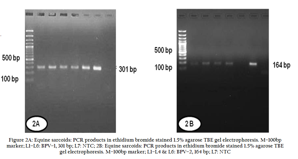
DNA extraction was performed from tissues using DNeasy Blood & Tissue kit (Qiagen, Hilden, Germany) following manufacturer’s protocol. BPVs were detected by PCR, targeting the L1 gene of BPV–1 and –2 with specific primers (Operon Biotechnologies, Genetix Biotech Asia Pvt. Ltd., Huntsville, AL, USA) and these were similar to the previous studies (Yaguiu et al., 2008; Pangty et al., 2010). Primers of BPV–1 (forward– 5'–gga gcg cct gct aac tat agg a–3'; reverse–5'–atc tgt tgt ttg ggt ggt gac–3') and BPV–2 (forward– 5'–gtt ata cca ccc aaa gaa gac cct–3'; reverse–5'–ctg gtt gca aca gct ctc ttt ctc–3') were expected to amplify the specific viral DNA template of 301 and 164 bp sizes, respectively. Amplified DNA fragments were run in 1.5% agarose gel containing ethidium bromide (0.5 g/ml) and visualized by transillumination under UV light (Geldoc, USA). Selective PCR products were directly sequenced commercially on ABI–PRISM dye terminator at DNA Sequencing Facility at Department of Biochemistry, Delhi University, South Campus, New Delhi using specific primers supplied from our laboratory. The generated sequences were blasted online on National Center for Biotechnology Information (NCBI) and analyzed by MegAlign (DNASTAR Inc.). The generated sequences were submitted to NCBI and EMBL data base.
RESULTS
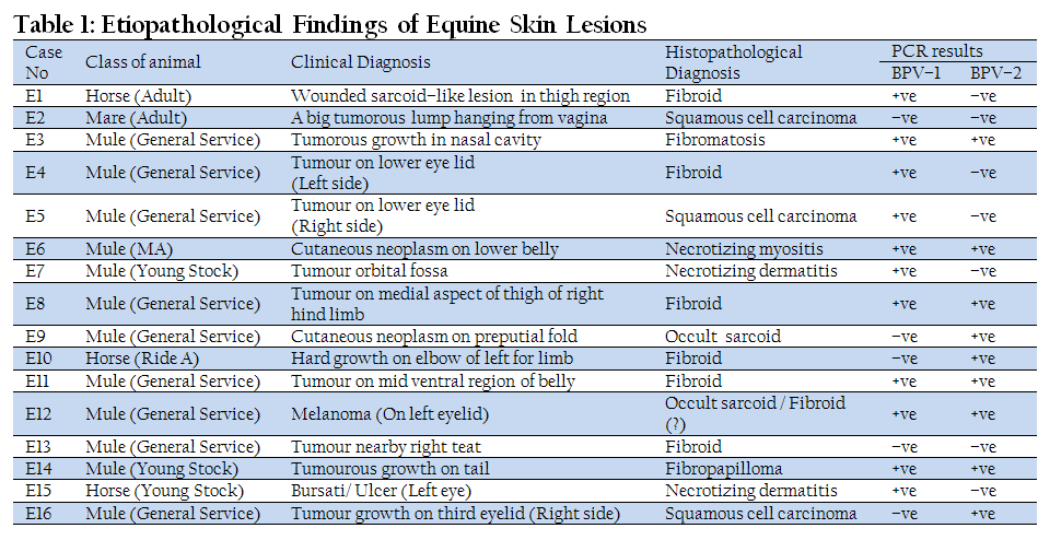
A total of 16 sarcoid–like cases were observed on different body parts of mules and horses. Grossly, the tumours were located on eyelids and various parts of the body with varied appearance (Figure 1A). The detailed observations are presented in Table 1. Histopathologically, these cases were categorized into three different types; neoplasms including benign and malignant tumours and non–neoplastic inflammatory conditions.
Neoplastic Conditions
Benign Tumours
Benign tumours were diagnosed histopathologically into different types including fibroid (6), occult sarcoid (2), fibromatosis (1) and fibropapilloma (1). The tumours diagnosed as fibroid revealed focal epidermal hyperplasia with a few rete pegs extending into the dermis and proliferation of fibrous connective tissue that was confluent with the epidermis. Proliferating fibroblasts were packed densely in spindle form and haphazardly arranged fascicles and interlacing bundles in whorls pattern with presence of small amount of collagen (Figure 1B). Occasionally, severe haemorrhages with engorgement of blood vessels, focal areas of necrosis with infiltration of neutrophils, mononuclear cells and marked eosinophils were also seen. Fibromatosis showed moderate proliferation of fibrous connective tissue with hyalinization of tissues and areas of infrequent engorgement of blood vessels with RBCs, haemorrhages and focal areas of lymphocytic infiltration. Duplicate sections stained with Masson’s Trichrome revealed proliferating fibrous connective tissues with abundant collagenous fibres that stained blue, while the nuclei stained black and other fibres, muscles stained red in variable shades, which was very less as compared to collagenous fibres (Figure 1C). Occult sarcoid showed mild thickening of epidermis with elongated basal cells. Lamina propria showed diffuse infiltration of mononuclear cells, mainly lymphocytes, as well as perivascular mononuclear cell infiltration (Figure 1D). Sebaceous glands were degenerated. Immature fibrous connective tissue was arranged in whirling pattern. In one case, areas of ulceration were seen while the non–ulcerated areas showed epidermal hyperplasia with prominent rete pegs extending into the dermal proliferation of fibroblasts. Melanosis was also evident with presence of scanty melanin pigment. Fibropapilloma revealed moderate degree of cornification with parakeratosis along with basket–weave appearance of stratum corneum. Hyperplasia of stratum spinosum with presence of koilocytes and islands of proliferating dermal connective tissue surrounded by hyperplastic epidermal cell layers with well developed rete pegs was seen. Basal cell layer was hyperplastic with hyperchromatic nuclei.
Malignant Tumours
Three tumor cases involving a growth in vagina and two on eyelids were histopathologically diagnosed as squamous cell carcinoma. The neoplastic squamous epithelial cells invaded deep down the dermis in the form of thick cellular cords, irregular sheets and independent cellular islands. In between the cellular islands, proliferation of fibrous connective tissue was seen. Aggressively proliferating neoplastic cells in large irregular sheets frequently exhibited nuclear indentations, mitotic figures and increased nuclear–cytoplasmic ratio (Figure 1 E&F). Necrotic areas with infiltration of inflammatory cells including polymorphonuclear and mononuclear cell and engorged blood vessels were also seen elsewhere.
Non–Neoplastic Conditions
Of three non–neoplastic conditions, one was diagnosed as necrotizing myositis and two were necrotizing dermatitis (Table 1). Microscopically the former was characterized by severe necrosis of muscle and connective tissue with infiltration of neutrophils and lymphocytes and presence of few isolated intact myocytes amidst necrotic debris. Necrotizing dermatitis was characterized by severe necrosis with infiltration inflammatory cell comprising of lymphocytes, neutrophils and eosinophils in addition to engorged blood vessels
PCR Results
All 16 DNA samples from sarcoid–like lesions from mules and horses were subjected to PCR. Results are presented in Table–1. Out of these, 5 samples were positive for BPV–1, 3 for BPV–2 and 6 showed mixed infection of both BPV–1 and –2. One each case of squamous cell carcinoma and fibroid were found negative for both BPV–1 and –2. The case of necrotizing myositis was positive for both BPVs while cases of necrotizing dermatitis were positive for BPV–1 only. Overall, 68.75% (11/16) and 56.25% (9/16) positivity was seen for BPV–1 and –2, respectively. Gel electrophoresis showed the specific PCR product size of 301 and 164 bp for BPV–1 and –2, respectively (Figure 2 A&B). Selective PCR products were sequenced and the generated sequences of BPV–1 and –2 were submitted to NCBI and EMBL database, respectively and accession numbers were assigned (KF114852, KF114854, KF114855 and KF148688). Partial BPV–1 sequences generated in the present study exhibited close homology among themselves. Similarly, generated BPV–2 sequences were also identical. On comparison, BPV–1 & –2 partial sequences generated in present study revealed close homology as well as with the earlier published BPV–1 & –2 sequences on NCBI.
DISCUSSION
Equine sarcoids and other tumors like squamous cell carcinomas seriously compromise the welfare of affected horses, donkeys and other equids and attributed to considerable financial losses to the owners. There are several different clinical manifestations of equine sarcoids, which have variable macroscopic appearances, growth patterns and behavior ranging from slow growing occult lesions or even, less frequently, spontaneous regressing lesions to locally aggressive fast growing and ulcerative fibroblastic lesions (McConaghy et al., 1994; Scott and Miller, 2003; Knottenbelt, 2005). In the present study, 16 skin lesions/growths of equines (Table 1) were studied. Macroscopically, these lesions/growths were small, solitary, variable in size and located on eye lids and different parts of the body. These findings are in accordance to earlier reports (Knottenbelt, 2005; Wobeser et al., 2010; Kainzbauer et al., 2012). Histopathologically, these cases were diagnosed as neoplastic (81.25 %) and non–neoplastic (18.75%) conditions. Among neoplastic conditions, benign tumours (62.50%) included fibroid (37.5%), occult sarcoid (12.5%), fibromatosis (6.25%) and fibropapilloma (6.25%) whereas, malignant tumours (18.75%) were all diagnosed as squamous cell carcinomas. Benign tumours in equines occurring in various body locations have been reported from different parts of the world (Martens et al., 2000; Chambers et al., 2003; Hallamaa et al., 2005). Similarly, occurrence of squamous cell carcinoma in the head and neck region, particularly in the periorbital, parotideal areas and in the jugular groove of a Connemara mare (Kainzbauer et al., 2012) has been described. In present study, remaining 18.75% were non–neoplastic conditions and diagnosed as necrotizing dermatitis. Cases of dermatitis associated with BPV infection have been described in horses (Yuan et al., 2007; Wobeser et al., 2012).
The skin biopsies tested for presence of BPV–1 and –2 DNA by PCR revealed that 31.25% were positive for BPV–1, 18.75% for BPV–2 and 37.5% for both types. One fibroid and one squamous cell carcinoma were found negative for BPVs while both cases of necrotizing dermatitis were positive for BPV–1. Earlier Campo (2006) reported that the bovine warts are a rich reservoir of BPV–1 virions and an equally high percentage of sarcoid affected horses show latent BPV infection in the normal skin. The relatively high occurrence of BPV infection in apparently healthy horses living in close contact with papilloma–affected cattle is indicative for a BPV cross–species transmission from bovines to equines. In horses living in direct contact with sarcoid affected horses, a 50% rate of latent infection of BPV has been observed (Bogaert et al., 2008). Our findings were more or less in corroboration with that of Martens et al., (2001) who also detected BPV DNA in 88% of swabs and 91% of scrapings from sarcoid–affected lesions and no BPV DNA could be detected from non–sarcoid lesions. BPV–1 and –2 DNA have been detected in sarcoids all over the world, either alone or as mixed infection of both types in the same horse (Teifke et al., 1994; Nasir and Reid, 1999; Bogaert et al., 2007). Our findings are in agreement with previous observations. In Europe, BPV–1 accounts for a vast majority of DNA detected with only a small number of sarcoids containing BPV–2 (Otten et al., 1993; Teifke et al., 1994). Studies in the United States have shown BPV–1 and BPV–2 in roughly equal proportions in the eastern United States (Teifke et al., 1994) while BPV–2 predominates in the western United States representing 63% of the amplified DNA (Carr et al., 2001). Equine sarcoids are most commonly associated with BPV–2 in western Canada (Carr et al., 2001; Wobeser et al., 2010). In horses with multiple tumours or same sarcoid sample, both types of BPV were demonstrated in the same animal (Martens et al., 2001<>; Bogaert et al., 2010; Wobeser et al., 2010). Despite the consistent detection of papillomavirus DNA in the sarcoid lesions, papillomavirus particles have not been demonstrated till date. BPV–1 and –2 infection is not uncommon in military dairy farms and cattle in the rural areas around Bareilly, Uttar Pradesh (Pangty et al., 2010; Kumar et al., 2013). Since horses and cattle are housed in the same premises and at times share pastures, the potential scope for cross–species transmission is always there. Systemic studies on equine skin lesions, particularly sarcoids were not conducted earlier in India and our findings suggest that skin biopsies should be analyzed for better formulation of treatment strategy in valuable equids.
CONCLUSION
Sarcoids are common lesion of the skin of equines and are associated with the BPVs. In India only limited field case studies and reports on this aspect is available. BPV–1 & –2 and their mixed infections were detected in histopathological confirmed cases of sarcoids/ squamous cell carcinomas. Further systemic research is needed to determine the importance of BPVs and/or other equine papillomaviruses as a cause of sarcoids and different clinical conditions.
ACKNOWLEDGEMENTS
We are thankful to Head, Division of Pathology and Director, IVRI, Izatnagar, UP, India for all necessary facilities for carrying out this research work.
AUTHORS DECLARATION OF INTERESTS
No conflicts of interest have been declared.
REFERENCES
Bogaert L, Martens A, De Baere and Gasthuys F (2005). Detection of bovine papillomavirus DNA on the normal skin and in the habitual surroundings of horses with and without equine sarcoids. Res. Vet. Sci. 79: 253–258.
http://dx.doi.org/10.1016/j.rvsc.2004.12.003
PMid:16054896
Bogaert L, Martens A, Depoorter P and Gasthuys F (2008). Equine sarcoids–association with bovine papillomavirus. Vlaams. Diergen. Tijdsch. 78: 131– 137.
Bogaert L, Martens A, Kast WM, Van Marck E and De Cock H (2010). Bovine papillomavirus DNA can be detected in keratinocytes from equine sarcoids. Vet. Microbiol. 146: 269–275.
http://dx.doi.org/10.1016/j.vetmic.2010.05.032
PMid:21095508
Bogaert L, Van Pouke M, De Baere C, Dewulf J, Peelman L, Ducatelle R, Gasthuys F and Martens A (2007). Bovine papillomavirus load and mRNA expression, cell proliferation and p53 expression in four clinical types of equine sarcoid. J. Gen. Virol. 88: 2155–2161.
http://dx.doi.org/10.1099/vir.0.82876-0
PMid:17622617
Campo MS (2006). Bovine papillomavirus: old system, new lessons? In: Campo MS, editors. Papillomavirus research: from natural history to vaccine and beyond, Caister Academic Press, Wymondham p. 373–383.
Carr EA (2009). New developments in diagnosis and treatment of equine sarcoids. 6th ed. St. Louis: Saunders, Elsevier.
Carr EA, Theon AP, Madewell BR, Griffey SM, Hitchcock ME (2001). Bovine papillomavirus DNA in neoplastic and non–neoplastic tissues obtained from horses with and without sarcoids in the western United States. Am. J. Vet. Res. 62:741–744.
http://dx.doi.org/10.2460/ajvr.2001.62.741
PMid:11341396
Chambers G, Ellsmore VA, O'Brien PM, Reid SWJ, Love S, Campo MS and Nasir, L (2003). Association of bovine papillomavirus with the equine sarcoid. J. Gen. Virol. 8:1055–1062.
http://dx.doi.org/10.1099/vir.0.18947-0
Culling CFA (1995). Hand book of histological techniques. 2nd ed. London: UK.
Dabas VS, Mistry JN, Chaudhary S and Rao, GS (2004). Intra–maxillary sarcoid and its surgical extirpation in a filly. Indian Vet. J. 81: 565–566.
Finlay M, Yuan ZQ, Burden F, Trawford A, Morgan IM, Campo MS and Nasir, L (2009). The detection of bovine papillomavirus type 1 DNA in flies. Virus Res. 144: 315–317.
http://dx.doi.org/10.1016/j.virusres.2009.04.015
PMid:19409942
Hallamaa RE, Saario E and Tallberg T (2005). Macroscopical and histopathological changes in regressing primary and recurrent equine sarcoids during active specific bio–immunotherapy. In vivo 19:761–768.
PMid:15999546
Kainzbauer C, Rushton J, Tober R, Scase T, Nell B, Sykora S and Brandt S (2012). Bovine papillomavirus type 1 and Equus caballus papillomavirus 2 in equine squamous cell carcinoma of the head and neck in a Connemara mare. Equine Vet. J. 44:112–115.
http://dx.doi.org/10.1111/j.2042-3306.2010.00358.x
PMid:21668491
Knottenbelt DC (2005). A suggested clinical classification for the equine sarcoid. Clin. Tech. Equine Prac. 4: 278–95.
http://dx.doi.org/10.1053/j.ctep.2005.10.008
Kumar P, Nagarajan N, Kumar D, Bind RB and Somvanshi R (2013). Detection and quantification of bovine papillomaviruses (BPVs) in cutaneous warts of cattle and buffaloes. Adv. Amin. Vet. Sci. 1 (2): 53–58.
Lepage MF, Carstanjen B and Von Tscharner C (1998). Equine sarcoid (part I): Causes, diagnosis, differential diagnosis. Prakt Tierarz 79: 627–36.
Luna LG. (1968). Manual of histological staining methods of the Armed Force Institute of Pathology. 3rd ed. Mc Graw– Hill Book Co.: London; p. 124–125.
Martens A, De Moor A and Ducatelle R (2001). PCR detection of bovine papilloma virus DNA in superficial swabs and scrapings from equine sarcoids. Vet. J. 161: 280–286.
http://dx.doi.org/10.1053/tvjl.2000.0524
PMid:11352485
Martens A, De Moor A, Demeulemeester J and Ducatelle R (2000). Histopathological characteristics of five clinical types of equine sarcoid. Res. Vet. Sci. 69:295–300.
http://dx.doi.org/10.1053/rvsc.2000.0432
PMid:11124103
McConaghy FF, Davis RE, Reppas GP, Rawlinson RJ, McClintock SA, Hutchins DR and Hodgson DR (1994). Management of equine sarcoids. New Zealand Vet. J. 42:180–184.
http://dx.doi.org/10.1080/00480169.1994.35816
PMid:16031776
Nasir L and Reid SWJ (1999). Bovine papillomaviral gene expression in equine sarcoid tumours. Virus Res. 61:171–175.
http://dx.doi.org/10.1016/S0168-1702(99)00022-2
Nel PJ, Bertschingerb H, Williamsc J and Thompsonb PN (2006). Descriptive study of an outbreak of equine sarcoid in a population of Cape mountain zebra (Equus zebra zebra) in the Gariep Nature Reserve. S. Afr. Vet. Ver. 77(4): 184–190.
Otten N, von Tscharner C, Lazary S, Antczak DF and Gerber H (1993). DNA of bovine papillomavirus type 1 and 2 in equine sarcoids: PCR detection and direct sequencing. Arch. Virol. 132:121–131.
http://dx.doi.org/10.1007/BF01309847
PMid:8394687
Pangty K, Singh S, Saikumar G, Goswami R and Somvanshi R (2010). Detection of BPV–1 and –2 and quantification of BPV–1 by real–time PCR in cutaneous warts in cattle and buffaloes. Transbound. Emer. Dis. 57(3): 185–196.
http://dx.doi.org/10.1111/j.1865-1682.2009.01096.x
PMid:20113447
Pratap K, Hoque M, Amarpal S, Pawde AM and Charan K (2000). Phalangeal sarcoid in a mare. Indian Vet. J. 77: 55–56.
Scott DW and Miller WH (2003). Equine Dermatology: sarcoid. Philadelphia: Saunders, Elsevier Science.
http://dx.doi.org/10.1016/B0-72-162571-1/50006-4
http://dx.doi.org/10.1016/B0-72-162571-1/50016-7
http://dx.doi.org/10.1016/B0-72-162571-1/50015-5
http://dx.doi.org/10.1016/B0-72-162571-1/50004-0
http://dx.doi.org/10.1016/B0-72-162571-1/50014-3
http://dx.doi.org/10.1016/B0-72-162571-1/50010-6
http://dx.doi.org/10.1016/B0-72-162571-1/50011-8
http://dx.doi.org/10.1016/B0-72-162571-1/50002-7
http://dx.doi.org/10.1016/B0-72-162571-1/50013-1
http://dx.doi.org/10.1016/B0-72-162571-1/50008-8
http://dx.doi.org/10.1016/B0-72-162571-1/50012-X
http://dx.doi.org/10.1016/B0-72-162571-1/50007-6
http://dx.doi.org/10.1016/B0-72-162571-1/50009-X
http://dx.doi.org/10.1016/B0-72-162571-1/50003-9
http://dx.doi.org/10.1016/B0-72-162571-1/50005-2
Singh CDN, Prasad LN, Tiwari SN, Jha SN and Thakur HN (1986). Some observation on animal tumours. Indian Vet. J. 63: 419.
Sudarsanam G and Christopher KJ (1992). Sarcoid in a horse. Indian Vet. J. 69: 349–50.
Teifke JP, Hardt M and Weiss E (1994). Detection of bovine papillomavirus DNA in formalin–fixed and paraffin–embedded equine sarcoids by polymerase chain reaction and non–radioactive in situ hybridization. European J. Vet. Pathol. 1: 5–10.
Wobeser BK, Davies JL, Hill JE, Jackson ML, Kidney BA, Mayer MN, Townsend, HGG, Andrew L and Allen AL (2010). Epidemiology of equine sarcoids in horses in western Canada. Canadian Vet. J. 51: 1103–1108.
PMid:21197201 PMCid:PMC2942047
Wobeser BK, Hill JE, Jackson ML, Kidney BA, Mayer MN, Townsend HGG and Allen AL (2012). Localization of Bovine papillomavirus in equine sarcoids and inflammatory skin conditions of horses using laser microdissection and two forms of DNA amplification. J Vet. Diag. Inves. DOI: 10.1177/1040638711425952.
http://dx.doi.org/10.1177/1040638711425952
Yaguiu A, Dagli MLZ, Birgel EH, Alves Reis BCA, Ferraz OP, Pituco EM, Freitas AC, Becak, W and Stocco RC (2008). Simultaneous presence of bovine papillomavirus and bovine leukemia virus in different bovine tissues: In situ hybridization and cytogenetic analysis. Gen. Mol. Res. 7 (2): 487–497.
http://dx.doi.org/10.4238/vol7-2gmr436
PMid:18561382
Yuan Z, Philbey AW, Gault EA, Campo MS and Nasir L (2007). Detection of bovine papillomavirus type 1 genomes and viral gene expression in equine inflammatory skin conditions. Virus Res. 124(1–2):245–249.
http://dx.doi.org/10.1016/j.virusres.2006.10.012
PMid:17140693


