Advances in Animal and Veterinary Sciences
Short Communication
Advances in Animal and Veterinary Sciences. 1 (6): 191 – 193Detection of Infectious Canine Hepatitis Virus in Vaccines by PCR
Vishal Chander*, Sukdeb Nandi, RishendraVerma
-
Centre for Animal Disease Research and Diagnosis (CADRAD), Izatnagar, U.P. India 243122
*Corresponding author:drvishal1@gmail.com
ARTICLE CITATION:
Chander V, Nandi S, Verma R (2013). Detection of infectious canine hepatitis virus in vaccines by PCR. Adv. Anim. Vet. Sci. . 1 (6): 191 – 193.
Received: 2013–07–16, Revised: 2013–08–16, Accepted: 2013–08–18
The electronic version of this article is the complete one and can be found online at
(
http://nexusacademicpublishers.com/table_contents_detail/4/119/html
)
which permits unrestricted use, distribution, and reproduction in any medium, provided the original work is properly cited
ABSTRACT
Canine adenovirus 1 (CAV– 1) and CAV– 2 cause infectious canine hepatitis (ICH) and infectious canine laryngo– tracheitis (ICLT) in dogs, respectively. The disease is characterized by fever, anorexia, increased thirst and abdominal pain with swollen liver and, in case of CAV–2, respiratory symptoms are also seen. The corneal opacity (blue eye) and interstitial nephritis may occur 1– 3 weeks after the clinical recovery as a consequence of the deposition of circulating immune complexes after CAV– 1 infections in dogs. CAV– 1 is genetically and antigenically distinct from canine adenovirus 2 (CAV– 2). Both viruses are shed in faeces and urine of the infected or recovered dogs, thus urine and faeces are important sources of infection to healthy dogs. A galaxy of immunoprophylactic agents based on CAV– 2 is available in the market for use in dogs to control infectious canine hepatitis in dogs. In spite of vaccination against CAV infections in dogs, the outbreak has been reported occasionally in dogs in India. In this study, PCR has been employed to amplify the genomic DNA of CAV in the vaccines and it has been found that CAV– 2 strains are present in 4 of 7 vaccines tested.
Infectious canine hepatitis (ICH) is a systemic disease of Canidae and Ursidae caused by canine adenovirus 1 (CAV– 1). The disease is characterized by fever, apathy, anorexia, increased thirst, abdominal pain with swollen liver, diarrhea and frequently dyspnoea (Appel, 1987). In addition, ocular and nasal discharges are also prominent (Appel and Carmichael, 1979). CAV– 1 is genetically and antigenically distinct from canine adenovirus 2 (CAV– 2) and is mainly associated with respiratory symptoms such as paroxysmal cough with variable expectoration and nasovascular discharge (Benetka et al., 2006; Schaer, 2010). CAV– 1 replicates in vascular endothelial cells and hepatocytes and produces an acute necrohemorrhagic hepatitis with a more severe clinical course in young than adult dogs (Greene, 1990) whereas CAV– 2 replicates in respiratory epithelium (Decaro et al., 2007). Corneal opacity (blue eye) and interstitial nephritis may occur 1–3 week after clinical recovery due to deposition of circulatory immune complexes in CAV– 1 infection (Wright, 1976). Both viruses are shed in faeces and urine of the infected or recovered dogs, thus urine and faeces are important sources of infection to healthy dogs (Macartney et al., 1988; Tham et al., 1998). Current methods of detection of CAV infections are based on hematological findings (lymphopenia and neutropenia)(Mosallanejad et al., 2010; Schaer, 2010), virus isolation (Decaro et al., 2007; MaCartney et al., 1988; Schaer, 2010), serological assays (Ditchfield et al., 1962; Jacobs et al., 2007; Pratelli et al., 2001), histopathology (Boomkens et al., 2004; Chvala et al., 2007; Chouinard et al., 1998; Mosallanejad et al., 2010; Park et al., 2007; Schaer, 2010; Yoon et al., 2010), immunohistochemistry (Chvala et al., 2007; Chouinard et al., 1998; Rodriguez–Tovar et al., 2007; Schaer, 2010; Yoon et al., 2010) and indirect haemagglutination assay (Ditchfield et al., 1962). The serological tests are usually laborious and take 2– 3 days to be performed and show higher titre after infection with virulent virus in contrast with modified live virus vaccines (Chouinard et al., 1998). Again, CAV– 1 and CAV– 2 can be difficult to differentiate in the laboratory by serological tests (Hu et al., 2001; Parthiban et al., 2009). With the advent of polymerase chain reaction (PCR), not only the detection but also differential identification of CAV–1 and CAV– 2 infections has become possible (Hu et al., 2001).
Since the use of modified live CAV– 1 vaccines has been found to cause adverse reactions, either a killed or an attenuated CAV– 2 vaccine has been used to protect dogs from CAV– 1 infection and found to be effective and more safe (Yin and Liu, 1985). Widespread vaccination has reduced very effectively the circulation of CAV– 1 in the canine population (Appel, 1987). However, recent report of some outbreaks of ICH in dogs in India has raised the doubt about the efficacy of vaccine in protecting against ICH. In this study, a rapid PCR based detection based on E3 region of genome of the virus responsible for variability among diverse adenoviruses has been reported. This also aids in differentiation of CAV– 1 and CAV– 2 depending on the degree of virulence and host specificity of the virus. The assay can be used to test the vaccines available in the market for the presence of CAV strain in order to find out the cause of vaccine failure and disease outbreak.
2 μL each of ICH vaccines namely Vanguard– 5 (Pfizer, USA ) containing canine distemper, canine adenovirus 2, canine parvovirus, canine parainfluenza; Megavac– 6 (1) (Indian Immunologicals, India) containing live attenuated canine distemper, canine adenovirus 2, canine parvovirus; Megavac– 6 (2)(Indian Immunologicals, India) containing inactivated Leptospira canicola and L. icterohaemorrhaegiae; Duramune PC (Ford Dodge USA ) containing canine corona virus and canine parvovirus; Canigen DHPPi (Virbac Carros, France) containing canine distemper, canine adenovirus 2, canine parvovirus, canine parainfluenza; Biocan– DHPPi (Bioveta, Czech Republic) containing canine distemper virus, canine adenovirus 2, infectious laryngotracheitis virus, canine parvovirus and parainfluenza; Biocan– L (Bioveta, Czech Republic) containing L. canicola, L. icterohaemorrhaegiae and L. grippotyphosa were used directly in the PCR. As a negative control cell culture medium was used in the reaction The cell culture supernatant of the CAV 1 and CAV 2 adapted in MDCK cell line were respectively used as a positive controls.
In the present study, it has been observed that out of 7 commercially available vaccines tested, 4 namely, Megavac– 6 (1), Canigen DHPPi, Biocan DHPPi and Vangurad– 5 contain the CAV– 2 as vaccine strain to provide protection against both the CAV– 1 and CAV– 2 which is in accordance with the results of Decaro et al. (2007). On the other hand, Megavac– 6 (2), Duramune PC and Biocan L did not contain any CAV as there was no amplification of DNA in PCR.
CAV infections occur occasionally in dogs in India (Parthiban et al., 2009). CAV– 1 and CAV– 2 infections in dogs in most cases are differentiated clinically from each other, but they have the same morphological features under the EM and same CPE on cell cultures. CAV– 2 can also infect the intestinal tract, one of the major target organs for CAV– 1 (Hamelin et al., 1986; Macartney et al., 1988).
The diagnosis of CAV infections is usually based on serological tests (Ditchfield et al., 1962; Jacobs et al., 2007; Pratelli et al., 2001) and virus isolation (Decaro et al., 2007; MaCartney et al., 1988; Schaer, 2010).
PCR detection of CAV– 1 in livers of infected dogs has also been reported (Kiss et al., 1996; Chouinard et al., 1998). However a number of diseases CDV, CPV, CCoV having similar type of symptoms often make it difficult to detect the specific pathogen accurately. In India, a large proportion of a total of about 25 million dogs mostly stray dogs are not vaccinated against the disease and harbour the pathogen to be transmitted to other susceptible dogs. The CAV– 1 is still circulating in the dog populations and occasionally it is responsible for a severe often fatal disease especially in animal shelters and breeding kennels when virus spread is ensured by close contact between the animals. Further, vaccinations are often carried out on pups with high titres of maternally derived antibodies that prevent an active immune response. This PCR based assay was applied to differentiate and cross validate whether vaccine strain is CAV– 1 or CAV– 2 as mentioned on the vaccine labels and to validate this test for future field investigations as well. From the study, it is clear that out of 7 vaccines tested 4 namely Megavac– 6 (1), Canigen DHPPi, Biocan DHPPi, Vanguard –5 contain CAV– 2 whereas Megavac– 6 (2) and Duramune does not contain any CAV– 2 as revealed in the products (Fig. 1). So, it is recommended that owners should check the product/vaccine for the presence of CAV– 2 before being used it in dogs against CAV infections. The PCR was performed in 200 μL thin layered PCR tubes (Axygen, USA) with a reaction volume of 50 μL. The forward (5’ CGCGCTGAACATTACTACCTTGTC 3’) and reverse (5’ CCTAGAGCACTTCGTGTCCGCTT 3’) primer set (Imperial Biomedics, India) (Kiss et al., 1996; Hu et al., 2001) specific for E3 gene of CAV– 1 and CAV– 2 were used in this study. The reaction mixture contained 200 μM of dNTPs, 10 pmol of each primer, 1X PCR reaction mixture containing 15 mM MgCl2 and 2 μL of each of the 7 vaccines as source of template DNA. Amplification was performed in a thermocycler (Applied Biosystems, USA). 1 μL of DNA polymerase was added to above reaction mixture after initial denaturation at 94 oC for 5 minutes in the thermocycler. The cyclic condition was denaturation at 94 0 C for 30 sec, primer annealing at 58oC for 1 min and extension at 72oC for 1 min. The cyclic condition was repeated for 30 times and a final extension at 72oC was given for 10 minutes. After PCR, the amplified products were analyzed on 1 % agarose gel containing ethidium bromide to a final concentration of 0.5 μg/ml. 10 μL of amplified product was mixed with 2 μL of bromophenol (6X) dye and loaded into the well and run along with 100 bp to 1 Kb DNA ladder in 1X TAE electrophoresis buffer at 5 volts/cm2. At the end of the electrophoresis, the gel was visualized under the UV transilluminator and results were recorded.
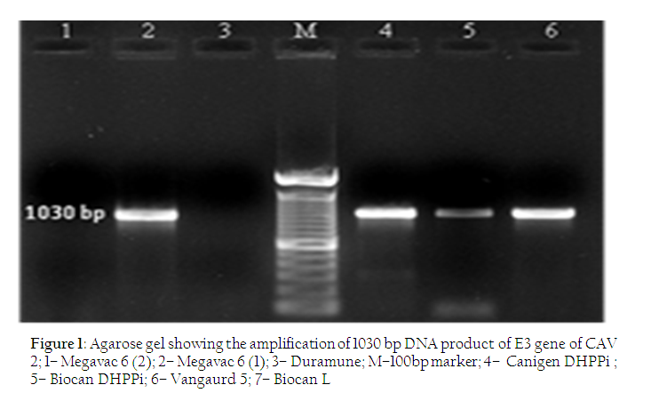
The cell culture adapted positive controls revealed 508 bp and 1030 bp DNA products in case of CAV 1 and CAV 2 respectively. (Figure 1 and 2) The sensitive PCR assay reported here revealed the amplified product of 1030 bp from 4 vaccines and the findings are in accordance with results of Chaturvedi et al., (2008) (Figure 2). There was no amplified product in the agarose gel in the negative control. This PCR assay is based on E3 gene present in the genome of mammalian adenovirus (Mastadenovirus and some Siadenovirus). E3 gene is present in all genera as compared to E1 and E4 regions present in some and causes variability among diverse adenoviruses and show evidence of different virulence degree and host specificity (Harrach and Benko, 2007). E3 region is responsible for weakening host defense mechanisms and therefore severe damage or death can also occur if normal inflammatory response is not suppressed by E3. Also E3 gene is not essential for growth in cells in culture (Flint et al, 2004).
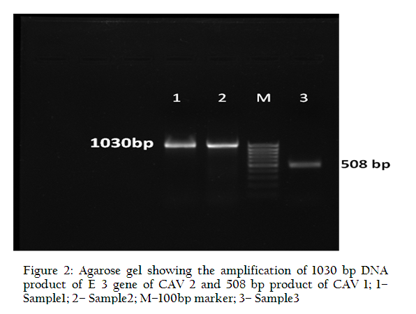
Figure 2: Agarose gel showing the amplification of 1030 bp DNA product of E 3 gene of CAV 2 and 508 bp product of CAV 1
The polymerase chain reaction has been applied for the diagnosis of canine adenoviral infections in clinical samples and shown to be rapid, sensitive and specific diagnostic method (Boomkens et al., 2004, Chaturvedi et al., 2008; Decaro et al., 2007; Hu et al., 2001; Park et al., 2007; Parthiban et al., 2009; Schaer, 2010; Yoon et al., 2010). But no report has been available to detect the presence of CAV strain present in the vaccines available in the market for use in dogs. Since the use of modified live CAV– 1 vaccines has been found to cause adverse reactions, CAV– 2 vaccines are usually used as an alternative in the prevention of ICH that are still effective but more safe (Decaro et al., 2007). In pups, maternally derived antibodies (MDA) represent the main protection against CAV infections but at the same time they may interfere with CAV vaccination (Appel, 1987). Further, a large pool of stray dogs in India is not covered under the vaccination umbrella and urine or faeces of infected or recovered dogs or dogs with sub– clinical form of infection form an important source of CAV– 1 and CAV– 2 (Macartney et al., 1988; Tham et al., 1998). With the reporting of CAV– 1 infections in dogs, it is of obvious suspicion whether the vaccine strain used in the vaccine is effective in protecting against ICH in dogs. Another reason of vaccine failure or ineffectiveness of the vaccination is that as the CAV– 2 is present in the vaccine along with other virus and bacteria and there may be competitive interference /suppression of one antigen by other potent antigen to be processed by the immune system in eliciting the protective immune response. Further, it is to be noted that field veterinarians do not follow either the uniform strategy in vaccinating the pups at the particular age or prior estimating the MDA.
In the present study, it has been observed that out of 7 commercially available vaccines tested, 4 namely, Megavac– 6 (1), Canigen DHPPi, Biocan DHPPi and Vangurad– 5 contain the CAV– 2 as vaccine strain to provide protection against both the CAV– 1 and CAV– 2 which is in accordance with the results of Decaro et al. (2007). On the other hand, Megavac– 6 (2), Duramune PC and Biocan L did not contain any CAV as there was no amplification of DNA in PCR.
CAV infections occur occasionally in dogs in India (Parthiban et al., 2009). CAV– 1 and CAV– 2 infections in dogs in most cases are differentiated clinically from each other, but they have the same morphological features under the EM and same CPE on cell cultures. CAV– 2 can also infect the intestinal tract, one of the major target organs for CAV– 1 (Hamelin et al., 1986; Macartney et al., 1988).
The diagnosis of CAV infections is usually based on serological tests (Ditchfield et al., 1962; Jacobs et al., 2007; Pratelli et al., 2001) and virus isolation (Decaro et al., 2007; MaCartney et al., 1988; Schaer, 2010).
PCR detection of CAV– 1 in livers of infected dogs has also been reported (Kiss et al., 1996; Chouinard et al., 1998). However a number of diseases CDV, CPV, CCoV having similar type of symptoms often make it difficult to detect the specific pathogen accurately. In India, a large proportion of a total of about 25 million dogs mostly stray dogs are not vaccinated against the disease and harbour the pathogen to be transmitted to other susceptible dogs. The CAV– 1 is still circulating in the dog populations and occasionally it is responsible for a severe often fatal disease especially in animal shelters and breeding kennels when virus spread is ensured by close contact between the animals. Further, vaccinations are often carried out on pups with high titres of maternally derived antibodies that prevent an active immune response. This PCR based assay was applied to differentiate and cross validate whether vaccine strain is CAV– 1 or CAV– 2 as mentioned on the vaccine labels and to validate this test for future field investigations as well. From the study, it is clear that out of 7 vaccines tested 4 namely Megavac– 6 (1), Canigen DHPPi, Biocan DHPPi, Vanguard –5 contain CAV– 2 whereas Megavac– 6 (2) and Duramune does not contain any CAV– 2 as revealed in the products (Figure 1). So, it is recommended that owners should check the product/vaccine for the presence of CAV– 2 before being used it in dogs against CAV infections.
ACKNOWLEDGEMENT
Authors are grateful to the Director, Indian Veterinary Research Institute, Izatnagar, UP for providing necessary facilities to carry out this work.
CONFLICT OF INTEREST
The authors have no conflict of interest including any financial, personal or other relationships with other people or organizations.
REFERENCES
Appel MJ (1987). Canine Adenovirus type 1 (Infectious canine hepatitis virus) . In :Appel, M.J. (Ed.), Virus infections of carnivores. Elsevier Science Publishers, Amsterdam, pp.29– 43.
PMid:2887435
Appel MJ and Carmichael LE (1979). In: Catcott, E.J. (Ed.), Canine Medicine, Vol. 1. Am. Vet. Pub. Inc., CA, p.25.
Benetka V, Weissenbock H, Kudielka I and Pallan C (2006). Canine adenovirus type 2 infection in four puppies with neurological signs. Vet. Record 158 (3): 91– 94.
http://dx.doi.org/10.1136/vr.158.3.91
PMid:16428663
Boomkens SY, Penning LC, Egberink HF, van den Ingh TSGAM and Rothuizen J (2004). Hepatitis with special reference to dogs. A review on the pathogenesis and infectious etiologies, including unpublished results of recent own studies, Vet. Quarterly. 26(3): 107-114. DOI: 10.1080/01652176.2004.9695174Boomkens SY, Slump E, Egberink HF, Rothuizen J and Penning LC (2005). PCR screening for candidate etiological agents of canine hepatitis. Vet. Microbiol. 108(1-2): 49-55.
Chaturvedi U, Tiwari AK, Ratta B, Ravindra PV, Rajawat YS, Palia SK and Rai A (2008). Detection of canine adenoviral infections in urine and feaces by polymerase chain reaction. J. Virol.Methods 149: 260– 263.
http://dx.doi.org/10.1016/j.jviromet.2008.01.024
PMid:18329729
Chouinard L, Martineau DM, Celine F and Christiane G (1998). Use of polymerase chain reaction and immunohistochemistry for detection of canine adenovirus type 1 in formalin fixed paraffin embedded liver of dogs with chronic hepatitis or cirrhosis. J Vet. Diag.Investigation 10: 320– 325.
http://dx.doi.org/10.1177/104063879801000402
PMid:9786518
Chvala S, Benetka V, Mo¨stl K, Zeugswetter F, Spergser J and Weissenbo¨ck H (2007). Simultaneous Canine Distemper Virus, Canine Adenovirus Type 2, and Mycoplasma Cynos Infection in a Dog with Pneumonia. Vet. Pathol. 44: 508–512. Decaro N, Campolo M, Elia G, Buonavoglia D, Colaianni ML, Lorusso A, Mari V and Buonavoglia C (2007). Infectious canine hepatitis : an old disease reemerging in Italy. Res. Vet. Sci.83: 269– 273.
Ditchfield J, Macpherson LW and Zbitnew A (1962). Association of a Canine adenovirus (Toronto A 26/61) with an outbreak of laryngotracheitis ("kennel cough"). Can. Vet. J. 3(8): 238-246, 247.
PMid:17421510 PMCid:PMC1585919
Flint SJ, Enquist LW, Racaniello VR and Skalka AM (2004).Principles of Virology: Molecular Biology, Pathogenesis, and Control of Animal Viruses, 2nd ed. ASM Press, Washington, DC.
Greene CE (1990). Infectious canine hepatitis.In : Greene, C.E. (Ed.), Infectious diseases of dog and cat. W.B. Saunders, Philadelphia, pp. 242– 251.
Hamelin CP, Jouvenne P and Assaf R (1986). Genotype characterization of type 2 variants of canine adenoviruses. Am. J. Vet. Res. 47: 625– 630.
PMid:3963563
Harrach B and Benko M (2007). Phylogenetic Analysis of Adenovirus SequencesIn: Adenovirus Methods and Protocols: Ad Proteins and RNA, Lifecycle and Host ed William S. M. Wold, Ann E. Tollefso DOI: 10.1007/978– 1– 59745– 277– 9_22; pp 299– 334.
Hu RL, Huang G, Qiu W, Zhong ZH, Xia XZ and Yin Z (2001). Detection and differentiation of CAV– 1 and CAV– 2 by polymerase chain reaction. Vet. Res. Comm. 25: 77– 84.Jacobs AA, Bergman JG, Theelen RP, Jaspers R, Helps JM, Horspool LJ and Paul G (2007). Compatibility of a bivalent modified-live vaccine against Bordetella bronchiseptica and CPiV, and a trivalent modified-live vaccine against CPV, CDV and CAV-2. Vet. Rec. 160(2): 41-45.
Kiss I, Matiz K, Bajmoci E, Rusvai M and Harrach B (1996). Infectious canine hepatitis : detection of canine adenovirus type 1 by polymerase chain reaction. ActaVeterinariaHungarica 44(2): 253– 258.
PMid:8908749
MaCartney L, Cavanagh HMA and Spibey N (1988). Isolation of canine adenovirus type 2 from the faeces of dogs with enteric disease and its unambiguous typing by restriction endonuclease mapping. Res. Vet. Sci. 44: 9– 14.
PMid:2836923
Mosallanejad B, Esmailzadeh S and Avizeh R (2010). A Diarrhoeic Dog with Clinical and Histopathologic Signs of ICH (Infectious Canine Hepatitis). Iranian J. Vet. Sci. Tech. 2(2): 123-128.Park NY, Lee MC, Kurkure NV and Cho HS (2007). Canine Adenovirus Type 1 Infection of a Eurasian River Otter (Lutra lutra). Vet. Pathol. 44:536–539. Parthiban M, Kumanan K, Sunder K, Senthil Kumar S and Kathiresan D (2009). Molecular detection of canine adenovirus using polymerase chain reaction and sequencing. Tamil Nadu J. Vet. Ani. Sci. 5(4): 140142.
Pratelli A, Martella V, Elia G, Tempesta M, Guarda F, Capucchio Mt, Carmichael Le and Buonavoglia C (2001). Severe Enteric Disease in an Animal Shelter Associated with Dual Infections by Canine Adenovirus Type 1 and Canine Coronavirus J. Vet. Med. B 48: 385- 392.
http://dx.doi.org/10.1046/j.1439-0450.2001.00466.x
Rodriguez-Tovar LE, Ramírez-Romero R, Valdez-Nava Y, Nevárez-Garza AM, Zárate-Ramos JJ and López A (2007). Combined distemper-adenoviral pneumonia in a dog. Can. Vet. J. 48(6): 632-634.
Schaer M. (2010). Adenovirus 1 infection (Infectious Canine Hepatitis) In: Viral infections, Infectious diseases, Clinical Medicine of the Dog and Cat. 2nd ed. Manson Publishing, London, UK. ISBN 9781-8407-6111-5.pp 96.
Tham KM, Horner GW and Hunter R (1998).Isolation and identification of canine adenovirus 2 from upper respiratory tract of a dog. New Zealand Vet. J. 46: 102– 105.
http://dx.doi.org/10.1080/00480169.1998.36068
PMid:16032028
Wright NG (1976). Canine adenovirus : its role in renal and ocular disease : a review. J. Small Ani. Prac.17 : 25– 33.
http://dx.doi.org/10.1111/j.1748-5827.1976.tb06543.x
PMid:175219
Yin Z and Liu JH (1985). Animal Virology. China Science Press, Beijing, 838– 840.
Yoon Soon-Seek, Byun Jae-Won, Park Young-Il, Kim, Min-Jeong, Bae You-Chan and Song Jae-Young (2010). Comparison of the diagnostic methods on the canine adenovirus type 2 infection. Basic and App. Pathol. 3: 52–56. doi:10.1111/j.1755-9294.2010.01073.


