Advances in Animal and Veterinary Sciences
Research Article
Advances in Animal and Veterinary Sciences 1 (5): 148 – 150Distribution Frequency of Avian Rotaviruses in India, 2011–2013
Jobin Jose Kattoor1, Kuldeep Sharma1, Naveen Kumar1, Munish Batra2, Naresh Jindal3, Ajit Singh Yadav4, Yashpal Singh Malik1*
- Division of Biological Standardization, Indian Veterinary Research Institute, Izatnagar 243122, Uttar Pradesh, India
- Department of Veterinary Pathology, G.B. Pant University of Agriculture and Technology, Pantnagar, Uttarakhand, India
- Department of Epidemiology, College of Veterinary Sciences, LALRUVAS, Hisar 125 001, Haryana, India
- Central Avian Research Institute, Izatnagar 243 122, Uttar Pradesh, India
*Corresponding author:malikyps@gamil.com
ARTICLE CITATION:
Kattoor JJ, Sharma K, Kumar N, Batra M, Jindal N, Yadav ASand Malik YS (2013). Distribution frequency of avian rotaviruses in India, 2011–2013 . Adv. Anim. Vet. Sci.. 1 (5): 148 – 150.
Received: 2013–09–26, Revised: 2013–10–05, Accepted: 2013–10–07
The electronic version of this article is the complete one and can be found online at
(
http://nexusacademicpublishers.com/table_contents_detail/4/105/html
)
which permits unrestricted use, distribution, and reproduction in any medium, provided the original work is properly cited
ABSTRACT
The present epidemiologic study was attempted to discern the existing status of avian rotavirus prevailing in different states of India. The frequency and dissemination of avian group A and D rotaviruses in northern and southern states of India was ascertained by using polyacrylamide gel electrophoresis with silver staining (PAGE–ss) and RT–PCR assays. A total of 215 faecal/intestinal contents of chickens (1–8 weeks old) collected during July 2011 to January 2013 were examined for the presence of rotaviruses. Of the 215 diarrheic faecal samples, 7 (3.25%) were detected positive in PAGE–ss, while 25 (11.6%) revealed amplicons of expected size in RT–PCR for group A rotaviruses. Three of the cases were positive for both group A and D rotaviruses in RT–PCR. In RNA–PAGE, rotavirus positive samples showed typical mammalian group A specific observable 4:2:3:2 genomic pattern. Additional studies are essential to corroborate the notable surveillance of rotaviruses in avian species.
INTRODUCTION
Rotaviruses have been documented as one of the main etiological agents of gastroenteritis in mammals and birds (Savita et al., 2008a, 2008b; Jindal et al., 2012; Malik et al., 2012, 2013). The virus constitutes a genome made up of 11 segments of double stranded RNA (Estes and Kapikian, 2007). The group specific VP6 protein possessing specific antigenic determinants categorize the virus into seven different groups (A to G) (Attoui et al., 2012). Among different groups, Group A rotaviruses affects both mammals and birds, while type D, F, and G have been reported only from the birds. Of these, group A rotaviruses are found to be the major cause of viral gastroenteritis in all the species of animals (Estes and Kapikian, 2007; Dhama et al., 2009; Kusumakar et al., 2010; Malik et al., 2012, 2013). The virus has been isolated from a wide variety of avian species, including turkeys, chickens, and pheasants (Gough et al., 1985, 1986; Reynolds et al., 1987a, 1987b; Theil et al., 1986; McNulty, 2003) causing Runting and Stunting syndrome (RSS) in chicken (Otto et al., 2006) and Poult enteritis syndrome (PES) in turkeys (Jindal et al., 2009; 2010). Rotavirus infections in poultry may induce subclinical manifestations, or may be connected with enteritis, dehydration, anorexia, low weight gain, and increased mortality (McNulty, 2003; Tamehiro et al., 2003). Symptoms of rotavirus infection may vary from a mild disease in young chickens to a more severe manifestation in 12 to 21–day–old chickens, characterized by unrest, litter ingestion, watery faeces, wet litter, and severe diarrhea (Barnes, 1997). The mixed aetiology of diarrhea due to avian rotavirus, Escherichia coli and Salmonella has also been reported (Savita et al., 2008a).
Reports describing the presence of group A avian rotaviruses in northern and central part of India are limited (Wani et al., 2003; Minakshi et al., 2004; Savita et al., 2008b, Niture et al., 2011). Recently, few reports came forward linking the poultry enteritis with group D avian rotaviruses across the world (Ahmed and Ahmed, 2006, Otto et al., 2012), but India far lags behind in this regard (Savita et al., 2008b, Niture et al., 2010). The present study was designed to know the distribution frequency of avian rotaviruses in northern and southern states of India using RNA–polyacrylamide gel electrophoresis (RNA–PAGE) and reverse–transcription–polymerase chain reaction (RT– PCR) detection systems.
MATERIALS AND METHODS
Clinical Sample Collection and Processing
During the period of July, 2011 to January, 2013, intestinal contents from 1 – 8 weeks old diarrheic and post mortem enteritis cases of chickens (n=215) were collected from northern (Uttar Pradesh, Haryana, Uttarakhand) and southern (Kerala, Tamil Nadu) states of India (Table 1). Intestinal contents from chickens of same flock and same farm collected aseptically at the same time were pooled and transported on ice to the Enteric Virus Laboratory, Indian Veterinary Research Institute, Mukteswar, India. The enteric contents were processed as 10% suspensions in phosphate buffer saline (0.01 M pH 7.2), and clarified at 12,000 x g for 30 min at 4ºC to get clear supernatant containing the virus, which was archived at –80oC until further use.
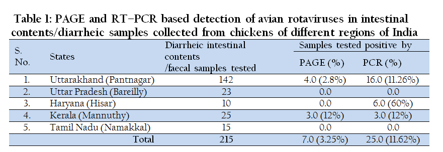
Table 1: PAGE and RT–PCR based detection of avian rotaviruses in intestinal contents/diarrheic samples collected from chickens of different regions of India
Viral RNA Extraction and RNA–Page
Extraction of viral RNA, polyacrylamide gel electrophoresis (PAGE) and silver staining was performed as described in our previous studies (Malik et al., 2012).
Two Step RT–PCR Assay
For detection and confirmation of the group A and D avian rotaviruses, two–step RT–PCR assay was executed. In the first step, reverse–transcription for cDNA synthesis from viral RNA was performed using 1.0 µl (100 ng/µl) random hexamer (Fermentas), 100 ng of viral RNA, and 2 µL of dimethyl sulphoxide (DMSO) were added to PCR tube containing sufficient volume of nuclease free water (NFW), followed by incubation of the reaction mixture at 70°C for 5 min to melt secondary structures within the template. The mixture was immediately snap chilled on ice followed by the addition of 4 µL of 5X RT buffer, 2 µL of 10 mM dNTPs (Fermentas, Lithuania), 40 U RNase Inhibitor (Ambion, USA), and 200 U MMLV–RT (Promega) and kept at 37°C for 90 minutes. The enzyme was denatured at 80⁰C for 3 min at the end of the incubation step to inactivate residual moloney murine leukemia virus–reverse transcriptase (MMLV–RT). The cDNA thus obtained was used for PCR and the remaining was kept at –20ºC until further use.
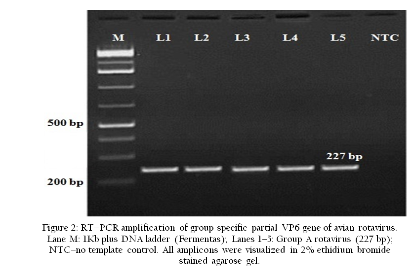
In the second step, for the detection of group A avian rotavirus, VP6 gene based PCR was implemented using the newly designed primers i.e. (sense 5´TTTGATCACTAAYTATTCACC 3´ and anti–sense 5´GGTCACATCCTCTCACTA3´) with the optimized reaction conditions (initial denaturation at 95°C for 5 min, cyclic denaturation at 95°C for 30 sec, annealing at 50°C for 30 sec and extension at 72°C for 30 sec for repeated 35 cycles and a final extension temperature–time combination of 72°C for 10 min). For the detection of group D avian rotaviruses, separate VP6 gene based diagnostic primers were used (sequences not shown) with the same amplification conditions described for group A rotaviruses. The specific PCR amplicons of 227 bp for group A and 185 bp for group D rotaviruses were visualized in ethidium bromide stained 2% agarose gel and documented using Transilluminator–UV®300 (UVP Inc., Upland, USA).
RESULTS AND DISCUSSION
The gastrointestinal infections associated with viral aetiology which occur in chickens tend to preponderate more the young birds. In the field conditions, these infections are convoluted by other infectious agent’s viz. bacteria or virus, making it problematic to measure the factual role of these agents in naturally befalling gastrointestinal diseases (Savita et al., 2008a). Thus, an understanding of the viruses that cause gastrointestinal tract disease in poultry, along with an understanding of their epidemiology is necessary for development of be–fitting rheostat measures. Initial works for classification of rotaviruses in to different groups (A–G types) was based on migration patterns of RNA segments in the PAGE (Pedley et al., 1986; Estes and Kapikan, 2007)
In this study, initial screening by RNA–PAGE for the presence of rotaviruses revealed typical genome segments migration pattern of 4:2:3:2 (Figure 1) explicit of mammalian group A rotaviruses in 3.25% cases (7/215). Avian rotavirus with an electropherotype similar to described here for group A rotaviruses was also detected in 4% (3/75) of diarrheic adult chickens for the first time in India (Wani et al., 2003). Interestingly, in contrast to avian group A rotavirus migration pattern of 5:1:3:2 (Schumann et al., 2009), migration of segment 5 close to 6 and triplet of segments 7, 8 and 9 in all the positive samples clearly indicated mammalian group A rotaviruses. Since, the first detection of avian group A rotaviruses a decade ago in India, several other reports successively recorded presence of rotavirus infection in the range of 7.84% to 22.2% (Wani et al., 2003; Minakshi et al., 2004; Savita et al., 2008b; Niture et al., 2011), suggestive of consistent commonness of rotavirus infection in the birds. In addition to more common group A avian rotaviruses, group D avian rotaviruses from central (77.7%) and western (7.84%) region of India have also been testified (Savita et al., 2008b; Niture et al., 2010). The RT–PCR assay, being the much more sensitive, detected 11.6% samples (25/215) positive for group A rotavirus (Figure 2) and mixed infection of both group A and D rotaviruses in 1.4% (3/215) samples. Our results are in compliance with the earlier findings where higher prevalence of group A compared to group D avian rotaviruses was recorded (Wani et al., 2003; Minakshi et al., 2004). At the same, in one of our previous study, the prevalence rate of group A rotaviruses was less (22.2%) compared to group D rotaviruses (77.8%) in the chicken flock of central India (Savita et al., 2008b). In another report from Maharashtra, Niture et al. (2010) also showed higher prevalence rate of group D than group A avian rotavirus.

Figure 1: Mammalian–like genomic segments migration pattern (4:2:3:2) of avian group A rotaviruses in silver stained RNA–PAGE
To the best of our knowledge, report of co–infection with different types of avian rotavirus in chickens are lacking so far. The three samples showing mixed infection of group A and D rotaviruses in RT–PCR assay did not show any positive presence of rotavirus dsRNA in conventional RNA–PAGE, which could be due to lower copy number of the group D rotavirus particles which failed its detection due to lower sensitivity limit of RNA–PAGE. The regional distribution of avian rotaviruses disclosed the high prevalence in Uttarakhand (7.44%, 16/215) followed by Haryana (2.8%, 6/215) and Kerala (1.3%, 3/215). None of the sample from Uttar Pradesh and Tamil Nadu yielded positive result, but before concluding absence of avian rotavirus in these regions, extensive epidemiological studies on more number of enteritis/diarrheic samples needs to be assessed.
In conclusion, the present study reports the occurrence of mammalian type group A avian rotaviruses from the southern and northern states of India using PAGE and RT–PCR assays. The existence of more than one virus group within a single host may not only complicate the severity of enteritis cases in poultry flocks but also could lead to emergence of reassortants having more pathogenic potential. Further studies will be essential to understand the evolving epidemiology of rotaviruses in avian species.
REFERENCES
Ahmed MS and Ahmed MU (2006). Detection of avian rotavirus–like virus in broiler chickens in Bangladesh. Bangl. J. Vet. Med. 4(2): 73–77.
Attoui H, Mertens PPC, Becnel J, Belaganahalli S, Bergoin M, Brussaard CP, Chappell JD, Ciarlet M, del Vas M, Dermody TS, Dormitzer PR, Duncan R, Fcang Q, Graham R, Guglielmi KM, Harding RM, Hillman B, Makkay A, Marzachì C, Matthijnssens J, Milne RG, Mohd Jaafar F, Morei H, Noordeloos AA, Omurea T, Patton JT, Rao S, Maan M, Stoltz D, Suzuki N, Upadhyaya NM, Wei C and Zhou H (2012). Reoviridae. In: King A, Adams M, Carstens E and Lefkwitz E. Virus Taxonomy, Ninth Report of the International Committee on Taxonomy of Viruses. Elsevier Academic Press; Waltham, MA, USA: 497–650.
Barnes HJ (1997). Virology of enteric infections. Diseases of poultry. 10th edn., Iowa state University press, Ames, 685–686.
PMid:9195624
Dhama K, Chauhan RS, Mahendran M, Malik SV (2009). Rotavirus diarrhea in bovines and other domestic animals. Vet. Res. Commun. 33(1):1–23
http://dx.doi.org/10.1007/s11259-008-9070-x
PMid:18622713
Estes M and Kapikian A (2007). Rotaviruses. In Fields Virology, 5th edn, vol.2, pp.1917–1974. Edited by D. M. Knipe, P. M. Howley, D. E. Griffin, R. A. Lamb, M. A. Martin, B. Roizman and S. E. Straus. Philadelphia, PA: Kluwer/Lippincott, Williams and Wilkins.
Gough RE, Wood GW and Spackman D (1986). Studies with an atypical avian rotavirus from pheasants. Vet. Rec. 118: 611–612.
http://dx.doi.org/10.1136/vr.118.22.611
PMid:3014708
Gough RE, Wood GW, Collins MS, Spackman D, Kemp J and Gibson LAC (1985). Rotavirus infection in pheasant poults. Vet. Rec. 116: 295.
http://dx.doi.org/10.1136/vr.116.11.295
PMid:3992834
Jindal N, Patnayak DP, Chander Y, Ziegler AF and Goyal SM (2009). Experimental reproduction of poult enteritis syndrome: Clinical findings, growth response, and microbiology. Poult. Sci. 88: 949–958.
http://dx.doi.org/10.3382/ps.2008-00490
PMid:19359682
Jindal N, Patnayak DP, Chander Y, Ziegler AF and Goyal SM (2010). Detection and molecular characterization of enteric viruses from poult enteritis syndrome in turkeys. Poult. Sci. 89: 217–226.
http://dx.doi.org/10.3382/ps.2009-00424
PMid:20075272
Jindal N, Chander Y, Patnayak DP, Mor SK, Ziegler AF and Goyal SM (2012). A multiplex RT–PCR for the detection of astrovirus, rotavirus, and reovirus in turkeys. Avian Dis. 56: 592–596.
http://dx.doi.org/10.1637/9958-100911-ResNote.1
PMid:23050480
Kusumakar AL, Savita, Malik YPS, Minakshi and Prasad G (2010). Genomic diversity among group A rotaviruses from diarrheic children, piglets, buffalo and cow calves of Madhya Pradesh. Indian J. Microbiol. 50(1): 83–88.
http://dx.doi.org/10.1007/s12088-010-0016-y
PMid:23100812 PMCid:PMC3450293
Malik YPS, Sharma K, Vaid N, Chakravarti S, Chandrashekar KM, Basera SS, Singh R, Minakshi, Prasad S, Gulati BR, Bhilegaonkar KN and Pandey AB (2012). Frequency of group A rotavirus with mixed G and P genotypes in bovines: predominance of G3 genotype and its emergence in combination with G8/G10 types. J. Vet. Sci. 13(3): 271–278.
http://dx.doi.org/10.4142/jvs.2012.13.3.271
PMid:23006956 PMCid:PMC3467402
Malik YPS, Kumar N, Sharma K, Sharma R, Kumar HB, Kusumakar A, Kumari S, Shukla S and Chandrashekar KM (2013). Epidemiology and Genetic Diversity of Rotavirus Strains Associated with Acute Gastroenteritis in Bovine, Porcine, Poultry and Human Population of Madhya Pradesh, Central India, 2004–2008. Adv. Anim. Vet. Sci. 1(4): 111 – 115.
McNulty MS (2003). Rotavirus infections. In: Saif YM, Barnes HJ, Glisson JR, FadlyAM, McdougaldLR, Swayne DE (eds.), Diseases of Poultry. 11th edn., Ames, IowaState University Press. 308–317.
http://dx.doi.org/10.1016/S0163-4453(03)00014-8
http://dx.doi.org/10.1053/jinf.2002.1095
Minakshi, Prasad G, Verma S and Dahiya S (2004). Detection of avian rotaviruses from diarrhoeic poultry in India. Indian J. Microbiol. 44: 205–209.
Niture GS, Karpe AG, Prasad M, Bhonsle AV and Patil SV (2010). Detection of group D avian rotaviruses among layer poultry from western India. Int. J. Poult. Sci. 9(1): 72–76.
http://dx.doi.org/10.3923/ijps.2010.72.76
Niture GS, Kapre AG, Prasad M and Zade NN (2011). Electrophoretic pattern analysis of rotaviruses of buffalo, poultry and human. J. Vet. Pub. Hlth. 1: 7–10.
Otto PH, Ahmed MU, Machnowska P, Reetz J, Roth B, Trojnar E and Johne R (2012). Detection of avian rotaviruses of groups A, D, F and G in diseased chickens and turkeys from Europe and Bangladesh. Vet. Microbiol. 156: 8–15.
http://dx.doi.org/10.1016/j.vetmic.2011.10.001
PMid:22079218
Otto PH, Liebler–Tenorio EM, Elschner M, Reetz J, Lohren U and Dille R (2006). Detection of rotaviruses and intestinal lesions in broiler chicks from flocks with runting and stunting syndrome (RSS). Avian Dis. 50: 411–418.
http://dx.doi.org/10.1637/7511-020106R.1
PMid:17039842
Pedley S, Bridger JC, Chasey D and Mc–crae MA (1986). Definition of two new groups of atypical rotaviruses. J. Gen. Virol. 67: 131–137.
http://dx.doi.org/10.1099/0022-1317-67-1-131
PMid:3003232
Reynolds DL, Saif YM, and Theil KW (1987a). A survey of enteric viruses of turkey poults. Avian Dis. 31: 89–98.
http://dx.doi.org/10.2307/1590779
PMid:3034232
Reynolds DL, Theil KW, and Saif YM (1987b). Demonstration of rotavirus and rotavirus–like virus in the intestinal contents of diarrheic pheasant chicks. Avian Dis. 31: 376–379
http://dx.doi.org/10.2307/1590889
PMid:3039968
Savita, Kusumakar AL, Malik YPS, Minakshi and Prasad G (2008a). Co–occurrence of avian rotavirus and bacterial pathogens in diarrheic poultry. Indian J. Anim. Sci. 78(5): 478–479.
Savita, Kusumakar AL, Malik YPS, Minakshi and Prasad G (2008b). Detection and characterization of group A and D avian rotaviruses in India. Indian J. Biotech. 7: 554–556.
Schumann T, Hotzel H, Otto P and Johne R (2009). Evidence of interspecies transmission and reassortment among avian group A rotaviruses. Virology 386(2) 334–343.
http://dx.doi.org/10.1016/j.virol.2009.01.040
PMid:19249805
Tamehiro CY, Alfieri AF, Medici C and Afieri AA (2003). Segmented double–stranded genomic RNA viruses in faecal samples from broiler chicken. Brazilian J. Microbiol. 34: 344–348
http://dx.doi.org/10.1590/S1517-83822003000400013
Theil KW, Reynolds DL and Saif YM (1986). Genomic variation avian rotavirus–like viruses detected by polyacrylamide gel electrophoresis. Avian Dis. 30: 829–834.
http://dx.doi.org/10.2307/1590594
PMid:3028359
Wani SA, Bhat MA, Ishaq SM, Ashrafi MA, Buchh AS and Haq M (2003). Detection of a mammalian– like group A rotavirus in diarrhoeic chicken. Vet. Microbiol. 94: 13–18
http://dx.doi.org/10.1016/S0378-1135(03)00079-8


