Research Journal for Veterinary Practitioners
Case Report
Research Journal for Veterinary Practitioners 1 (3): 36 – 38Case of Canine Parvovirus Infection in a Pointer Pup Treated Symptomatically
Arslan Tariq1*, Asim Shahzad2
- Department of Clinics Medicine and Surgery, Faculty of veterinary sciences, University of Agriculture Faisalabad, Pakistan
- Department of Pathology, Faculty of veterinary sciences, University of Agriculture Faisalabad, Pakistan
*Corresponding author:dr.arslantariq3418@live.com
ARTICLE CITATION:
Tariq A and Shahzad A (2013). Case of canine parvovirus infection in a pointer pup treated symptomatically. Res. J. Vet. Pract. 1 (3): 36 – 38.
Received: 2013–09–07, Revised: 2013–09–18, Accepted: 2013–09–19
The electronic version of this article is the complete one and can be found online at
(
http://nexusacademicpublishers.com/table_contents_detail/13/93/html
)
which permits unrestricted use, distribution, and reproduction in any medium, provided the original work is properly cited
ABSTRACT
Canine parvovirus infection is one of the biggest causes of death in canines due to hemorrhagic enteritis. A pup showing related clinical signs of this infection was seen in outdoor clinic at department of Clinic Medicine and Surgery (CMS), University of Agriculture Faisalabad, Pakistan. Anorexia, vomiting, bloody diarrhea, swollen lymph nodes and high fever provided initial base to diagnose the disease as parvovirus infection. Further confirmation was done with complete blood count (CBC) in which leukopenia was seen. Disease was treated for 7 day symptomatically. Clinical sign of disease started vanishing from 5th day of treatment. CBC was again done after 10 days to check the immune status of animal. Increase in leukocyte count confirmed that animal was going towards recovery.
Rarely fatal but infectious disease of canines called as Canine parvovirus infection is known to cause high rate of morbidity and mortality in dogs mostly in young pups. This disease is distributed all over the world (Decaro et al., 2005; Litster et al., 2012). In 1978, Canine parvovirus was first recognized as non–enveloped, negative sense ssDNA virus containing 2 structural and 2 nonstructural proteins, icosahedral in shape and 26 nm in diameter. Antigenic property of this virus was found due to substitutions of amino acid in a structural protein VP2 (Desario et al., 2005; Truyen, 2006; Decaro et al., 2005).
The most common cause of enteritis in dogs is CPV–2. Approx. 3–10 days after transmission by Fecal–oral route, animal start showing signs like high rise in temperature, anorexia, vomiting and bloody diarrhea whereas high titer can be seen in feces of infected dogs on 8–10 days. Leukopenia is also an indication in this disease (Desario et al., 2005; Decaro et al., 2007).
Disease can be diagnosed on the basis of clinical signs and hematological examination. Other test includes like ELISA and HA tests can be used but all are less sensitive. Fecal sample from diarrheic dog is used for these tests. Instead of these a sensitive test known as virus isolation can be used but is not feasible because it consumes a lot of time. Real time PCR, which is very specific and sensitive, is developed to diagnose CPV–2 (Desario et al., 2005; Decaro et al., 2005).
This disease is major threat to canine population and cause 91% mortality in untreated cases so need to be control (Prittie and Jennifer, 2004). For this purpose modified live virus vaccines are now available to protect this infection. Vaccination at proper time is helpful to control the disease. The aim of this case report is to show the diagnosis on clinical basis, symptomatic treatment and importance of vaccination in canine parvovirus infection.
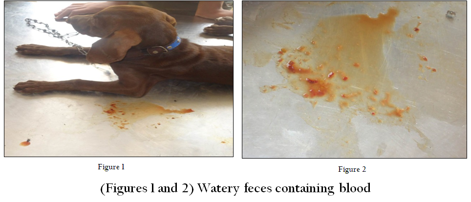
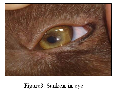
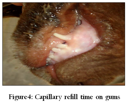
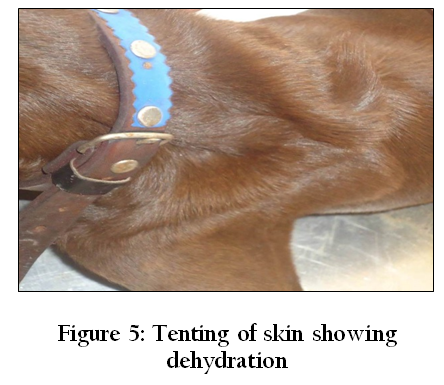
Approximately 3 month old pointer pup visited the outdoor clinic at Department of Clinics, Medicine and Surgery (CMS), Faculty of veterinary sciences (FVS), University of Agriculture Faisalabad (UAF) on April. 2013. Present complains included anorexia, vomiting and bloody diarrhea (Figure 1 and 2) from 3 days. Vaccination was not done until now but deworming with syrup combantrin (Active ingredient: Pyrantel Pamoate) was done 15 days before. Clinical examination revealed high fever, increase heart beat and respiration as 1040F, 120/min and 45/min respectively. Eyes were sunken in (Figure~3) and lymph nodes were palpable. CRT>4 indicated that animal was severely dehydrated (Figure~4 and 5). Fecal examination showed no parasitic infestation but CBC reveled leukopenia (Table 1). Clinical signs and hematology provided the clues for diagnosing the disease as parvovirus infection.
Infusion of 0.9% normal saline solution @ 20ml/kg b.w, as supportive and Aminopentamide @ 0.4mg/kg b.w, Metoclopramide @ 1–2mg/kg b.w/day, ketoprufen @ 2mg/kg b.w and enrofloxacin @2.5 mg/kg b.w was recommended as symptomatic treatment for 7 days. Vitamin B–complex was also added in treatment. Injectable were replaced by Syrups after 3 days of start of treatment. The animal was found pretty well in condition after 10 days of treatment with increase in leukocyte count (Table 2). From many different parts of the world parvovirus infection has been reported as major threat of gastroenteritis in canine population (Panda et al., 2009). During first 6 weeks of life the maternal antibodies are known to protect the pup from CPV infection but from 6–12 weeks of age the dogs are more susceptible (Desario et al., 2005; Truyen, 2006).
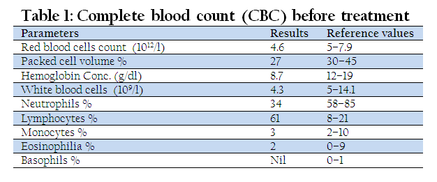
Infection was diagnosed on the basis of clinical signs and blood profile. Anorexia, vomiting, bloody diarrhea and dehydration were seen in animal on clinical examination which provided the 1st important clue for this disease. But in Complete blood count (CBC) leukopenia was seen and as also reported by Decaro et al (2007) confirmed the disease. Usually animal become lethargic first and then bloody diarrhea and vomiting cause dehydration and electrolytes imbalance. Anemia can also occur due to blood loss in feces. In case of severe dehydration animal may go into shock and may lead to death if not treated in early stages (/Canine%20 parvovirus %2 0 –% 20 Wikipedia, % 20the%20free%20encyclopedia.htm).
The animal was treated symptomatically because according to Panda et al (2009), in absence of specific treatment the symptomatic treatment is only way to decrease mortality. But now a day’s feline interferon (omega type) has been known to provide good results. In under develop countries like Pakistan such interferon are not available if available then expensive and out of reach for owner so only symptomatic treatment is feasible. Mortality can be decrease to 5–20% from 91% by aggressive therapy. If treatment is done at right time the survival chances may reach to 80–95% (Prittie and Jennifer, 2004). After 10 days again blood sample was taken from animal to check the WBC’s level. A marked increase is seen in WBC’s count shows the effectiveness of treatment (Table~2).

According to Truyen, (2006), potent modified live virus vaccines are developed to protect animal from canine parvovirus (CPV) infection, which are actually derived from original type of virus which was first recognized in late 1970’s. By vaccination we can protect our animals from large number of deaths in young dogs until feline interferon (omega type) are not easily available in market at affordable price.
ACKNOWLEDGEMENT
Special thanks to Dr. Tanveer Ahmad Assitant Professor at Department of clinics, medicine and surgery (CMS), Faculty of Veterinary Sciences (FVS), University of Agriculture Faisalabad (UAF) for helping me out in respective case report and reviewing of manuscript.
REFERENCES
Decaro N, Elia G, Martella V, Desario C, Campolo M, Trani LD, Tarsitano E, Tempesta M and Buonavoglia C (2005). A real–time PCR assay for rapid detection and quantitation of canine parvovirus type 2 in the feces of dogs. Veterinary Microbiology 105: 19– 28.
http://dx.doi.org/10.1016/j.vetmic.2004.09.018
http://dx.doi.org/10.1016/j.vetmic.2005.05.014
Decaro N, Martella V, Elia G, Desario C, Campolo C, Lorusso E, Colaianni ML, Lorusso A and Buonavoglia C (2007). Tissue distribution of the antigenic variants of canine parvovirus type 2 in dogs. Veterinary Microbiology 121: 39– 44.
http://dx.doi.org/10.1016/j.vetmic.2006.11.005
PMid:17169509
Desario C, Decaro N, Compolo M, Cavalli A, Cirone F, Elia G, Martella V, Lorusso E, Camero M and Buonavoglia C (2005). Canine parvovirus infection: Which diagnostic test for virus? Journal of Virological Methods 126: 179– 185.
http://dx.doi.org/10.1016/j.jviromet.2005.02.006
PMid:15847935
Litster AL, Pressler B, Volpe A and Dubovi E (2012). Accuracy of a point–of–care ELISA test kit for predicting the presence of protectivecanine parvovirus and canine distemper virus antibody concentrations. The Veterinary Journal 193: 363– 366.
http://dx.doi.org/10.1016/j.tvjl.2012.01.027
PMid:22381707
Panda D, Patra RC, Nandi S and Swarup D (2009). Oxidative stress indices in gastroenteritis in dogs with canine parvoviral infection. Research in Veterinary Science 86: 36– 42.
http://dx.doi.org/10.1016/j.rvsc.2008.05.008
PMid:18572211
Prittie and Jennifer (2004). "Canine Parvoviral Enteritis: A Review of Diagnosis, Management, and Prevention". J. Vet. Emerg. Crit. Care. 14 (3): 167– 176.
http://dx.doi.org/10.1111/j.1534-6935.2004.04020.x
Truyen U (2006). Evolution of canine parvovirus a need for new vaccines?" Veterinary Microbiology 117: 9– 13
http://dx.doi.org/10.1016/j.vetmic.2006.04.003
PMid:16765539
http://www.merckmanuals.com/vet/appendixes/reference_guides/hematologic_reference_ranges.html


