Research Journal for Veterinary Practitioners
Research Article
Research Journal for Veterinary Practitioners 1(1) 1–4Surveillance of Poultry Diseases in Punjab Province, Pakistan; Special Reference to Newcastle Disease
Habib–ur–Rehman1*, Naseem Fawad1, Ghulam Abbas1, Ghazala Naheed1, Bushra Siddique1, Farhan Afzal1, Jawad Munawar1, Mian Aurangzeb1, Muhammad Atif, Imtiaz–ul–Haq2, Jamila Shafi3, Muhammad Sabir Farooq4, Zahid Ali Tahir5, Muhammad Qamar6, Abid Hussain7, Muhammad Saleem8, Muhammad Tahir Naseem9, Muhammad Javed10
- Poultry Research Institute, Rawalpindi–Pakistan
- Poultry Disease Diagnostic Laboratory–Jhelum
- Poultry Disease Diagnostic Laboratory–Samundri
- Poultry Disease Diagnostic Laboratory–Gujar Khan
- Poultry Disease Diagnostic Laboratory– Kamalia
- Poultry Disease Diagnostic Laboratory– Bahawal Nagar
- Poultry Disease Diagnostic Laboratory–Ghakkar
- Poultry Disease Diagnostic Laboratory–Jhang
- Poultry Disease Diagnostic Laboratory–Arifwala
- Deputy District livestock officer Sargodha
*Corresponding author:habib.rehman71@hotmail.com
ARTICLE CITATION:
Rehman H, Fawad N, Abbas G, Naheed G, Siddique B, Afzal F, Munawar J, Aurangzeb M, Atif M, Haq I, Shafi J, Farooq MS, Tahir ZA, Qamar M, Hussain A, Saleem M, Naseem MT, Javed M (2013). Surveillance of poultry diseases in Punjab province, Pakistan; special reference to Newcastle disease. Res. j. vet. pract. 1 (1): 1 – 4.
Received: 2013–03–21, Revised: 2013–04–25, Accepted: 2013–04–25
The electronic version of this article is the complete one and can be found online at
(
http://nexusacademicpublishers.com/table_contents_detail/13/33/html
)
which permits unrestricted use, distribution, and reproduction in any medium, provided the original work is properly cited
ABSTRACT
Prevalence of poultry diseases in Punjab province of Pakistan was investigated by analyzing data from 8 regional and one central disease diagnostic laboratory of the directorate of Poultry Research Institute, Rawalpindi. The post mortem– examinations were conducted during June 2011 to July 2012 and data is presented quarterwise starting from July 2011. For important diseases like Newcastle (ND), avian influenza (AI), salmonellosis, mycoplasmosis postmortem findings were supported by laboratory investigations in certain cases. Avian Influenza and Newcastle disease viruses were cultrured on embroynated eggs from tissue samples of the suspected cases and then confirmed by using specific antisera. Serum samples were also processed for haemaglutination inhibition (HI) test to monitor antibody titers against ND and AI. Salmonella were isolated on differential media and identified by either biochemical tests or through PCR. In day old chicks or chicks of fewer than 10 days, serum was tested with known antigen of Mycoplasma gallisepticum, M. synoviae and Salmonella pullorum/gallinarum for vertical transimission testing. Mycoplasmosis or salmonellosis in adult birds was either identified by culturing on specific medium or using serum plate agglutination tests. Coccidiosis and mycotoxicosis were at peak from July to September (14.4% & 8.9% respectively). Prevalence of ND increased significantly (P=0.005) from 5.9% in quarter 1 to 15% in quarter 4. Gumboro Disease persisted at 5.9% with no significant difference (P≤0.05) among quarters. High pathogenic AI was not reported during the year whereas low pathogenic AI remained around 1% level with a mean of 0.8%. Mean prevalence of hydropericardium syndrome (2.5%), Mycoplasma gallisepticum infection (8.7%), pullorum disease (2.7%), cholera (1.4%), typhoid (7.1%), colibacillosis (10.1%), coryza (4.4%), endo–parasitism (3.0%), ecto–parasitism (0.7%), coccidiosis (13.1%), mycotoxicosis (6.1%) and others (17.6%) was no significantly different among four quarters of the study year (P≤0.05).
INTRODUCTION
Punjab is the largest province with reference to human as well as poultry population in Pakistan. Commercial poultry developed here successfully by virtue of agricultural base and availability of cheap labour. During fiscal year 2010–11, 709.5 million poultry were raised in Punjab which included 631.97 million broilers, 27.75 million layer, 12.55 million breeder and 37.23 million backyard poultry (Punjab Poultry Statistical Report 2011–12). The poultry industry at present comprises of open farm houses of 1000 birds rearing capacity to environmentally controlled houses of 20.000 to 30.000 birds. Both systems run side by side, and different diseases break out time to time. Presence of different variants of infectious bronchitis virus identified through sero–surveillance in the major poultry producing areas of Punjab and neighboring province of KPK in the North West region of Punjab/Pakistan was found to be an important cause of mortality and morbidity (Ahmed et al., 2007).
Occurrence of bacterial, viral and other nutritional/environmental diseases in relation to seasons has been reported by Yunus et al. (2008) in the Chakwal district of Punjab. Ahmed et al. (2012) surveyed important poultry diseases in neighboring high altitude areas of Azad Kashmir. Presence of avian influenza virus in wild birds living around commercial poultry areas is also a serious concern (Khawaja et al., 2005). Many studies provide important information about consistent presence of variety of diseases in the standing poultry population along with surge of different viral outbreaks from time to time (Brown, 2010; Alam et al., 2012).
Since poultry diseases impose severe economic and production losses, it is important to remain updated about prevailing health issues of poultry in the areas of concern. The present study was conducted to know the prevalence of diseases in poultry population of the province. So that forecasting and better control measures could be adopted by the farmers and local clinicians.
MATERIALS AND METHODS
Necropsy
The study was based generally on postmortem examinations conducted during June 2011 to July 2012 at 8 regional and one central disease diagnostic laboratory of the directorate of Poultry Research Institute, Rawalpindi Pakistan. The data were recorded upon history, clinical signs and postmortem lesions for individual diseases.
Virus Isolation and Identification
Clinical samples (serum, tissues, swabs) from cases requiring differential diagnosis or for the sake of confirmation of diagnosis, were submitted to the disease section laboratory of the directorate from regional laboratories for virus isolation {Newcastle disease (ND) and Avian Influenza (AI)} and/or for virus identification through specific antisera (Alexander, 2003; Anonymous, 2008).
Bacterial Isolation and Identification
Isolation of Salmonella and Escherichia coli (E.coli) was done on MacConkey –/Xylose Lysine Deoxycolate agar and identified by biochemical tests through commercially available kit (API 20–ETM,Biomerieux). Serum plate agglutination tests for Mycoplasma gallisepticum/synoviae (Mg/Ms) and Salmonella pullorum/gallinarum (Spg) were performed using commercially available antigen (Lohmann Animal Health Int., Charles River Laboratories USA (Muhammad et al., 2002). The data were compiled among four quarters (Q1, Q2, Q3, Q4) of the year starting from 1st July 2011. Average of percent occurrence for one quarter (three months) were calculated from monthly data (n=3) from each laboratory and means of percent occurrence at the Punjab level were calculated through statistical software SPSS 16.0 taking one average figure of the quarter from each laboratory (n=9). The data were further subjected to analysis of variance (ANOVA) and Least Significant Difference Test (LSD). Differences of means were considered significant at 95% confidence level and when these were significant, actual value of probability (P) is described.
RESULTS AND DISCUSSION
The LSD test revealed no significant difference among mean prevalence of diseases observed during the four quarters of the year except for ND. Maximum level of prevalence of coccidiosis and mycotoxicosis was observed during July to September as compared to other quarters of the year. Relatively high prevalence of coccidiosis and mycotoxicosis from July to September is obvious because of high humidity in air during this time of the year. This is in agreement with the findings of Awais et al. (2012) who reported that prevalence of coccidiosis was highest in autumn. However, comparison among broiler and layer flocks reflects a steady prevalence of coccidiosis in broilers throughout the four quarters of the year with just a variation of 0.5%. This may be due to poor litter management and a constant brooding phase for broilers whereas layer flocks are brooded once in a year and get immune with advancing age. The percent prevalence of coccidiosis in layers during July to December was double than broilers (Table 1). This is in agreement with the observation of Yunus et al. (2008) who reported significantly higher prevalence of coccidiosis in layers than in broilers in Chakwal district. But when data for all types of commercial poultry in the province were pooled, LSD test revealed no significant difference of means among four quarters of the year (Table 2).
It may be interpreted that coccidiosis in laying poults or broilers may be seen any time of the year and clinicians should expect same level of threat of coccidiosis during hot & dry, hot & humid, humid winter or dry winter months of the year. This interpretation is also supported with the data figure of least percent prevalence of coccidiosis in broilers during monsoon season of 2003–04 (Yunus et al., 2009) in the Chakwal district. Ahmed et al. (2012) reported just a difference of 2% in prevalence of coccidiosis in broiler or layer flocks reared at an altitude less than 4000 feet in Punch district over a study period from 2008–2011. Mustafa and Ali (2005) reported much higher (10.66%) prevalence of coccidiosis in backyard layers of indigenous and Fayoumi breeds. Relatively higher prevalence of coccidiosis in backyard layers may be because of poor medical cover.
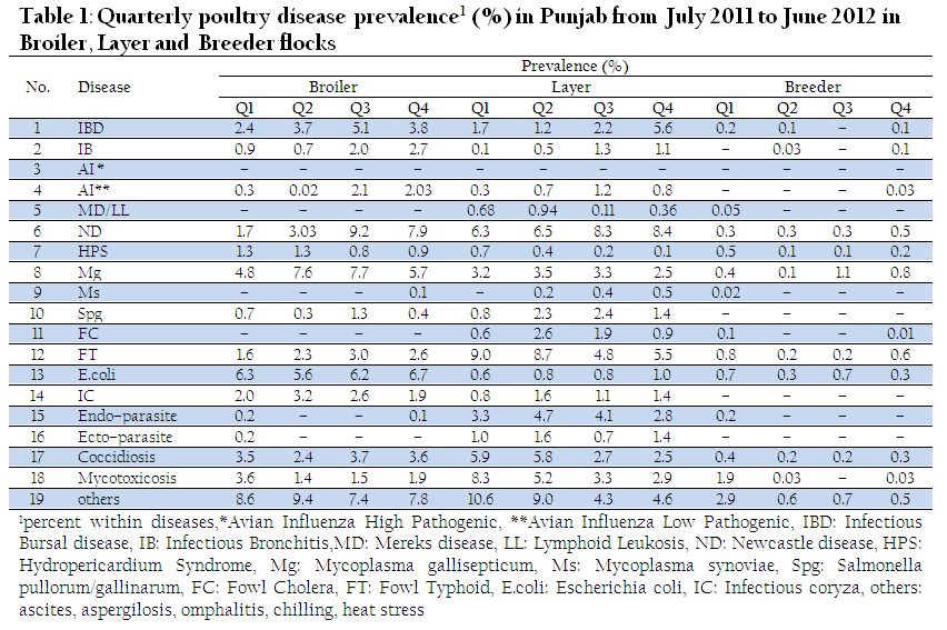
Table 1: Quarterly poultry disease prevalence1 (%) in Punjab from July 2011 to June 2012 in Broiler, Layer and Breeder flocks
Prevalence of mycotoxicosis was at its peak (8.9%, Table 2) in Punjab during quarter 1 (July to September) and then declined to it’s almost half during all three consequent quarters. However this highest figure in Q1 was not significantly higher from other three figures of the latter quarters. In depth observation of the data revealed that two of the laboratories did not reported prevalence of mycotoxicosis in any of the quarter of the year. Thus the data were re–analyzed excluding these two laboratories. Now LSD test revealed significantly higher (11.5%) mean of prevalence of mycotoxicosis during Q1 than that of during Q3 (5.4%) and Q4 (5.5%), (P=0.037 & P=0.04 respectively). The prevalence of mycotoxicosis (6.4%) in post rainy season (Q2) `was not significantly lower than in Q1. This is indicative of mycotoxin contaminated feed materials were being consumed during October to December and depleted in latter quarters.
Newcastle disease outbreaks remained havoc throughout the study year in commercial layer and broiler flocks (Table 1 & 2). Cumulative prevalence of ND was at record high and persistent during January 2012 to June 2012. The prevalence was significantly higher in Q4 than in Q1 (P=0.005) and Q2 (P=0.022). The ND outbreaks started during Q1 of 2011 and kept increasing in Q2 with no significant difference. The percentage of ND outbreaks further rose in Q3 but difference remained non–significant in comparison to Q1 & Q2. However, the difference of ND prevalence between Q1 and Q3 was not significant just with a slight increase in probability level (P=0.054). The authors consider it significant keeping in view that a careful data collection could have been resulted in significant difference. Clinical and laboratory results showed it to be velogenic–viscerotropic type, caused high mortality and no available vaccine showed satisfactory prevention or cure. The ND outbreaks were predicted by Numan et al. (2005) upon finding low circulating ND antibody titers in commercial broilers in Punjab. Latter Ahmed et al. (2008) and Siddique et al. (2012) isolated velogenic forms of ND virus from commercial poultry in the province. Breeder flocks stood healthy which is indicative of biosecurity/management laps at commercial layer and broiler farm level towards ND outbreaks.
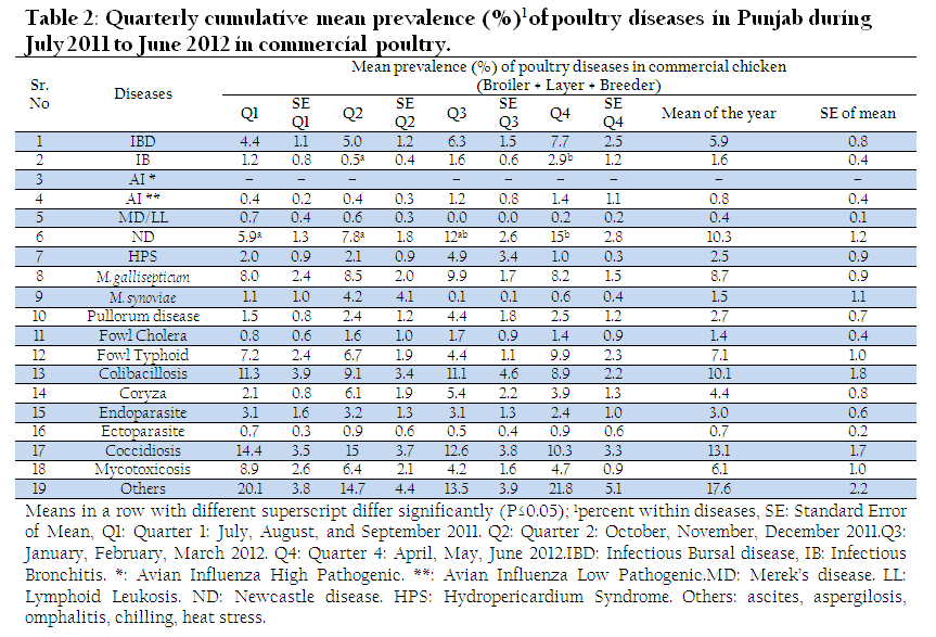
Table 2: Quarterly cumulative mean prevalence (%)1 of poultry diseases in Punjab during July 2011 to June 2012 in commercial poultry
Prevalence of IB was found very low (1.6% mean of the year) in present study which is not in agreement with a report on IB prevalence by Ahmed et al. (2007). The authors reported high percentage of variants of IBV in broiler and layer population in important poultry rearing areas of Punjab. They found about 30% enhancement in rate of IBV isolation after treatment of lungs/tracheal tissues with phospholipase C. They further concluded reverse transcription PCR as more reliable tool for pinpoint diagnosis. By comparing the data on IB from our study and Ahmed et al. (2007), it can be commented that IB was overlooked in postmortem lesions at field stations. Moreover laboratories were not equipped for sero–diagnosis or virus isolation. There is an immediate need to improve diagnosis of IB at both levels.
Low pathogenic AI was also reported at very low or at zero level in broiler breeder population. AI–H9N2 is pandemic in Pakistan and this reporting seems to be due to limited access of government veterinarians to large entrepreneurs for data and sample collection. However, figure of low pathogenic AI among broiler and layer flocks is quiet realistic (Table 1 & 2) and in agreement with Naeem et al. (2007).
Fowl typhoid (FT) and avian mycoplasmosis were the top most prevailing bacterial diseases. Fowl typhoid remained persistent. In broiler flocks it was two to four times lesser than in layer flocks (Table 1, 2& 3). This difference of high prevalence of salmonellosis in layers is obvious because of their longer duration of life and more likelihood to get infected with salmonella from poorly managed farm houses. High rate of prevalence of salmonellosis in layer flocks in the Faisalabad region was also reported by Majid et al. (1991).
Salmonellosis is one of the major zoonotic diseases. Worldwide 0.155 million human deaths and 93.8 million human cases of gastroenteritis are estimated annually because of this organism (Majowicz et al., 2010). In general, galliform birds are highly susceptible to salmonella and commercially raised poultry are particular source of food born salmonellosis as well as impose direct public health issue. Salmonella enteritidis, S. typhimurium, S. kentucky, and S. heidelberg are common isolates of commercial and backyard chicken in healthy, as well as in sick conditions. Mean prevalence (7.1%) of FT through the year in all types of commercial poultry in Punjab with no significant difference among the quarters is a matter of serious concern particularly when non motile salmonella viz. Gallinarum biovar Gallinarum and Pullorum have been eradicated from North America and most of Western Europe. Pullorum disease was not reported in breeder flocks which reveals strict culling policy at grandparent level.
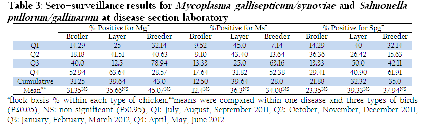
Table 3: Sero–surveillance results for Mycoplasma gallisepticum/synoviae and Salmonella pullorum/gallinarum at disease section laboratory
Infection of Mycoplasma gallisepticum (Mg) was persistent showing no significant difference of prevalence among quarters (Table 2). In broilers it rose about 2% during cold weather of Q2 and Q3 but effect of temperature was not much evident in layer flocks and prevalence remained around 3% in layers in all four quarters with no significant difference between layer and broiler flocks. Among flocks showing respiratory distress, Siddique et al. (2012) found 80% flocks were sero–positive for M. gallisepticumin in different districts of Punjab whereas Mukhtar et al. (2012) reported around 50% of the flocks showing respiratory distress were sero–positive for Mg and showed about 20% higher prevalence in winter months than in summer in layer flocks in and around Faisalabad district. However in present study, sero surveillance of broiler, layer and breeding flocks was done regardless of presence or absence of respiratory distress and prevalence of Mg/Ms differed non significantly among broiler, layer and breeding flocks (Table 3).
During analysis, it was realized that the data should also be presented on flock basis to be considered as single entity case. Number of healthy flocks and their respective population during the month should also be included in the monthly reports.
REFERENCES
Ahmed A, Hanif A, Shahid A, Najeeb MI and Ahmad MD (2008). Study of disease outbreak in layer flocks in and around Sammundri area. Pak. J. Life & Soci. Sci. 6(1): 59-62.
Ahmed I, Anjum MS and Hanif M (2012). Prevalence of poultry diseases at high altitudes of district Poonch Aazad Jammu& Kashmir. Pak. J. Sci. 64(4): 334-336.
Ahmed Z, Naeem K and Hameed A (2007). Detection and seroprevalence of infectious bronchitis virus strains in commercial poultry in Pakistan. Poult. Sci. 86(7): 1329-1335.
http://dx.doi.org/10.1093/ps/86.7.1329
PMid:17575179
Alam J, Muhammad F, Siddiqui MU, Khan SA, Rehmani S and Ahmad A (2012). Dot-ELISA for Newcastle Disease, Infectious Bursal Disease and Mycoplasmosis. Pak. J. Zool. 44(5): 1301-1305.
Alexander DJ (2003). Newcastle disease, other avian paramyxoviruses and pneumovirus infection. Disease of poultry. 12th Ed. Saif YM, Barnes HJ, Glisson JR, Fadly AD, McDougald LR, Swayne DE (eds). Iowa State University Press, Ames, USA. pp: 64-87.
Anonymous (2008). Newcastle disease. Manual of diagnostic tests and vaccines for terrestrialanimals, 6thed, OIE Press, Paris, France, pp: 576-589.
Awais MM, Akhtar M, Iqbal Z, Muhammad F and Anwar MI (2012). Seasonal prevalence of coccidiosis in industrial broiler chickens in Faisalabad, Punjab, Pakistan. Tropical Anim. Health & Production. 44 (2): 323-328.
http://dx.doi.org/10.1007/s11250-011-0024-x
PMid:22102015
Brown IH (2010). Summary of avian influenza activity in Europe, Asia and Africa 2006-2010. Avian Dis. 54:187–193.
http://dx.doi.org/10.1637/8949-053109-Reg.1
PMid:20521631
Khawaja JZ, Naeem K, Ahmed Z and Ahmad S. 2005. Surveillance of avian influenza viruses in wild birds in areas adjacent to epicenter of an outbreak in federal capital territory of Pakistan. Int. J. Poult. Sci. 4 (1): 39-43.
http://dx.doi.org/10.3923/ijps.2005.39.43
Majid A, Siddique M and Khan MZ (1991). Prevalence of salmonellosis in commercial chicken layers in and around Faisalabad. Pak. Vet. J. 1: 37-41.
Majowicz SE, Musto J, Scallan E, Angulo FJ, Kirk M, O'Brien SJ, Jones TF, Fazil A and Hoekstra RM (2010). The global burden of non typhoidal Salmonella gastroenteritis. Clin. Infect. Dis.50(6):882-889.
http://dx.doi.org/10.1086/650733
PMid:20158401
Muhammad K, Rabbani M and Siddiq M (2002). Isolation and identification of veterinary bacterial pathogens. 1st ed. Ferozan Publishing Co. Kabir St.Urdu Bazar. Lahore. Pakistan.
Mukhtar M, Awais MM, Anwar MI, Hussain Z, Bhatti N and Ali S (2012). Seroprevalence of Mycoplasma gallisepticum among commercial layers in Faisalabad, Pakistan. J. Basic & Appl. Sci. 8: 183-186.
Mustafa MY and Ali SS (2005). Prevalence of infectious diseases in local and fayoumi breeds of rural poultry (gallusdomesticus). Punjab Univ. J. Zool. 20 (2): 177-180.
Naeem K, Siddique N, Ayaz M and Jalalee MA (2007). Avian Influenza in Pakistan: Outbreaks of Low- and High-Pathogenicity Avian Influenza in Pakistan during 2003-2006. Avian Dis. 51(1): 189-193.
http://dx.doi.org/10.1637/7617-042506R.1
PMid:17494552
Numan M, Zahoor MA, Khan HA and Siddique M (2005). Serologic status of Newcastle disease in broilers and layers in Faisalabad and surrounding districts. Pak. Vet. J. 25(2):55–58.
Punjab Poultry Statistical Report (2011-12). Directorate of Poultry Research Institute, Rawalpindi Pakistan. (http://poultry.punjab.gov.pk/download/PUNJAB%20POULTRY%20STATISTICS%20.2011-12.pdf)
Siddique AB, Rahman SU, Hussain I and Muhammad G (2012). Frequency distribution of opportunistic avian pathogens in respiratory distress cases of poultry. Pak. Vet. J. 32(3): 386-389.
Yunus AW, Nasir MK, Farooq U and Böhm J (2008). Prevalence of poultry diseases and their interaction with mycotoxicosis in district chakwal: 1. Effects of age and flock size. J. Anim. Pl. Sci. 18(4): 107-113.
Yunus AW, Nasir MK, Aziz T and Böhm J (2009). Prevalence of poultry diseases in district chakwal and their interaction with mycotoxicosis: 2. Effects of season and feed. J Anim. Pl. Sci. 19(1): 1-5.


