Research Journal for Veterinary Practitioners
Case Report

Management of Vaginal Prolapse in Late Gestation Associated with Difficult Birth in a Young Bitch
Ashutosh Basera*, Asif Pasha, Brijesh Kumar, Jitendra K Agrawal, Atul Saxena
Department of Veterinary Gynaecology and Obstetrics, College of Veterinary Science and Animal Husbandry, U.P. Pandit Deendayal Upadhyay Pashu Chikitsa Vigyan Vishwavidhyala evam Go-Anusandhan Sansthan, Mathura, Uttarpradesh, India.
Abstract | An eight month, French Pitbull (weighing 10.5kg) bitch was presented at VCC, DUVASU, Mathura with the history of vaginal prolapse for 4 days. The Animal was 52 days pregnant. Transabdominal ultrasonography revealed the presence of live fetuses as indicated by heartbeats of fetuses. Prolapse mass was edematous and hard. Reduction and reposition of prolapsed mass was done. Prolapse re-occurred at 62nd days of pregnancy. X-ray examination revealed a minimum of 7 fetuses. Prolapse mass and narrow birth canal hindered normal parturition. Cesarean section was performed and 9 fetuses (8 live and 1 dead) were delivered and prolapse mass was reposited using nylon in horizontal mattress suture pattern with tubing on both sides of vulvar region.
Keywords | Vaginal prolapse, Bitch, Fetus, Pregnancy, Cesarean
Received | March 31, 2020; Accepted | May 11, 2020; Published | July 08, 2020
*Correspondence | Ashutosh Basera, Department of Veterinary Gynaecology and Obstetrics, College of Veterinary Science and Animal Husbandry, Uttarpradesh, India; Email: ashutoshbasera327@gmail.com
Citation | Basera A, Pasha A, Kumar B, Agrawal JK, Saxena A (2020). Management of vaginal prolapse in late gestation associated with difficult birth in a young bitch. Res J. Vet. Pract. 8(3): 26-28.
DOI | http://dx.doi.org/10.17582/journal.rjvp/2020/8.3.26.28
ISSN | 2308-2798
Copyright © 2020 Basera et al. This is an open access article distributed under the Creative Commons Attribution License, which permits unrestricted use, distribution, and reproduction in any medium, provided the original work is properly cited.
Introduction
In the bitch, true vaginal prolapse or uterine prolapse is a rare condition, which mainly occurs during or shortly after parturition (Schaefers-Okkens, 2001) due to the decline of serum progesterone and the increase of serum estrogen (Johnston et al., 2001; Konig et al., 2004; Rani et al., 2004). Brachycephalic breeds appear predisposed to vaginal prolapse and may possess a hereditary weakness of the perivaginal tissue (Wykes, 1986). It has been recommended that affected bitches should not be rebred (Trager, 1970). Markandeya et al. (2004) reported that excess antepartum relaxation of pelvic tissues and increased intra-abdominal pressure may be the etiology of pre-partum prolapse. Chronic prolapse during pregnancy in the bitch has been reported (Alan et al., 2007) that was treated by hysteropexy, and subsequent cesarean followed by ovariohysterectomy. Furthermore, ovariectomy or ovariohysterectomy is strongly recommended in order to reduce the incidence of recurrence (Wykes, 1986), as the recurrence rate in affected bitches is high.
Case history and clinical observations
An eight-month-old intact female, French Pitbull (weighing 10.5 kg) was presented at the VCC, Mathura with the history of prolapse mass coming out from the vulvar region for 4 days (Figure 1). Bitch was mated 52 days back with 2year aged dog of the same breed. The tissue protruding from the vulvar lips was edematous but there was no laceration or necrosis on its surface. Abdominal palpation and trans-abdominal ultrasonography revealed live fetuses in the uterine horns.On X-ray examination minimum of 7 fetuses were counted. On per-vaginal examination, the prolapsed mass on its ventral side was hard on palpation and the dorsal surface was soft on palpation. Temperature, heart rate, respiration rate was within the normal range. The Appetite was normal.
Treatment
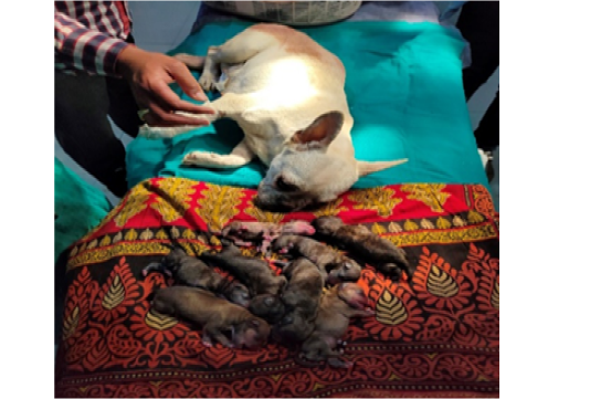
The protruded mass was cleaned with cold freshwater then icepacks were used for the reduction of prolapsed mass, topical antibiotic and lignocaine jelly applied in prolapsed mass. After reposition of prolapsed mass, retention was done by using nylon suture with a horizontal suture pattern with tubing done on both lateral walls of the vulva (Figure 2). Nine days later sutures were removed. Two days later after removal of suture, prolapsed mass again came out. At 62nd day of pregnancy animal treated with DNS 25% (100 ml ) i/v, Ca sandoze 5 ml i/v, oxytocin 5 I.U. i/v. the Animal was taking straining but unable to deliver fetuses (Figure 3). Because of the re-occurrence of prolapse mass, the owner was advised for ovariohysterectomy of the bitch but the owner did not agree for ovariohysterectomy. Therefore, cesarean section was performed by midline incision 1 inch behind the umbilicus ventrally. An Incision in large curvature of the uterine horn was applied separately in each horn, 9 fetuses were removed (8 live and 1 dead) (Figure 4). Suturing of uterus done with the polygalactin 910 suture no.1 with lambert followed by cushing suture pattern. After closing peritoneum, muscle and skin,that prolapsed mass was washed with normal saline and lignocaine jelly applied to the prolapsed mass than prolapsed mass was reposited and vulva was sutured in horizontal mattress suture pattern using nylon. Post-operative treatment was done with cefotaxime 250 mg bid, meloxicam 0.4ml, pheniramine maleate 1 ml, vitamin B complex 2ml along with fluid therapy DNS 5% (100 ml) and metronidazole 30 ml for 5 days.

Figure 1: Prolapse mass at 52nd day after mating


Figure 3: Exposed uterus during caesarean section
Result and Discussion
This case was unusual because most of the vaginal fold prolapse mainly occurs inproestrus/estrus but in the present case, vaginal fold prolapse was noticed 48 days after mating which was reported in the brachycephalic breed (French pitbull) and the animal was mated in the first estrus, as the age of the animal was 8 months. Vaginal fold prolapse was diagnosed as third degree. Eleven days later after removal of vulvar suture prolapse mass comes out again. As prolapsed mass and narrow birth canal parturition could not take place and for the survival of fetuses caesarean section was performed and 8 live fetuses were recovered. The owner of the dog complained that there was no milk let down of bitch so he advised giving milk replacer to the newborn puppies but the owner could not give milk replacer and all the puppies died on the next day.
Vaginal prolapse is a rather uncommon condition in the bitch. Little is known about hereditary aspects of vaginal prolapse in the bitch. It most commonly occurs in young bitches (2 years or younger) during their first three estrous cycles (Hedlund, 2002). It appears that the incidence of the condition is higher in brachycephalic breeds (Schaeferes-Okkens, 2001). They may possess a hereditary weakness of the perivulvar tissue (Wykes, 1986). Because the vaginal prolapse always originates from the floor of the vagina, cranial to the urethral papilla, the external urethral orifice is seen on the ventral surface of the prolapsed mass (Johnston et al., 2001). In the present case also, vaginal prolapse mass was originated from the floor of the vagina in the last third of pregnancy and re-occurred a few days later. The case was followed for two months and there was no re-occurrence of the condition as pressure due to pregnancy was relieved.
Conclusion
Occurrence of the vaginal prolapse in a pregnant bitch in the late gestation should be managed properly. If prolapse mass reoccurs at the time of parturition and hinder normal parturition process and can result into dystocia.
Acknowledgement
Authors are highly thankful to the Head of the department for providing necessary facilities to carry out the work
Conflict of interest
There is no conflict of interest.
authors contribution
Ashutosh Basera: Main Surgeon and manuscript writing
Asif Pasha: Assistance surgeon. Brijesh kumar: Assistance during operative procedure. Jitendra Agrawal: Case Proceeding and manuscript writing and Atul Saxena: Case Diagnosis, Proceeding, correction, editing.
References


