Research Journal for Veterinary Practitioners
Case Report

Case of Canine Parvovirus Infection
Muhammad Ahmad1*, Abdul Waheed2
1Department of Veterinary and Biosciences, Shaheed Benazir Bhutto University of Veterinary and Animal Sciences Sakrand, Pakistan; 2Veterinary Physician and Surgeon, Mphil Animal Surgery (Scholar) UVAS, DVM (BZU), RVMP (ISBD) Callvet SMC Pvt. Ltd. Lahore, Pakistan.
Abstract | Canine parvovirus (CPV) is an infectious disease that attacks puppies at the age of 1-6 month. The hemorrhagic enteritis is the main cause of death in canines conditions. Infected dogs shed viruses in their dung. The present case was seen at Callvet pet care clinic centre at Lahore, Pakistan. The pups (Labrador Retriever, German Shepherd) of three and four months age was suffering from bloody diarrhea, loss of appetite (anorexia), dehydration, vomiting and subnormal temperature. Based on clinical signs and symptoms, it was symptomatically diagnosed CPV infection. These dogs were adopted for treatment on their physical and clinical diagnoses. During treatment 3 dogs were expired and remaining recovered in a ~5-7 days. After completing the recovery process of infected puppies, the owner was advised to clean the living area and vaccinate puppies regularly. The aim of the present case report is to review tentative diagnose approaches and treatment(s) of parvovirus disease in 1-6 month old puppies.
Keywords | Parvovirus, Canine, CPV, Diagnose, Supportive therapy
Received | June 01, 2020; Accepted | June 04, 2020; Published | June 21, 2020
*Correspondence | Muhammad Ahmad, Department of Veterinary and Biosciences, Shaheed Benazir Bhutto University of Veterinary and Animal Sciences Sakrand, Pakistan; Email: mahmad118@yahoo.com
Citation | Ahmad M, Waheed A (2020). Case of canine parvovirus infection. Res J. Vet. Pract. 8(2): 23-25.
DOI | http://dx.doi.org/10.17582/journal.rjvp/2020/8.2.23.25
ISSN (Online) | 2307-8316; ISSN (Print) | 2309-3331
Copyright © 2020 Ahmad and Waheed. This is an open access article distributed under the Creative Commons Attribution License, which permits unrestricted use, distribution, and reproduction in any medium, provided the original work is properly cited.
Introduction
Canine parvovirus (CPV) is a highly contagious viral disease of dogs that was first observed in 1978. CPV is associated with myocarditis and enteritis in dogs. CPV remained as the endemic pathogen of dogs in 1978 and became a cause of millions of clinical diseases, and many died although vaccination also protected puppies as often as possible. Aroundthe globe, CPV infected > 80% of the canine family during 1978 and 1979 (Parrish and Kawaoka, 2005). Parvovirus is a small, non-enveloped, single-stranded DNA genome and recombination can occur during tissue culture and has happened between some rodents viruses (Truyen, 2006). Parvovirus needs the host for replication, specifically, cell nucleus and binds itself to the host cell with double-stranded ends of the genome and it replicates only in rapidly dividing cells such as precursor cells of bone marrow (Ganaie and Qiu, 2018). Canine parvovirus has two types called minute virus (CPV1) and CPV2. Canine parvovirus type-2 second parvovirus that observed the second time in dogs, initially, it has high morbidity and mortality in the canine population, but after the introduction of the vaccine, outbreaks were less. In 1980, a new strain of canine parvovirus-2 appeared that was named CPV-2a but this virus mutated rapidly and in 1984, new stain emerged that was named CPV-2b. Nowadays, CPV-2a and CPV-2b both are common viruses which cause disease worldwide in canines. In 2008 a new stain CPV-2c has been reported in Italy and soon appeared in Spain, United States, Vietnam, Germany, South America, Portugal and the United Kingdom. This stain is reported as highly virulent with high morbidity and mortality rates. CPV-2 can be seen in any breed, age and sex of dogs but puppies are specifically more susceptible to this strain. Puppies are protected against infection through maternal antibodies and half-life of maternal antibodies has approximately 10 days for parvovirusin the first few weeks of life. Factors that cause the parvovirus infection in puppies are low protective immunity, overcrowded of dogs at kennel, stress and intestinal parasites. Some breeds are highly susceptible against CPV such as Labrador Retriever, German Shepherd and American Pit Bull Terrier. CPV-2 spreads through the fecal-oral route and virus replicates in lymphoid tissues. Initial clinical signs are non-specific such asanorexia, depression, fever and weakness (Gerlach et al., 2020). Later on, typical signs such asvomiting and watery bloody diarrhea are repoted. Due to a large amount of protein(s) and fluid loss from gastrointestinal tract, dehydration and hypovolemic shock often develop quickly (Goddard and Leisewitz, 2010). This disease is usually occurring in non-vaccinated dogs due to unawareness of owner, high cost of vaccines and poor insecurity practice. The constant presence of the pathogen at a particular area becomes endemic.
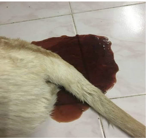
Figure 1: Bloody Diarrhea
Table 1: Show the clinical history of puppies
| Animal no. | History at clinic at 1st day |
| Dog1 | Diarrhea, Vomiting, fever 103 ˚F, vaccinated, dewormed, dull |
| Dog2 | Bloody diarrhea, Vomiting, fever 105 ˚F, vaccinated, dewormed |
|
Dog3 |
Diarrhea, Vomiting, fever 104 ˚F, vaccinated, dewormed, dull |
| Dog4 | Abdominal pain, low body temperature 99 ˚F, loss of appetite, bloating, dehydration, bloody diarrhea, dewormed |
| Dog5 | Bloody diarrhea, Vomiting, fever 103 ˚F, vaccinated, dewormed |
| Dog6 | Abdominal pain, Bloody diarrhea, Vomiting, fever 103 ˚F, vaccinated, dewormed |
| Dog7 | Abdominal pain, low body temperature 98 ˚F, loss of appetite, dehydration, bloody diarrhea |
| Dog8 | Abdominal pain, low body temperature 97 ˚F, loss of appetite, bloating, dehydration, bloody diarrhea |
History
Ten dogs (Labrador retriever and German shepherd) of three to four-month age was suffering from bloody diarrhea, abdominal pain, bloating, vomiting, loss of appetite, dehydration, high temperature (103-105˚F) and pulse rate at same kennel in Lahore, Pakistan as shown in Table 1 and Figure 1. Therefore, first shot of vaccination was carried out to start the therapeutic process, but the sudden attack of CPV caused death of 2 dogs immediately. Other dogs were also exhibited the severe conditions. Drenching was also done before the attack of CPV after vaccination.
Diagnose
Fecal examination showed no parasitic infection. Based on history, clinical signs, symptoms and hematology provide the clues for diagnosing as a canine parvovirus infection as shown in Figure 2.
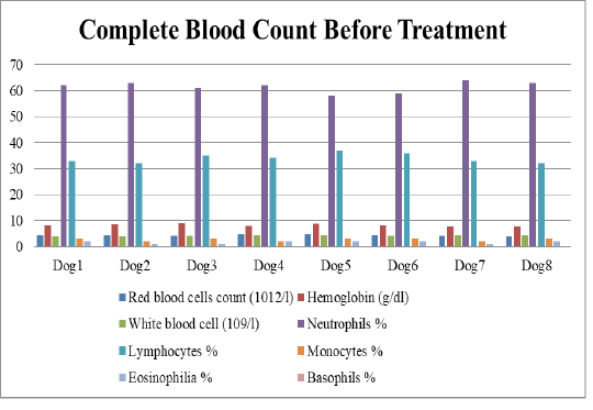
Figure 2: Complete blood count test show low amount of red blood cells, low haemoglobin and low amount of white blood cells.
Treatment
Symptomatic and supportive treatment were performed by using 5% Ringer lactate infusion along with Ranitidine (Anzole) @ 2ml/IV , Metamex (Metoclopramide) at 0.5mg/kg body weight. For diarrhea, Flagyl (Metronidazole) drip of 100 mL used daily for each puppy. Antibiotic Oxidil (Ceftriaxone Sodium) @ 2ml/IV used on daily basis for 5 days, Transamine (Traneximinic acid) 5mg/kg body weight, Injection (Inj.) Onset 8mg intravenous (IV) carried outfor three days, Haemoccel drip (20mL for each puppy) used for anemia on 3rd to 5th day in severe bloody diarrhea associated dogs (Singh and Sharma, 2008). For boost up the immunity against the parvovirus injection conglobe DHLaPPi used. But in these 5 days of treatment, 3 more dogs died and 5 dogs recovered as shown Figure 3. At 6th day of treatment, the owner(s) was advised to keep the unhealthy/infected dogs separatefrom the healthy dogs followed bycleaning the living area of these pups (Greene, 2012).
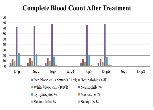
Figure 3: Complete blood count test after treatment of 5 days show great improvement in health status of puppies. Dog 4, 7, 8 has been expired due to severe condition.
Prognosis
The prognosis of the presented case was satisfactory because the owner followed the treatment regime as per standard protocol directed by Callvet (SMC) Pvt. Ltd. i.e., continuation of prescribed treatment.
Discussion
CPV is a highly contagious disease that is common in 1-6month age pups. The CPV infected dogs exhibited clinical signs such as anorexia, vomiting, dehydration, bloody diarrhea and paleness of mucosa.Initiation of therapeutic process with fluid and antibiotic showed significantimprovements in first 4 days and affected dogs with respective virus returned towards normal life in 6-8 days (Dongre et al., 2015). The results of the present case report are similar to the results of Munibullah et al. (2017) and Singh et al. (2008). The highest rate of CPV occurrence in young pups may be due to viral attraction for rapid multiplying intestinal crypt cells with the highest mitotic catalogue due to alternation in bacterial flora (Deka et al., 2013). CPV can be controlled by providing good nutrition, hygiene environment, reduce overcrowd and vaccinate the dogs timely (Odueko, 2019).
Conclusion
About 70-80 % of dogs can survive if the parvovirus disease detected as early as possible and start the proper treatment.
acknowledgements
Authors want to acknowledge Dr. Majid Hussain Somroo (Assistant Professor, Department of Veterinary Parasitology, Faculty of Veterinary Sciences, Shaheed Benazir Bhutto University of Veterinary and Animal Sciences, Sakrand-67210, Sindh, Pakistan) for his inspiring and encouraging support.
References


