The Journal of Advances in Parasitology
Research Article

Epidemiology of Tick-Borne Infection in Ruminants in Peshawar
Khurshaid Anwar
Veterinary Research & Disease Investigation Center Balogram, Swat, Pakistan.
Abstract | Haemoprotozoans are responsible for considerable economic losses in livestock in Pakistan. This study was carried out to determine the presence and distribution of tick-borne haemoprotozoan parasites (Theileria, Babesia and Anaplasma) in apparently healthy ruminants. In the present study, a total of 1101 blood samples (690 cattle (Bos indicus), 243 Buffaloes (Bubalus bubalis), 108 sheep, and 60 goats) were examined for blood parasites from June 2013 to June 2016. Microscopic examination of Giemsa-stained peripheral blood smears exhibited an overall prevalence of theileriosis in three years to be 67.39, 15.63, 15.74, and 0 %, babesiosis 15.79, 51.44, 14.81 and 41.66 %, anaplasmosis 2.9, 13.99, 29.63, and 1.66 % in cattle, buffaloes, sheep and goats respectively. The prevalence of theileriosis and babesiosis was significantly higher in cattle, buffaloes and sheep while goats showed low prevalence percentage of anplasmosis and Zero % of theileriosis. The present work emphasized that theileriosis was the most prevalent disease in cattle and need to be eradicated.
Keywords | Haemoprotozoans, Livestock, Buffaloes, Theileriosis, Cattle, Anaplasmosis
Editor | Muhammad Imran Rashid, Department of Parasitology, University of Veterinary and Animal Sciences, Lahore, Pakistan.
Received | March 01, 2018; Accepted | March 25, 2018; Published | March 28, 2018
*Correspondence | Khurshaid Anwar, Veterinary Research & Disease Investigation Center Balogram, Swat, Pakistan; Email: drkkhan06@yahoo.com
Citation | Anwar K (2018). Epidemiology of tick-borne infection in ruminants in peshawar. J. Adv. Parasitol. 5(1): 6-10.
DOI | http://dx.doi.org/10.17582/journal.jap/2018/5.1.6.10
Copyright © 2018 Anwar. This is an open access article distributed under the Creative Commons Attribution License, which permits unrestricted use, distribution, and reproduction in any medium, provided the original work is properly cited.
Introduction
Parasitic infections are one of the major constraints for profitable dairy industry and are of considerable economic importance in tropic and subtropics and bring losses to animals (Wright, 1989; Fadraga et al., 1991).
Haemoprotozoan infections are considered the most important blood parasites of cattle and buffaloes in Pakistan. Sporadic cases of haemoprotozoan diseases are seen throughout the year (Ashfaq et al., 1983; Muhammad et al., 1999). However, their Outbreaks in exotic and crossbred cattle are mostly reported during the hot and humid months (July, September) of the year. Occurrences of these parasites have been reported by Eren et al. (1998) and ElMetenawy (2000) in apparently healthy cattle. Muhammad et al. (1999) recorded 112 cases of theileriosis between March, 1993 and September, 1998 at Faisalabad and recommended further work on its sub-clinical infections.
Ruminants’babesiosis is caused by Babesia bigemina, Babesia bovis, Babesia divergens and Babesia major. Babesia species have the potential for wide distribution wherever their tick vectors are encountered. Two species, B. bovis and B. bigemina, have a considerable impact on cattle health and productivity in tropical and subtropical countries Uilenberg (1995).
Babesia is intraerythrocytic parasites of domestic animals and is the cause of anemia and haemoglobinuria. They are transmitted by ticks in which the protozoan passes trans-ovarially, via the egg from one tick’s generation to the next. Hard ticks of the family Ixodidae in which trans-ovarian infection ensures that Babesiais transmitted by stages of the next generation of ticks Urquhart et al. (1996). The acute cases of piroplasmosis may cause parasitemia and death in 20% of the infected animals Urquhart (1998).
Bovine anaplasmosis or gall sickness caused by the intraerythrocytic Rickettsia, Anaplasma marginale is transmitted biologically by infected ticks or mechanically by biting flies and contaminated fomites Ristic (1981). Acute anaplasmosis is characterized by fever, progressive hemolytic anemia, weight loss, abortion and even death resulting in significant loss to meat and milk production Alderink & Dietrich (1981). The economic loss due to infections caused by Babesia and Anaplasma infections in India was estimated US$57 million Tick Cost Version (1999). The genus Anaplasma also includes Anaplasma bovis, formerly referred as Ehrlichia bovis Dumler et al. (2001).
T.parvaand T.annulata sometimes T.mutan are important spp. responsible for theileriosis in cattle and small calf Ali et al. (2013). The diseases caused by haemoprotozoan are known as anaplasmosis, babesiosis, and theileriosis, respectively Ashfaq et al. (1983). These diseases are tick-borne because most of the transmission and dissemination of these diseases occurs via numerous genera of tick vectors Merk. Vet. Manual. (1998). Occurrence of these three haemoparasites are noted sporadically and cause disease throughout the year (Ashfaq et al.,1983; Muhammad et al., 1999; Eren et al., 1998; El-Metenawy 2000). The present study, therefore, was designed to determine the prevalence of haemoprotozoans in cattle, buffaloes and small ruminants in District Peshawar Pakistan.
Materials and Methods
A total of 1101 samples were examined in Veterinary Research Institute, Peshawar’s parasitology laboratory to determine the prevalence of haemoprotozoans in ruminants from June 2013 to June 2016.
The samples of whole blood (in EDTA) or blood smears of ruminants with symptoms such as anorexia, pyrexia, enlarged superficial lymph nodes, trembling in standing, pale conjuctival mucus membrane, haemoglobinuria, nasal discharge, coughing and grinding of teeth were screened for haemoprotozoan parasites and in the laboratory, the blood smears were fixed in methanol for 5 min and stained for 30 min in Giemsa stain diluted with 5% buffer. Slides were examined for intra-erythrocytic forms of Theilerai, Babesia and Anaplasma spp. under 100x objective magnification. Approximately 20,000 erythrocytes per slide were examined for the calculation of percentage of infected erythrocytes. The smears were recorded as negative for piroplasms if no parasites were detected in 50 oil-immersion fields.
Statistical Analysis
The data was analyzed by simple percentage method.
Results and Discussion
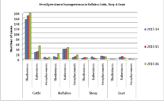
Among 1101 smears of (Cattle, Buffaloes, Sheep and Goats) screened by Giemsa’s stained blood smear, 882 (80.01%) were found positive for haemoprotozoan parasites (Table 5). Further, it was observed in the present study that the occurrence of these haemoprotozoan diseases were found to be high among the cattle followed by buffaloes, sheep and goats respectively during the course of the study from 2013-2016 (Figure 1).
The overall prevalence percentage of theileriosis in three years was found to be 67.39, 15.63, 15.74 and 0% in cattle, buffaloes, sheep and goats respectively. The prevalence of babesiosis in three years was found to be 15.79, 51.44, 14.81 and 41.66 % in cattle, buffaloes, sheep and goats respectively (Table 1-4). The prevalence of anaplasmosis in three years was found to be 2.9, 13.99, 29.63 and 1.66% in cattle, buffaloes, sheep and goats respectively (Table 1-4). The lowest prevalence in three years was recorded for theileriosis in goats which was zero percent (Table 4). This study does not agree with the findings of Mayzyad et al. (2002), they recorded 7.1 and 7.5% babesiosis and theileriosis in sheep and goats. In the same study they recorded 9.6 % and 8.1% babesiosisis and theleriosis in cattle. It might be concluded that the findings of our study disagree with the study of Mayzyad et al. (2002) may be due to their restricted area of consideration for the animals observed in North Sinai Governorate, Egypt and some epidemiological factors important for haemoprotozoan diseases.
The present findings revealed that ticks carrying sheep had higher prevalence (29.63%) of the parasite than buffaloes (13.99%) (Table 3 & 2). Because cattle are the main hosts of Anaplasma parasite Kuttler (1984) and buffaloes are susceptible to most of the diseases. Our study’s findings showed more positive cases in buffaloes than cattle for anaplasmosis. Water buffaloes are generally considered as healthier animal in comparison to cattle Johan (2001-2002). Our findings showed ticks-infestations in cattle and buffaloes are 2.9% & 13.99% (Table 1 & 2) which is not in accordance with the findings of Haider and Bilqees (1988), as their result was 61% in Karachi and adjoining areas. This difference may be due to the moderate climate and high relative humidity in Karachi that favor ticks-infestation. Ocaido et al. (2005) found 11.8% prevalence of A. marginale in cattle of Sorti District, Uganda, while in the present study it was 2.9% (Table-1).
This study is in partial agreement with the study conducted by Zabita et al. (2005), they reported 8.06% and 8.77% anaplasmosis and babesiosis only in cattle spp. respectively. Although microscopy is widely accepted and cost effective technique for the diagnosis of haemoprotozoans and haemorickettsial organisms, the technique lacks the higher sensitivity. The low prevalence observed in the study carried by Ristic and his team may be due to the fact that carrier cattle usually do not reveal inclusions in their blood films Ristic (1981). The findings in the study of Gale indicated the higher sensitivity and specificity of the molecular methods (Figueroa et al., 1993; Gale et al., 1996) compared to the conventional methods especially in detecting low level rickettsaemia seen in carrier animals. Microscopy could not detect a single case of A. bovis in the study of Sreekumar even though Sounderrajan & Rajavelu (2006) could detect 0.53 per cent out of 150 blood smears collected from Chennai. Sreekumar et al. (2000) couldn’t detect A. bovis in a blood smear of an infected buffalo and later identified the etiology by culturing the organism in blood mononuclear cells. All the above studies are in contradiction with our findings in which 0-34% A. bovis was detected in blood smears of infected animals. This might be due to high infestation and on time detection of the haemoprotozoans under the microscope and might be due to our year wise survey work on anaplasmosis in cattle as compare to their studies’ findings.
Table 1: Prevalence of Different Haemoprotozoans in Cattle in District Peshawar.
| Cases Positive for Haemoprotozoans | |||||
| Year | Total Cases | Theileriosis | Babesiosis | Anaplasmosis | Total Prevalence |
| 2013-14 | 195 | 115 (58.97%) | 27 (13.85%) | 8 (4.1%) | 76.92% |
| 2014-15 | 235 | 170 (72.34%) | 30 (12.77%) | 4 (1.7%) | 86.81% |
| 2015-16 | 260 | 180 (69.23%) | 52 (20%) | 8 (3.08%) | 92.30% |
| Total | 690 | 465 (67.39%) | 109 (15.79%) | 20 (2.9%) |
86.08% |
Table 2: Prevalence of Different Haemoprotozoans in Buffaloes in District Peshawar.
| Cases Positive for Haemoprotozoans | |||||
| Year | Total Cases | Theileriosis | Babesiosis | Anaplasmosis | Total Prevalence |
| 2013-14 | 77 | 10 (12.99%) | 39 (50.65%) | 7 (9.09%) | 72.73% |
| 2014-15 | 63 | 7 (11.11%) | 40 (63.49%) | 10 (15.87%) | 90.48% |
| 2015-16 | 103 | 21 (20.39%) | 46 (44.66%) | 17 (16.50%) | 81.55% |
| Total | 243 | 38 (15.63%) | 125 (51.44%) | 34 (13.99%) |
81.06% |
Table 3: Prevalence of Different Haemoprotozoans in Sheep in District Peshawar.
| Cases Positive for Haemoprotozoans | |||||
| Year | Total Cases | Theileriosis | Babesiosis | Anaplasmosis | Total Prevalence |
| 2013-14 | 37 | 4 (10.81%) | 9 (24.32%) | 12 (32.43%) | 67.57% |
| 2014-15 | 49 | 7 (14.29%) | 5 (10.20%) | 11 (22.45%) | 46.94% |
| 2015-16 | 22 | 6 (27.27%) | 2 (9.09%) | 9 (40.90%) | 77.27% |
| Total | 108 | 17 (15.74%) | 16 (14.81%) | 32 (29.63%) |
60.18% |
Table 4: Prevalence of Different Haemoprotozoans in Goat in District Peshawar.
| Cases Positive for Haemoprotozoans | |||||
| Year | Total Cases | Theileriosis | Babesiosis | Anaplasmosis | Total Prevalence |
| 2013-14 | 21 | 0 (0%) | 8 (38.1%) | 1 (4.76%) | 42.86% |
| 2014-15 | 23 | 0 (0%) | 11 (47.83%) | 0 (0%) | 47.83% |
| 2015-16 | 16 | 0 (0%) | 6 (37.5%) | 0 (0%) | 37.5% |
| Total | 60 | 0 (0%) | 25 (41.66%) | 1 (1.66%) |
43.33% |
The study was conducted for three years to observe the seasonal prevalence as well, a considerable seasonal variation was found with the occurrence of haemoprotozoan disease in animals. Most of the animals suffered during monsoon months, which might be due to more number of ticks in monsoon which were developed during summer months. This is in accordance with the observations made by (Roy et al., 2004; Ananda et al., 2009) they found highest prevalence in monsoon months. In the Kenya highlands (Deem et al., 1993; Gitau et al., 1994; Gitau et al., 1997; O’Callaghan, 1998; Perry et al., 1990; Gitau et al., 2000) have demonstrated that the prevalence of Theileria parva infections can vary significantly by zones and grazing system and that these differences have important implications for both the impact and control of East Coast Fever.
Table 5: Overall Prevalence of Haemoprotozoans in Buffaloes, Cattle, Sheep and Goats in District Peshawar, and Periphery, Khyber Pakhtunkhwa, (Pakistan).
| Haemoprotozons (Theileria, Babesia and Anaplasma) in Buffaloes, Cattle, Sheep and Goats | |||
| Year | Total Examined Cases | Total Positive Cases | Overall Prevalence |
| 2013-2016 | 1101 | 882 | 80.01% |
Our findings are in partial agreement with the epidemiological survey conducted by Kokab (1996) in cloven footed animals in Khyber Pakhtunkhwa; who reported 44.27 and 27.11 % babesiosis and theileriosis and in our study it was 67.39 15.79 % for cattle respectively. The aggregate infection percentage for babesiosis and anaplasmosis was 51.44 and 13.99 %, in buffaloes while it was 15.63% for theilariosis. In sheep the aggregate percentage of infection prevalence recorded fortheilariosis was 15.74 %, babesiosis 14.81 % and anaplasmosis was 29.63 % respectively, which was the highest prevalence percentage of infection in all the spp. for anaplasmosis. The aggregate infection percentage of babesiosisand anaplasmosis in goats was 41.66 and 1.66 %, respectively (Table 4). The incidence of babesiosis infection in goats was second to highest in buffaloes (41.66 %) as shown in (Table 4). The findings of the study revealed that, the prevalence of different blood protozoan infection was different in all the four animal species and there is no co-relation of infection prevalence among the different species.
The lowest prevalence of theileriosis recorded in goats may be due to the facts that most of the flock owners do not care for their small ruminants and the animals expire without any laboratory diagnosis. Another reason might be that the low presence of vector ticks in goats may be responsible for low prevalence of theileriosis infection. The pattern of the present study was different from all other studies conducted so for in this regard. This study encircles four different animal species and three blood parasites, so the prevalence for different haemoparasitic infection cannot be correlated with the studies being conducted on one animal spp. or one blood protozoan.
Recommendations
This study recommends proper steps to be taken by the Livestock Department, including farmers’ awareness through mass media campaign for tick control measures, acaricidal spray through arranging field days, timely laboratory diagnosis of protozoan diseases and close contact between farmers and Veterinary Research Institutes.
Acknowledgements
The author is grateful to the staff who had contributed their valuable data for this study during their tenure in the Veterinary Research Institute, Peshawar and I also sincerely extend my gratitude to my father late Mr. Khuna Gul (Retired Principal, Education Department) for his constant guidance in compilation of this research work.
Conflict of interest
The author declares that he has no competing interests.
Authors Contribution
Dr. Khurshaid Anwar Senior Research Officer VR & DIC Balogram, Swat designed, carried out the experiment, performed the computation, read and approved the research article.
References


