The Journal of Advances in Parasitology
Research Article
The Journal of Advances in Parasitology 1 (2): 27 – 29Thyroxin Levels and Haematological changes in Dogs with Sarcoptic Mange
Bhavanam Sudhakara Reddy 1*, Karumuri Nalini Kumari 2, Sirigireddy Sivajothi3
2. Department of Veterinary Medicine, College of Veterinary Science, Sri Venkateswara Veterinary University, Tirupati – 517 502, Andhra Pradesh. India
3. Department of Veterinary Parasitology, College of Veterinary Science, Sri Venkateswara Veterinary University, Proddatur – 516360, Andhra Pradesh. India
*Corresponding author: bhavanamvet@gmail.com
ARTICLE CITATION:
Reddy BS, Kumari KN, Sivajothi S (2014). Thyroxin Levels and Haematological changes in Dogs with Sarcoptic Mange ge. J. Adv. Parasitol. 1 (2): 27 – 29.
Received: 2014–04–25, Revised: 2014–05–05, Accepted: 2014–05–05
The electronic version of this article is the complete one and can be found online at
(
http://dx.doi.org/10.14737/journal.jap/2014/1.2.27.29
)
which permits unrestricted use, distribution, and reproduction in any medium, provided the original work is properly cited
ABSTRACT
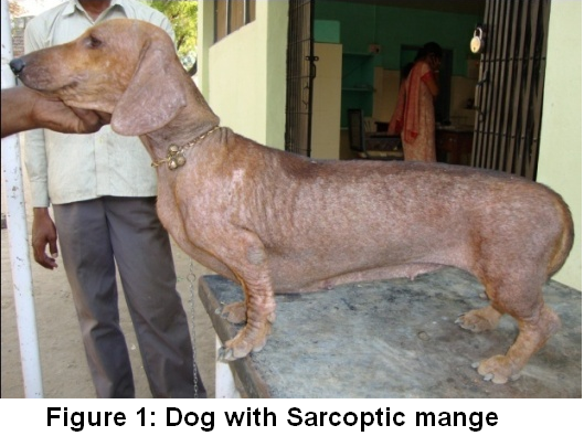
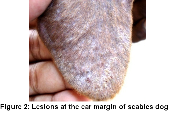
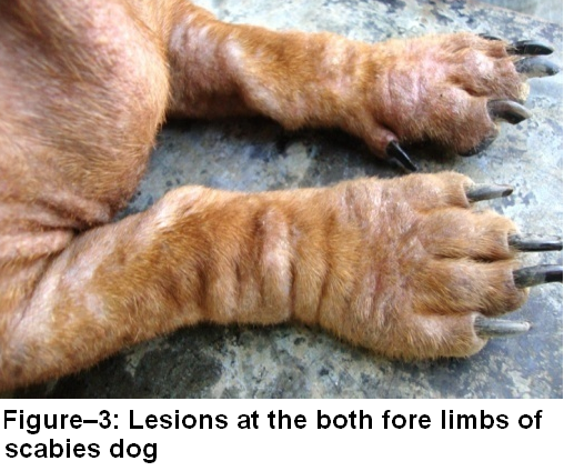
Sarcoptic mange is one of the common contagious skin diseases affecting all the domestic animals through host–adapted variants. In the present study sixteen dogs with scabies was noticed. All the dogs had intense itching, restlessness, frantic scratching, hair loss, reddened skin all over the body and scabs. Distributions of the lesions were noticed at the margins of the ears, elbows, face and legs. Haematological abnormalities include reduced total erythrocyte count and haemoglobin concentration levels. Affected dogs also showed leukocytosis with neutrophilia, eosinophilia and monocytosis. Biochemical changes included reduced mean total serum protein and serum albumin levels when compare with healthy dogs. Reduced total T4 (2.32±0.25µg/ dL) and free T4 (1.37 ±0.04 ng/dL) levels were also recorded in this study.
INTRODUCTION
Scabies is a transmissible and zoonotic ectoparasitic skin infection caused by tiny mites of the species Sarcoptes scabiei. It is transmitted readily among the animals, often even throughout an entire household, by skin–to–skin contact. The parasite commonly affects young dogs and dog with poor nutrition, but can affect healthy dogs who are exposed to the mites. Scabies in humans is most prevalent in overcrowded and poor hygienic conditions. Zoonotic importance of scabies was also recorded in Andhra Pradesh (Reddy and Kumari, 2013). It was felt to be a disease of overcrowding and poverty rather than a reflection of poor hygiene (Heukelbach, 2005). Scabies is normally an intensely uncomfortable skin disease due to itchiness caused by the parasite. This pruritus is thought to be due to a hypersensitivity to proteins found in the fecal matter produced by Sarcoptes mites as they burrow through the layers of the epidermis. Dogs with scabies were an intensely prurutic, scratches, rubs almost constantly and uncomfortable. In many animal species, the prevalence of scabies is very high and often confers the death of the untreated animals.Presence of Sarcoptic mange was also responsible for development of recurrent pyoderma in dogs (Reddy et al., 2014a). Literature was available on reports on Sarcoptic mange in different animals (Qianqian Xu et al., 2013; Sivajothi et al., 2013). But, no literature was available on haemato biochemical changes and thyroxin levels in dogs affected with scabies. So, present study was conducted to record the thyroxin changes along with other haemato–biochemical findings in dogs affected with scabies.
MATERIALS AND METHODS
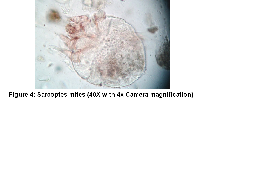
Present study was carried out from 2009 to 2012 at College Hospital of College of Veterinary Science, Tirupati. Dogs with dermatological problems were regularly screened for the Sarcoptic infestation. Superficial skin scrapings, deep skin scrapings, slide impression smears, hair plucks and tape impression smears were collected from the various sites of all the suspected dogs. Out of 200 dogs examined with dermatological problems 16 dogs were found to be affected with Sarcoptic mange mites. All the dogs with scabies had papules, scales, hyperpigmentation, erythema, alopecia and pustules in few cases. Eczematous lesions on the face, margins of ears, trunk, legs, elbows, hocks, chest and ventral abdomen were noticed (Figure 1, 2, 3). All the dogs of the ear flaps (pinnae) were gently rubbed and examined for their hind leg scratching motion, because the pinnae are extremely itchy in most of the infested dogs. All the dogs exhibited pinnal pedal reflex. Superficial skin scrapings from all the dogs revealed ova and adult live mites (Figure 4). Based on the clinical and microscopic examination of the scrapings dogs were identified as they are suffering with the scabies. From all the effected dogs and ten apparently healthy dogs’ blood was collected in 10% EDTA coated vials. Blood was processed for packed cell volume (PCV) by microhaematocrit method, haemoglobin (Hb) by Sahli’s method, total leucocyte count (TLC) and total erythrocyte count (TEC) by dilution method. The blood smears were directly prepared and stained by Leishman’s stain for differential count (DLC) with battlement method and absolute counts were also calculated. Serum was collected from all the dogs and stored at deep freezer (–20°C) until serum analysis was done. Serum total protein, albumin and total cholesterol were estimated by using Span diagnostics Ltd. Kits. Total T4 and free T4 levels were estimated by ELISA (as per manufacture’s procedure) using a kit obtained from United Biotech Inc. (Reddy et al., 2014b). The data in respect of each parameter was tabulated group wise and statistical analyses were carried out by using Statistical Package for Social Sciences (SPSS) using student “t” test to compare between infested and control dogs; 𝑃 value of 0.05 or lower was considered to be significant.
RESULTS AND DISCUSSIONS
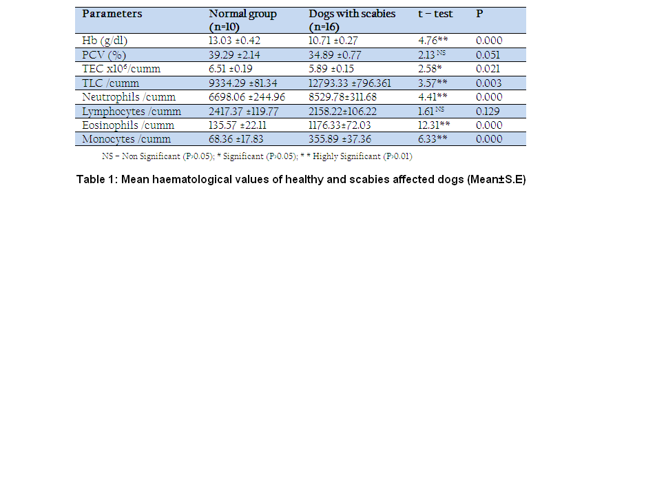
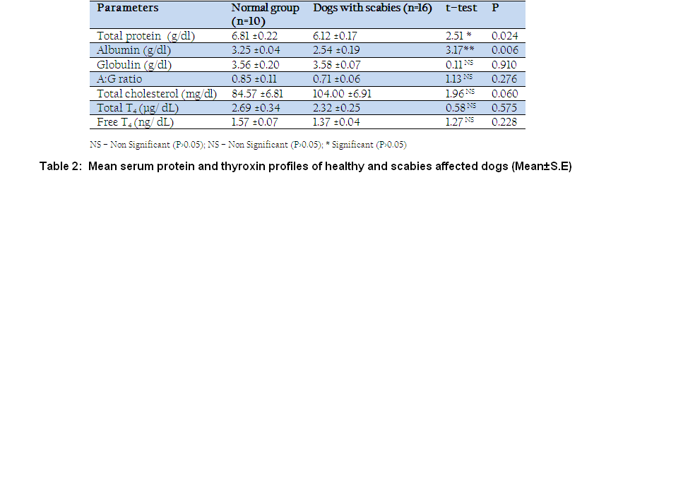
The mean haematology and serum biochemistry values of apparently healthy dogs and dogs with scabies are given in Tables 1 and 2. The haemogram of apparently healthy dogs was within the normal reference range as quoted by Villers (2005). Dogs with scabies had reduced mean Hb concentration (10.71 ±0.27g/dl) range from 9.8 g/dl to 12.2 g/dl. Mean PCV levels was 34.89 ±0.77 per cent range of 33–37 percent. TEC ranged from 5.62 ±0.15 x 10⁶/cumm to 6.15 x 10⁶/cumm with mean of 5.89 ±0.15 x 10⁶/cumm. Statistically significant difference was noticed in the decreased PCV levels. The decrease in the values of haemoglobin and TEC might be due to anaemia caused by the loss of skin protein and stress arising from the disease. Similar findings were reported by Gupta and Prasad (2001) and Biswas et al. (2002). Significant increased in mean total leucocyte count (12793.33 ±796.361), mean absolute neutrophil count (8529.78±311.68/cumm), mean absolute eosinophil count (1176.33±72.03/cumm), mean monocytes (355.89 ±37.36/cumm) was noticed. Reduction in the mean lymphocytes (2158.22±106.22/cumm) was noticed but, it was not significant in this study. Dadhich and Khanna (2008) also observed the same findings in their studies. Decreased levels of haemoglobin may be due to chronicity and generalized nature of the disease (Ferreira et al, 1987). It might also be due to the significant low erythrocyte count and erythrocyte fragility and low production of erythropoietin due to toxaemia caused by mites. Leucocytosis might be due to allergic reaction caused by mite or their products of inflammatory reaction (Shah, 1994). The serum biochemical values of apparently healthy animals recorded were within the normal range as reported by Villers (2005). Dogs with scabies revealed lowered mean total serum protein levels 6.12 ±0.17g/dl (range from 5.86 to 7.21 g/dl) and reduced mean serum albumin levels 2.54 ±0.19 with range from 2.14 g/dl to 3.24 g/dl. Serum globulin levels range from 3.14 g/dl to 4.10 g/dl with mean of 3.58 ±0.07 g/dl was recorded. Hypoproteinaemia which was in agreement with the observations of Biswas et al. (2002) and Solanki et al.(2007). The mean total T4 values of the normal and scabies affected dogs were 2.69 ±0.34 µg/dL and 2.32±0.25µg/dL. In this slight reduction in the mean levels was noticed, but there is no statistically significant was noticed when compare with healthy dogs. The mean free T4 value also (1.37±0.04 ng/dL) reduced than the normal group (1.57±0.07 ng/dL) without any significant levels. In this study dogs with scabies did not show any hypothyroid signs.All the dogs with scabies were treated with oral administration of ivermectin @ 200 µg/ kg body weight, along with supportive therapy. Previously different types of mange were treated with oral ivermectin at different dose rates in dogs and cats (Reddy and Kumai 2010, Sivajothi et al. 2014; Reddy and Sivajothi 2014c). Presence of severe itching due to the mites all over the body, there is a chance of proliferation of staphylococci which leads to severe damage to the skin. In severe cases ulcers can also observed due to secondary bacterial infections (Reddy BS et al., 2014a). In the present study the decreased levels of haemoglobin and protein were reported. It suggests that dogs with scabies should require high protein diet and heamatonics in their feeding schedule.
CONCLUSION
Dogs affected with scabies revealed significant reduction in total erythrocyte count, haemoglobin concentration. Leukocytosis with neutrophilia, eosinophilia was noticed. Reduced total serum protein and serum albumin levels were recorded.
ACKNOWLEDGEMENT
All the authors are thankful to Sri Venkateswara Veterinary University for providing facilities to carry out this work. Corresponding author would like to express his thanks to local field veterinarians.
CONFLICT OF INTEREST
No conflict of interest.
REFERENCES
Biswas L, Mukhopadhyay SK, Bhattacharya MK, Roy S (2002). Journal of Interacademicia. 6: 734–736.
Dadhich H, Khanna R (2008). Pathological, heamatobiochemical and immunological studies of cutaneous ectoparasitoses in dogs. Proceedings of the 15th Congress of FAVA, Bangkok, Thailand. Pp. 409–412.
Ferreira H, Figueiredo C, Burini RC, Curi PR (1987). Serum vitamin levels, erythrocyte and lymphocyte counts, packed cell volume and haemoglobin determinations in normal dogs. Dogs with scabies and dogs with demodicosis. Arq. Bras. Med. Vet. 39: 905–917.
Gupta N, Prasad B (2001). Clinico diagnosis and therapeutic management of acariasis in dogs. Indian. Vet. Med. 21:73–75.
Heukelbach J, Walton SF, Feldmeier H (2005) Ectoparasitic infestations. Curr. Infect. Dis. Rep. 7:373–380.
http://dx.doi.org/10.1007/s11908-005-0012-2
PMid:16107235
Qianqian Xu, Shijin Guo, Jichang Li, Yubo Wang, Zhiqiang Shen, Yanping Wang, Ying Zhang, Zhimei Zhang, Shijun Fu, Li Ma, Limei Yang, Jianjun Wang, Duanhui Ma (2013) The Efficacy of Milbemycin Oxime in the Treatment of Naturally Acquired Infestations of Sarcoptes Scabiei on Dogs. International Journal of Veterinary Health Science & Research 1:201.
Shah H (1994). Clinico–biochemical and therapeutic studies on mange in dogs. M.V.Sc thesis, IVRI, Izatnagar, India.
a name="Solanki2007"> Solanki JB, Hasnani JJ, Patel DM, Patel PV, Raval S.K. (2007). Canine demodicosis in Anand. J.Vet. Parasitol. 21(1): 79–80.
Reddy BS and Kumari KN (2010) Demodicosis and its successful management in dogs. Indian journal of field veterinarians, 2010, 6(2), 48-50.
Reddy BS, Kumari KN. (2013) Canine Scabies – Its Therapeutic Management and Zoonotic importance. Intas Polivet. Vol. 14 (II): 292–294.
Reddy BS, Kumari KN, Rao VV, Rayulu VC (2014a) Efficacy of Cefpodoxime with Clavulanic Acid in the Treatment of Recurrent Pyoderma in Dogs Hindawi Publishing Corporation ISRN Veterinary Science Volume 2014, Article ID 467010, 5 pages.
Reddy BS, Kumari KN, Sivajothi S (2014b) Haemato–biochemical findings and thyroxin levels in canine demodicosis. Comp Clin Pathol. DOI 10.1007/s00580–014–1893–y.
http://dx.doi.org/10.1007/s00580-014-1893-y
Reddy BS and Sivajothi S (2014c) Notoedric Mange Associated With Malassezia in Cats. International Journal of Veterinary Health Science & Research 2:101.
Sivajothi S, Reddy BS, Rayulu VC (2013) Management of Sarcoptic Mange in Rabbits. International Research Journal of Life Sciences. Vol. 1 (1) Sept' 13. 21 – 25.
Sivajothi S, Reddy BS, Rayulu VC, Sreedevi C. (2014). Notoedres cati in cats and its management. J Parasit Dis. DOI 10.1007/s12639-013-0357-7.
http://dx.doi.org/10.1007/s12639-013-0357-7
Villers E (2005). Introduction to haematology. In: BASAVA manual of canine and feline pathology" 2nd Edn, Villers E and Blackwood L, BASAVA, Gloucester, UK, pp:33–60.


