The Journal of Advances in Parasitology
Case Report
The Journal of Advances in Parasitology 2 (2): 21 – 23Ear Mite Otodectes Cynotis) Induced Otitis Externa and Complicated by Staphylococci Infection in a Persian cat
Muhammad Ahaduzzaman
*Corresponding author: zaman.cvasu@gmail.com
ARTICLE CITATION:
Ahaduzzaman M (2014). Ear mite (Otodectes cynotis) induced otitis externa and complicated by staphylococci infection in a persian cat. J. Adv. Parasitol. 2 (2): 21 – 23.
Received: 2014–02–12, Revised: 2014–04–03, Accepted: 2014–04–13
The electronic version of this article is the complete one and can be found online at
(http://dx.doi.org/10.14737/journal.jap/2014/2.2.21.23)
which permits unrestricted use, distribution, and reproduction in any medium, provided the original work is properly cited
Abstract
Ear mite induced otitis externa complicated with Staphylococci was determined in a two month old Persian cat. For treatment, two doses of ivermectin @ 0.2 mg / Kg body weight were given to 0.3% ciprofloxacin otic drop for seven days. After seven days post treatment clinical symptoms disappeared and no agents of Otodectes cynotis were identified by microscopic examination.
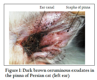
Otodectes cynotis commonly known as ear mite is non–burrowing, white and very active parasite belongs to the member of Psorptidae and the most common cause of ear infection in cats while it is less common in dogs (Maazi et al., 2010). The host’s range of this highly contagious mite is wide enough and includes cats, dogs, foxes, ferrets and infrequently in humans (Wiwanitkit, 2011). The cat plays a vital role in transmission of ear mite in adult dogs, rabbit and ferret (Sasikala et al., 2011). Cat infested with ear mite results in itching sensation and otitis externa which is manifested by erythematous ear canal and the presence of dark brown ceruminous otic exudates, not necessarily all cats will display symptoms of ear mites but often they will scratch at their ears or shake their heads (Wiwanitkit, 2012). Secondary bacterial (mainly Staphylococci) and fungal (Malassezia spp.) infections are also very common in cats affected by the ear mite as being most frequently isolated microorganisms in such cases (Roy et al., 2011). Evidences of risk factors such as age, sex, breed, shape of ear, type and length of hair effect are usually controversial but contact with other animals may act as potential risk factors (Souza et al., 2008). The degree and extent of damage depend on duration of illness, housing system, litter mates, secondary infection and physiological status of the cat (Khara, 2014). The diagnosis and management of the disease is laborious and efficacy is controversial in complicated cases as most of the owners are not aware about the ear canal of their pets incomparable to the body coat and also a burden for the veterinarian to treat due to unavailability of suitable veterinary product to combat the condition (Jacobs, 2000). Therefore, the current therapy was taken to find out the therapeutic outcome of otitis externa caused by O. cynotis complicated with secondary bacterial infection. Two months old male Persian cat was admitted to the Thana Veterinary Hospital (Metro), Boalia, Rajshahi, Bangladesh with a history of head shaking and ear scratching. Physical examination revealed bilateral excessive dark brown ceruminous exudates softly attached to the inner surface of pinnae. The exudates were crustier towards the ear canal than the scapha of pinnae (Figure 1). No offensive odor or other abnormalities were identified in the ear.
Three smears of ear discharge were made for both parasitological and bacteriological examinations by cotton swabs. Smears were stained with Gram’s staining for evaluation of bacteria as per the procedures described by Merchant and Packer (1969). On the other hand, direct microscopic examination of ear swab was also performed for parasitological examination under light microscope as described by Richard and David (2001). Audiometry test was done to test the hearing ability of the cat according to the protocol described by Thompson et al. (2013). The cat was treated with two doses of Ivermectin @ 0.2 mg / Kg body weight subcutaneously (IvertinR, 1%, Chemist Laboratories Ltd., Bangladesh). Second dose was given after 15 days interval to break re–infection cycle. Ciprofloxacin 0.3% ear drop (CiproxR; 10 ml, Opso Saline Ltd., Bangladesh) was used two times a day for seven days.
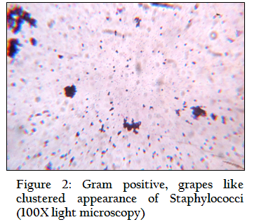
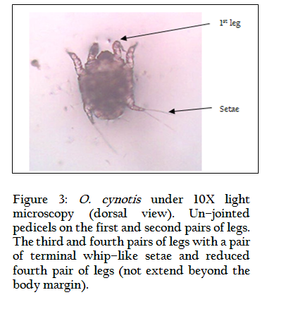
Figure 3: O. cynotis under 10X light microscopy (dorsal view). Un–jointed pedicels on the first and second pairs of legs. The third and fourth pairs of legs with a pair of terminal whip–like setae and reduced fourth pair of legs (not extend beyond the body margin).
Staphylococci were found in ear cerumen that was confirmed by culture morphology, appearance and property on Mannitol salt agar (selective media) followed by Gram’s staining visualization of the bacteria under the microscope (Figure 2). On microscopic examination of ear cerumen four parasites characteristics of adult O. cynotis was found (Figure 3). Following seven days post therapy cat ear became normal without exudation and irritation. No parasites were identified by re–examination. The cat noticed normal respond toward the source of sound and was interpreted as normal hearing ability
Otodectes cynotis mites are surface mite that posse’s otitis externa by harboring ear wax in cats and dogs manifested by intense itching in one or both ears, which in turn triggers scratching at the affected ear. During feeding of superficial epidermis it can cause a hypersensitivity reaction and pyogenic secondary infection. Bacterial infection particularly Staphylococci are common findings due to their normal inhabitant of the skin, nose and ear canal and become pathogenic due to concurrent infection with Otodectes cynotis as supported by Petrov et al. (2013). Salib and Baraka (2011) also found that specific and mixed infestation of Otodectes cynotis in cats was (24.56%) and (6.57%) respectively in otitis externa.
DISCUSSION
Age is an important parameter on which pathogenic involvement, prevalence, severity, stage and rate of infection depends on. Lefkaditis et al. (2009) described more susceptibility in kitten between 3–6 months of age, but Thompson (2011) found younger cats are more vulnerable to serious mite infestations than adults and this is because the immunity of cats towards mites develops and builds up with age that agreed with our findings as we studied cat was two months old.
There are a number of approved medications for ear mites in many countries of the world. Many of these products can be applied directly into the external ear canal depending on availability. Ivermectin is effective against a wide group of external and internal parasites. The pharmacokinetics of ivermectin in cats receiving a single dose of 0.2 mg / Kg by subcutaneous injection revealed a rapid absorption, high distribution, slow elimination and high possibility for the elimination as described by Chittrakarn et al. (2009). Use of topical otic antibiotic is a common practice among the veterinarian to treat ear infection with a good success rate. Mösges et al. (2011) stated that fluroquinolones (ciprofloxacin) showed a trend towards the superiority of cure rate in comparison to non quinolones. Ciprofloxacin is a drug of choice against Staphylococci in the developing country like Bangladesh, where antibiogram rate is high (Ahaduzzaman et al., 2014).
Cat ear mite infection is irritating and challenging clinical problem for its contagious and unwelcome disease nature. Systemic ivermectin was used in our study with a satisfactory outcome. And it may be recommended that systemic therapy with ivermectin can also be taken with the success rate instead of acaricidal otic drop where availability of this drug is inadequate.
ACKNOWLEDGEMENTS
Author grateful to the animal owner, livestock officer (TLO) and District Livestock Office, Rajshahi for providing laboratory support.
REFERENCES
Ahaduzzaman M, Hassan MM, Alam M, Islam SKMA and Uddin I (2014). Antimicrobial resistance pattern against Staphylococcus aureus in environmental effluents. Res. j. vet. pract. 2(1): 13–16.
Chittrakarn S, Janchawee B, Ruangrut P, Kansenalak S, Chethanond U, Kobasa T and Thammapalo S (2009). Pharmacokinetics of ivermectin in cats receiving a single subcutaneous dose. Res. Vet. Sci. 86(3): 503–507.
http://dx.doi.org/10.1016/j.rvsc.2008.08.005
PMid:18835001
Jacobs DE (2000). Selamectin – a novel endectocide for dogs and cats. Vet. Parasitol. 91:161–162.
http://dx.doi.org/10.1016/S0304-4017(00)00288-0
Khara K (2014). Ear mites in dogs. (http://www.ibuzzle.com/articles/ear-mites-in-dogs.html). Retrive: 23.02.2014.
Lefkaditis MA, Koukeri SE and Mihalca AD (2009). Prevalence and intensity of Otodectes cynotis in kittens from Thessaloniki area, Greece. Vet. Parasitol. 163: 374–375.
http://dx.doi.org/10.1016/j.vetpar.2009.04.027
PMid:19520513
Maazi N, Jamshidi SH and Hadadzadeh HR (2010). Ear Mite Infestation in Four Imported Dogs from Thailand; a Case Report. Iran J. Arthropod–Borne Dis. 4(2): 68–71.
PMid:22808403 PMCid:PMC3385556
Merchant IA and Packer RA (1969). Veterinary Bacteriology and Virology. 7th edn., The Iowa State University Press, Ames, Iowa, USA. 211–305.
Mösges R, Nematian–Samani M, Hellmich M and Shah–Hosseini K (2011). A meta–analysis of the efficacy of quinolone containing otics in comparison to antibiotic–steroid combination drugs in the local treatment of otitis externa. Curr. Med. Res. Opin. 27(10): 2053–2060.
http://dx.doi.org/10.1185/03007995.2011.616192
PMid:21919557
Petrov V, Mihaylov G, Tsachev I, Zhelev G, Marutsov P and Koev K (2013). Otitis externa in dogs: microbiology and antimicrobial susceptibility. Revue Méd. Vét. 164(1): 18–22.
Richard W and David S (2001). Veterinary Ectoparasites: Biology, Pathology and Control. Blackwell Science Ltd. 215–225.
Roy J, Bedard C and Moreau M (2011). Treatment of feline otitis externa due to Otodectes cynotis and complicated by secondary bacterial and fungal infections with Oridermyl® auricular ointment. Can. Vet. J. 52: 277–282.
PMid:21629420 PMCid:PMC3039897
Salib FA and Baraka TA (2011). Epidemiology, genetic divergence and acaricides of Otodectes cynotis in cats and dogs. Vet. World. 4(3): 109–112.
Sasikala V, Saravanan M, Ranjithkumar M, Sarma K, and Vijayakaran K (2011). Management of ear mites in cats. Indian Pet J. 11: 5–9.
Souza CP, Ramadinha RR, Scott FB and Pereira MJS (2008). Factors associated with the prevalence of Otodectes cynotis in an ambulatory population of dogs. Pesq. Vet. Bras. 28(8): 375–378.
http://dx.doi.org/10.1590/S0100-736X2008000800005
Thompson A (2013). How to test your dog's hearing; instructions–2. (http://www.ehow.com/how_5693288_test-dog_s-hearing.html). Retrive: 23.08.2013.
Thompson T (2011). Ear mites in cats: A study of diagnosis and symptoms (http://www.nativeremedies.com/petalive/articles/ear-mites-cats-study-diagnosissymptoms.shtml). Retrieved on 10.02.2014.
Wiwanitkit V (2011). Dog ear mite infestation: a possible problem in public health system. Iran J. Arthropod–Borne Dis. 5(2): 1.
PMid:22808412 PMCid:PMC3385579
Wiwanitkit V (2012). Role of Molecular Diagnosis for Dog Ear Mite Infestation. Int. J. Mol. Vet. Res. 2(2): 6–7.


