The Journal of Advances in Parasitology
Research Article
J. Adv. Parasitol. 8(3): 32-36
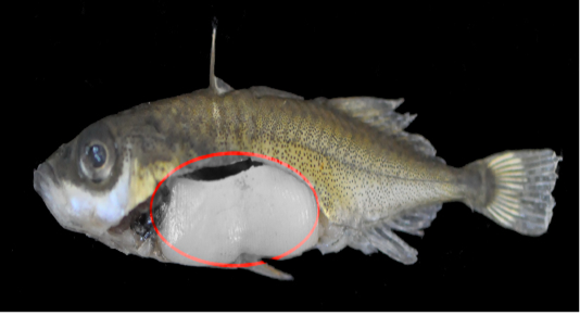
Figure 1
G. aculeatus individual infected with Schistocephalus solidus, 46.24 mm TL, May 2020 (S. solidus, circled in red.)
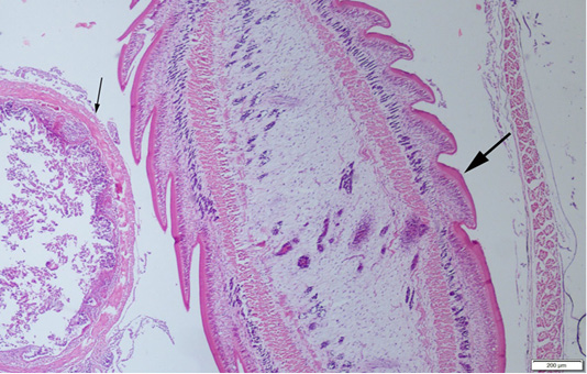
Figure 2
A plerocercoid (thick arrow) in abdominal cavity of the fish, near the intestine (thin arrow) of the host, HE, Bar=200µm.
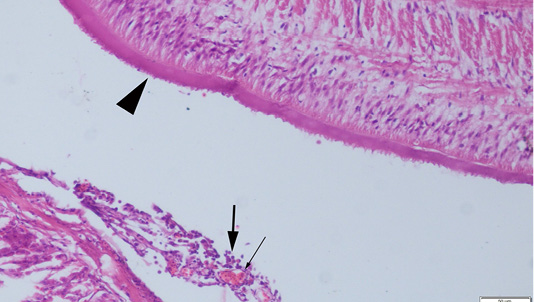
Figure 3
Serosal surface of the fish intestine, hyperemia of the vessels (thin arrow) and inflammatory cell infiltration (thick arrows) near the plerocercoid (arrow head), HE, Bar=50µm.


