The Journal of Advances in Parasitology
Research Article
J. Adv. Parasitol. 8(1): 9-12

Figure 1
Morphological features analyses (A): Visceral organs cut open to show the parasite load. (B): Whole mount of the parasite, Bars=2 mm.
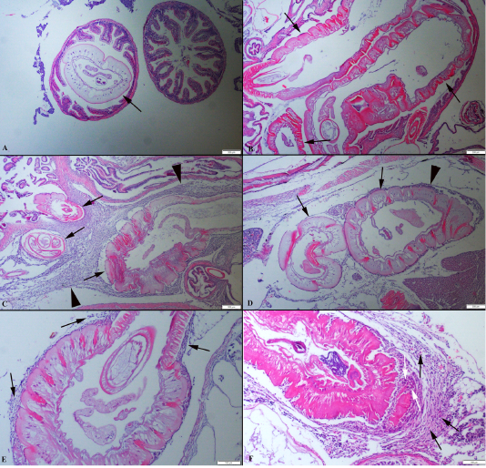
Figure 2
(A): Histopathological aspects of intestine one parasite localised in the lumen. (B): Three parasites (arrows) in an intestinal section. (C) Numerous parasites (arrows) localised abdominal cavity surrounded by severe inflammatory reaction (arrow head). (D) Two different parasites (arrows) near the hepatopancreas and moderate inflammatory reaction (arrow head), HE, Bars=200 µm. (E) Higher magnification of the inflammatory reaction around the parasite (arrows). (F) Higher magnification of the mononuclear cells (black arrows) and macrophages (white arrows), HE, Bars=100 µm.


