Advances in Animal and Veterinary Sciences
Research Article

Herd Report: Outbreak of Mixed Dermatophilosis and Pox Infection in Camels (Camelus dromedarius) in South Iraq
Muna Tawfeeq Abd
Veterinary Medicine College, Al-Muthanna University, Iraq.
Abstract | This report describes for the first time an outbreak of dermatophilosis that occurred simultaneously with pox infection in pastoralist camel herd heavily infested with tick in south of Iraq in April to May 2017. The infected animals were examined clinically and the outbreak was reported as mixed infection of camelpox and dermatophilosis. Hair matting particularly on the head, neck, and abdomen was the common skin lesion that observed on the infected animals. Lesions showed hairless brownish crusts with irregular sizes and pus exudation that left irregular border crusted lesions. In addition, evaluated camels were infected with pox papules that distributed over all parts of the body accompanied with fever and enlargement of lymph node. Some of the affected camels revealed systemic form of the camelpox. The lesions appeared on the mucous membranes of the mouth and respiratory tract and some case fatality recorded. However, dermatophilosis in camels has not previously been reported in Iraq. In conclusion this report focused on mixed infection of camelpox and dermatophilosis in herd of the local Iraqi camel. The author recommends to do a future study on these diseases, as dermatophilosis is reported for the first time in Iraq and need more investigation to understand its epidemiology because camel herds are moving continuously, and also veterinary service are not consulted for diagnosis and treatment.
Keywords | Dermatophilosis, Camels, Pox, Ticks.
Editor | Kuldeep Dhama, Indian Veterinary Research Institute, Uttar Pradesh, India.
Received | March 06, 2018; Accepted | May 28, 2018; Published | August 06, 2018
*Correspondence | Muna Tawfeeq Abd, Veterinary Medicine College, Al-Muthanna University, Iraq; Email: munatawfeeq87@mu.edu.iq
Citation | Muna Tawfeeq Abd (2018). Herd report: outbreak of mixed dermatophilosis and pox infection in camels (camelus dromedarius) in south Iraq. Adv. Anim. Vet. Sci. 6(8): 321-324.
DOI | http://dx.doi.org/10.17582/journal.aavs/2018/6.8.321.324
ISSN (Online) | 2307-8316; ISSN (Print) | 2309-3331
Copyright © 2018 Muna Tawfeeq Abd et al. This is an open access article distributed under the Creative Commons Attribution License, which permits unrestricted use, distribution, and reproduction in any medium, provided the original work is properly cited.
Introduction
Dermatophilus congolensis infection is an acute, subacute or chronic skin disorder (Zaria et al., 1993). Dermatophilosis affects wide host range including cattle, sheep and horse (Awad et al., 2008) and usually camel (Gitao,1992; Gitao et al., 1990) and cats (Kaya et al., 2000). Dermatophilosis worldwide and can be epizootic in tropical and subtropical areas of the world (Zaria et al., 1993; Radostits et al., 2007). Clinical findings are extensive skin matting with dark brown scabs or crusts over the abdomen and hind limbs on flanks, shoulders, and neck (Khodakaram-Tafti et al., 2012).The hair were matting together into small tufts forming a characteristic “paint brush” effect in the early lesions (Gitao et al., 1990). Most of the skin may be covered by powdery crust with varying degree of alopecia and young camels tend to be more severely affected (Gitao et al., 1998b), under the effect of stress and skin damage, tick infestation and increment in humidity and rainfall (Radostits et al., 2007) and malnutrition (Sanders et al.,1990). Dermatophilosis can be treated using long acting oxytetracycline, 2 doses for 3 days apart, in addition to oral administration of potassium iodide 10 gram daily for 10 days gave 100% cure rate also, vitamin A and mineral supplementation is necessary to obtain fast cure rate (Osman, 2014). Dermatophilosis can be affect people causing lesion appear as eczemoid cells, numerous pustules, or even furuncles on the hands and forearms. In most cases, the lesions heal spontaneously within two to three weeks (Krauss et al., 2003). The disease is acquired by direct contact between sick animals, but It can also be spread indirectly through the bite of flies and ticks or from of infected animals, as it has been proven that the bacteria can remain viable in scabs for long periods of time (Zaria et al., 1993). It has been reported in people with relevant contact (Harman et al., 2001) and without relevant contact (Dickson et al., 2010).
In Iraq there is no information available about the disease (OIE, 2005) and this study describes a skin disease of high prevalence was observed among herd of camels in a region of al-Muthanna province in south Iraq.
Herd Report
Apastoralist herd about 200 camel (Camelus dromedarius) in al-Muthanna desert province of Iraq which known as (badiyah al-samawah) has been reported with an outbreak of mixed infection of pox and dermatophilosis in April to May 2017. It was moved from al-Muthanna desert to al-Najaf desert in the southwest of Iraq. Clinically, infected camels with pox were affected with papules distributed over all parts of the body, fever and enlargement of lymph node. Also, there was systemic form of the disease; pox lesions appeared on the mucous membranes of the mouth and respiratory tract and some case fatality recorded.
The herd had been observed by the veterinarian and evaluated without treatment (Karima Al-Salihi, personal communication). In addition to pox infection there was another skin lesions affected the herd; Clinically, lesion appeared as hair matting particularly on the head, neck, and abdomen as in Figures 1 and 2. Lesions show hairless brownish crusts with irregular sizes and pus exudation as in Figure 3. The female were the most affected than male.The herd moved to al-Najaf desert without treatment. it had been observed a scab formation with coarse powdery texture of lesions in some animals after healing spontaneously as in Figure 1.Another observation made during physical examination of the animals where, camels had very high tick infestations. The disease was diagnosed as dermatophilosis according the clinical findings and case history.
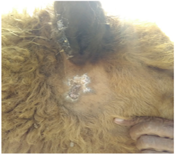
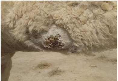
Figure 2: Dermatophilosis lesion on the abdomen
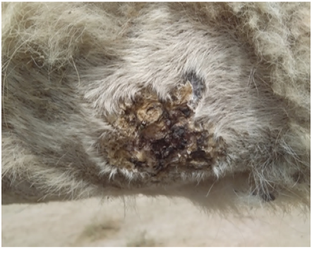
Figure 3: Dermatophilosis lesion in abdomen lesions show hairless brownish crusts with irregular sizes and pus exudation.
Discussion
Dermatophilus congolensis infection in camels has been reported in Saudi Arabia (Gitao et al., 1998b), Sudan (Gitao et al., 1998) and Iran (Khodakaram-Tafti et al., 2012). There is no report about dermatophilosis in Iraq. Camelpox is an important contagious skin disease of camels characterized by mild local skin infection and severe systemic infections. It causes economic loss in results of morbidity, mortality, loss of weight and reduction in milk produce (Bhanuprakash et al., 2010). Camelpox infection has been reported previously in Iraq in 1977 (Al Falluji et al.,1979).However, no mixed infection involving both D. congolensis and camelpox has been reported in Iraq to yet.
Clinically, both infections found in pastoralist camel herd in Al-Muthanna desert where dermatophilosis lesion characterized by hair matting, exudative dermatitis and brown scab formation which agreed with clinical findings in Sudan (Gitao et al., 1998), Saudi Arabia (Gitao et al., 1998b) and Iran (Khodakaram-Tafti et al., 2012). While camelpox characterized with mild skin lesion to sever systemic form with fever, lymphadenopathy and death which is characteristic of camelpox (Abu Elzein et al.,1999). In this case, camels also affected with heavily tick infestation which it may play an important role in the pathogenesis of dermatophilosis in camels (Khodakaram-Tafti et al., 2012; Gitao, 1993).Tick infestation of camels has been evaluated in al-Muthanna province in Iraq; the results were anemia, Skin inflammation, secondary infection of the skin were the main effects of tick infestation in addition to reduce animal production efficiency and reduce the market value of the skin and hides (Wajid, 2017).
Clinically, mixed infection of dermatophilosis with Microsporum gypseum have been recorded in camels (Gitao et al., 1998b). Complications due to bacterial Secondary infection to dermatophilosis has also been documented in camels (Dalis et al., 2010) .Also, an infection of dermatophilosis concurrently mixed with caseous lymphadenitis was reported in Jordan (Tarazi and Al-Ani, 2016). Dermatophilosis may be induced due to the stress of an infections and immune efforts.
However, various levels of immunity to dermatophilosis occur in spite of it is not highly pathogenic and infection is most important in immunocompromised hosts by skin injury and tick infestation where cattle can be resistant to ticks or immune against tick’s effects. Immune defenses might contribute to recovery from infection by isolating layers of infected keratinocytes, but they are also involved in the development of chronic lesions (Ambrose et al., 1999). Vitamin deficiency and tick infestation thought to be produce sever skin lesion of camel dermatophilosis (Gitao, 1993).
It has been reported that the disease was more prevalent in semi-arid areas in the wet season compared with its prevalence in the dry season (Gitao, 1993; Gitao et al., 1990)while the climate in al-Muthanna desert is dry, high temperature degrees from May to October with low humidity and rainfall.
In conclusion, there is a need for more research on camel dermatophilosis in Iraq, with focus on epidemiology, diagnosis and treatment with respect that the pastoralist conditions of camel herd are a major challenge in supply of the veterinary service.
Acknowledgments
The author is grateful to Dr. Karima Akool Al-Salihi, from the Department of Veterinary Internal and Preventive Medicine, Veterinary Medicine College, Al-Muthanna University.
Conflict of Interest
There is no conflict of interest.
Author contributions
The clinical examination of camels and taking pictures was done at the camel herd owner’s place by dr. Karima al-Salihi, near al-Samawah city, south of Iraq. The laboratory tests for identification of the causative agents of the outbreak were not achieved due to difficulties in detection of dermatophilus and moving of the herd from al-Muthanna desert to al-Najaf desert. The author only one was writing, revising, and approving the article.
References


