Advances in Animal and Veterinary Sciences
Research Article
Adv. Anim. Vet. Sci. 6(3): 113-120

Figure 1
Milk medium showing the classical stormy clot reaction of C. perfringens.The tube on the left extreme is uninoculated.
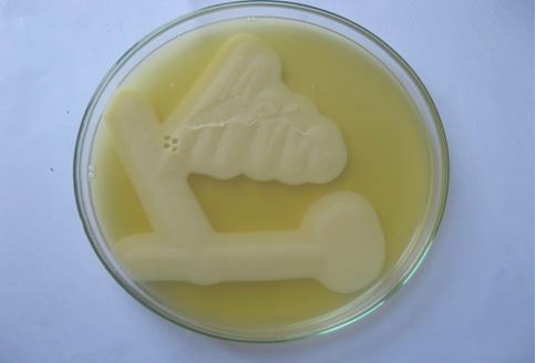
Figure 2
Lecithinase activity of C. perfringens in egg yolk agar after 24 hours of growth. Streaks showing opalescence production around C. perfringens.
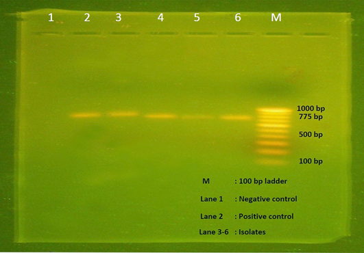
Figure 3
Agarose gel electrophoresis showing 775 bp amplicon of alpha toxin gene of C. perfringens. Lane M- 100 bp DNA ladder, Lane 1- Negative control, Lane 2- Positive control, Lane 3-6 – Isolates positive for C. perfringens.
Figure 4
Intestinal mucosa showing diffuse necrosis and pseudomembrane which forming a dirty turkey towel appearance.


