Advances in Animal and Veterinary Sciences

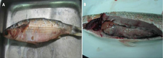
(A) Naturally infected Oreochromis niloticus fish showing skin darkness and fin and tail rot, (B): Naturally infected Oreochromis niloticus fish showing congested gills, pale and enlarged liver, (C): Naturally infected Clarias gariepinus fish showing hemorrhagic ulceration as well as fin and tail rot, and (D): Naturally infected Clarias gariepinus fish showing pale and enlarged liver and hemorrhages all over the body.
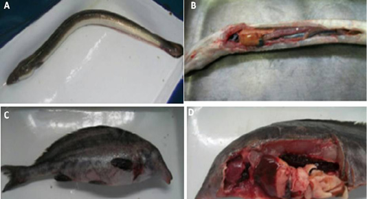
(A) Naturally infected Anguilla anguilla fish showing skin darkness and hemorrhages., (B): Naturally infected Anguilla anguilla fish showing pale and enlarged liver and congested spleen, (C ): Naturally infected Mormyrus kannume fish showing hemorrhages all over the body., and (D): Naturally infected Mormyrus kannume fish showing congestion in different organs (gills, liver and kidney)
(A) : Naturally infected Mugil cephallus fish showing hemorrhages all over the body, abdominal dropsy and scales detachment , (B): Naturally infected Mugil cephallus fish showing congested gills, pale liver and paleness of intestine.

Genotyping characterization of (A) Vibrio alginolyticus lanes (2-6), (737 bp), Lane (1) Ladder (100 bp) and (B) Vibrio harveyi lanes (2-6) (235 bp), Lane (1) Ladder (100 bp) using PCR specific primers targeting and collagenase and hemolysins (vhh) virulence encoding genes respectively.
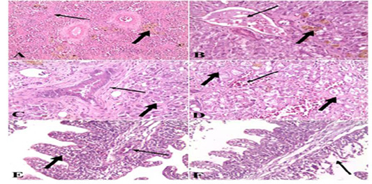
A). Spleen of naturally infected Clarias gariepinus with Vibrio species showed severe fibrosis (thin arrow) and hemosiderin-phages (thick arrow) (H&E x 10); B). Liver of naturally infected Clarias gariepinus with Vibrio species Showed congested portal vein (thin arrow), accumulation of bile pigment (thick arrow) and coagulative necrosis of hepatocytes (H&E x 20); C) Liver of naturally infected Clarias gariepinus with Vibrio species, showed bile duct hyperplasia, increased connective tissue around the bile duct (thin arrow) and coagulative necrosis of hepatocytes (thick arrow) (H&E x 20); D). Liver of naturally infected Clarias gariepinus with Vibrio species showed severe hemorrhages (thin arrow) and proliferation of connective tissue in between hepatocytes (thick arrow) (H&E x 20); E). Gills of naturally infected Clarias gariepinus with Vibrio species showed congested blood vessels (thin arrow), mononuclear cell infiltrations and thickening of secondary lamellae (thick arrow) (H&E x 20); F): Gills of naturally infected Clarias gariepinus with Vibrio species, showed necrosis and desquamation of the secondary lamellae epithelium (arrow) (H&E x 20).
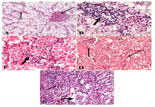
A). Liver of naturally infected Oreochromis niloticus infected with Vibrio Species Showed severely congested blood vessels and hepatic sinusoids (arrow) (H&E x 20); B). Liver of naturally infected Oreochromis niloticus infected with Vibrio Species Showed mononuclear cell infiltrations (arrow) (H&E x 40); C). Kidney of naturally infected Oreochromis niloticus infected with Vibrio Species, showed mononuclear cell infiltrations around renal tubules (arrow) (H&E x 40); D). Kidney of naturally infected Oreochromis niloticus infected with Vibrio Species showed severe hemorrhages (arrow) (H&E x 20); E). Kidney of naturally infected Oreochromis niloticus infected with Vibrio Species, showed congested glomerular capillaries (thin arrow), degeneration and necrosis of renal tubular epithelium (thick arrow) (H&E x 20).
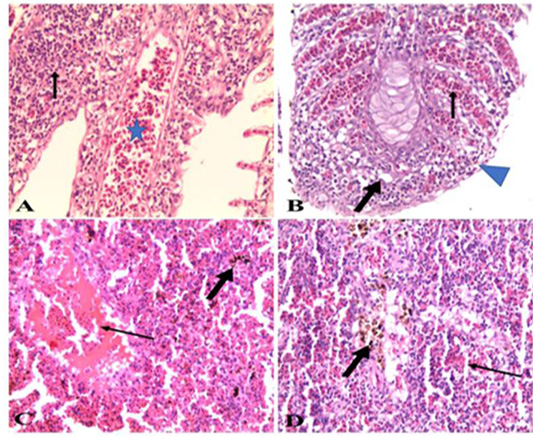
A). Gills of naturally infected Oreochromis niloticus with Vibrio Species showed severely congested blood vessels and mononuclear cell infiltrations (arrow) (H&E x 20); B). Gills of naturally infected Oreochromis niloticus with Vibrio Species, showed severely congested blood vessels (thin arrow), mononuclear cell infiltrations, vacuolation of epithelium (thick arrow) and adhesion between secondary lamellae (arrow head) (H&E x 20); C). Spleen of naturally infected Oreochromis niloticus with Vibrio Species showed severe hemorrhages (thin arrow) and hemosiderin-phages (thick arrow) (H&E x20); D). Spleen of naturally infected Oreochromis niloticus with Vibrio Species showed hemorrhages (thin arrow) and hemosiderin-phages (thick arrow) (H&E x 40).


