Advances in Animal and Veterinary Sciences
Research Article
Adv. Anim. Vet. Sci. 5(11): 463-467
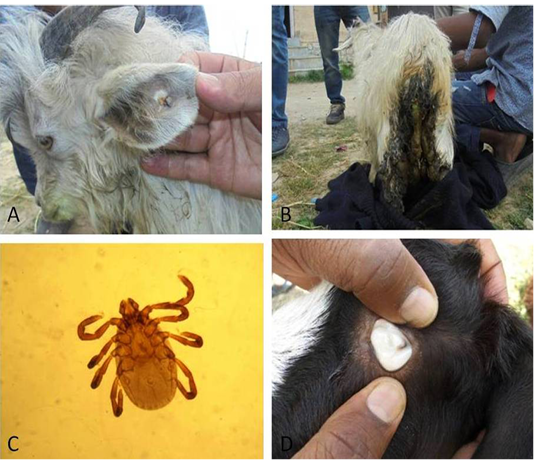
Figure 1
Animals affected with ticks on different body parts (A, B); Haemaphysalis spp. removed from different body parts (C); pale mucus membranes visible (D)
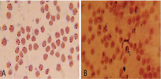
Figure 2
Microscopic examination of Giemsa-stained blood smears showing presence of intra-erythrocytic Babesia ovis (A) and Babesia motasi (B) (1000x)


