Advances in Animal and Veterinary Sciences
Case Report

Case Report on the Medical Management of Corneal Opacity in a Captive Elephant Tusker
P. Preena1, B. Aravind2
1Ph.D. Scholar, IVRI, Izatnagar, Bareilly, India (UP); 2Senior Veterinary Surgeon, Veterinary Hospital, Punthalathazham, Kollam, Kerala, India.
Abstract | A case of twenty-year-old captive elephant (Tusker) was presented with the history of photophobia, and profuse lacrimation on the right eye. Detailed clinical examination revealed complete corneal opacity without any apparent corneal ulcers. The patient was treated with 0.5% Moxifloxacin and 0.3% Tobramycin eye drops post-through washing with hyper saline. Subconjunctival injection of Placentrex was administered initially followed by Methylprednisolone at weekly intervals. The treatment was continued for 6 months with partial cure within 3 months and complete cure within 6 months. Thus, a case of corneal opacity was successfully managed with the complete recovery of the patient.
Keywords | Corneal opacity, Lacrimation, Elephant tusker, Moxifloxacin, Placentrex
Editor | Kuldeep Dhama, Indian Veterinary Research Institute, Uttar Pradesh, India.
Received | July 02, 2017; Accepted | August 08, 2017; Published | August 22, 2017
*Correspondence | P Preena, IVRI, Izatnagar, Bareilly, India (UP); Email: preenalinesh@gmail.com
Citation| Preena P, Aravind B (2017). Case report on the medical management of corneal opacity in a captive elephant tusker. Adv. Anim. Vet. Sci. 5(8): 342-345.
DOI | http://dx.doi.org/10.17582/journal.aavs/2017/5.8.342.345
ISSN (Online) | 2307-8316; ISSN (Print) | 2309-3331
Copyright © 2017 Preena and Aravind. This is an open access article distributed under the Creative Commons Attribution License, which permits unrestricted use, distribution, and reproduction in any medium, provided the original work is properly cited.
Elephants, the largest terrestrial mammal, are kept in captivity in Kerala, the southern most state of the Indian subcontinent, especially for timber hauling operations and temple festivals (Ajitkumar et al., 2010). Project elephant was launched in 1992 by the Government of India for the welfare of these animals as 60% of Asian elephants (Elephas maximus) are found in India. A total population of 29, 391 to 30, 711 is reported in India during 2012, out of which 5, 942-6, 422 elephants are in Kerala (Moef, 2017).
The cornea is the most powerful optical refractory surface of the eye. Being a transparent structure with appropriate curvature and position in front of the globe, it provides a straight path for the light to enter the eyes to reach the retina. Corneal transparency is maintained by numerous specialized anatomic and physiologic feature of the eye. Any factor that alters these specialized features like senility, UV radiations, corneal edema, injury, vitamin A deficiency, diabetes, infectious diseases, increased intraocular pressure, genetics etc. can lead to loss of transparency. Corneal opacity is a condition wherein the cornea becomes clouded or scarred leading to blurred vision, lacrimation and blepharospasms. If untreated, the condition can lead to permanent loss of vision and thereby the loss of the animal (Maggs, 2008; Ahmed and Doley, 2016).
In a retrospective study of ocular occurrence in domestic animals, corneal opacity showed a higher incidence compared to other affections like amaurosis,trauma, conjunctivitis,and tumor/dermoid (Tamilmahan et al., 2013). Among captive Indian elephants, the incidence of corneal opacity was 5-6%, which could be primarily due to vitamin A deficiency and injury (Ajitkumar et al., 2010; Chandrasekharan et al., 1995). Recently, a report of corneal corneal cloudiness with impaired vision in 5 Asian elephants was successfully managed medically with antibiotics and Vitamin A injection (Ahmed and Doley, 2016). Corneal opacity in domestic animals has been documented due to poor management like injuries or external trauma, foreign bodies and parasites (Pratap et al., 2003). Symptoms of corneal opacity depend on the inflammatory response due to the particular cause, which includes redness and swelling of the ocular tissues and eyelid, lacrimation, blurred vision, irritation, sensitivity to light, blepharospasms and lacrimation. Most importantly, the cornea turns milky or cloudy or “ground glass” appearance (Carola et al., 1990).
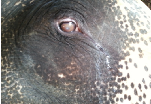
In this particular case, a twenty year old elephant Tusker (Figure 1) was presented with photophobia, profuse lacrimation, milky discoloration of the right cornea (Figure 2, 3 and 4), high stepping gait, especially notable during temple processions. The mahout has noticed these symptoms gradually over a period of 1 month. Upon detailed history taking, animal was under normal diet with green coconut palm leaves and dewormed with Albendazole (2.5mg/Kg PO). On detailed clinical examination, normal rectal temperature of 980F, pale roseate conjunctiva and trunk mucosa, no evidence of corneal ulcers, the milky nature of the cornea of the right eye with more discoloration in the center was observed.On bacteriological examination of conjunctival swab and blood smear examination, no pathology was revealed.
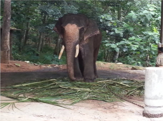
Figure 1: Twenty year old Tusker presented
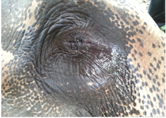
Figure 2: Profuse lacrimation from the right eye
The patient was thus diagnosed as corneal opacity probably due to injury inflicted by the mahouts. There are reports of traumatic injuries inflicted by mahouts hitting the elephant
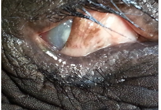
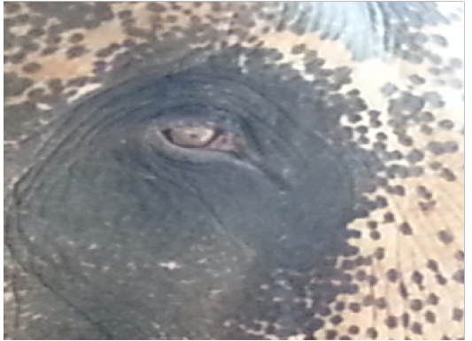
Figure 3: Blepharospasms and opacity of cornea
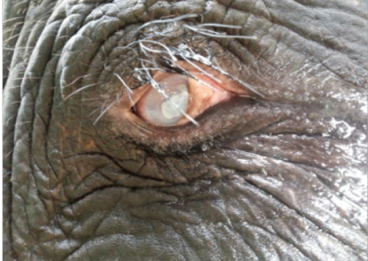
Figure 4: Milky nature of cornea with lacrimation
with a stick on the cheek. Sometimes, the stick hits the eye, causing an inflammatory reaction and opacity (Fowler and Mikota, 2008). The owner was advised to perform hypersaline eyewash three times daily, followed by administration of 0.5% Moxifloxacin eye drops and 0.3% Tobramycin eye dropsfor 2 weeks (10 drops tid). Subconjunctival injection of placentrex on the upper eyelid was administered for 1 week, followed by methyl Prednisolone at weekly intervals. There was considerable improvement in signs of lacrimation, photophobia by 3 months; however, the cloudy nature of the eye was not completely cleared (Figure 5). The patient had a clear cornea with regained vision by 6 months of proper treatment and management care (Figure 6).
In this case report, we have shown that unilateral corneal opacity in elephants can be addressed promptly with medical intervention using hypersaline eyewash, antibiotic drops, Placentrex and steroid therapy. Loss of clarity or opacification of the cornea is one of the basic pathologic lesion with corneal diseases. The normal turgesced state of the cornea is maintained by barrier function and metabolic pump associated between endothelium and epithelium. A Na+/K+ ATPase metabolic pump is present in the lateral endothelial cell membrane which pumps Na+ ions into the aqueous humour, creating an osmotic gradient that draws water out of the stream. Conditions, which damaged this mechanism, can cause stromal edema due to leak rate as a result of increased pumping rate. Opacity can be white, red or even pigmented based on the nature of the etiology (Martin, 2009).
Hypertonic saline was advised to reduce the corneal edema, which draws the fluid out of the cornea into the tears and also increases the tonicity of the tear film. However, for an osmotic gradient to occur, the epithelium has to be intact and should function as a semi-permeable membrane to water and not electrolytes. In addition, the hypertonic saline eye wash is most effective for epithelial edema, as stromal edema caused by endothelial dysfunction. It was reported that vision and comfort was improved in selected patients with months and occasionally years of this therapy (Costagliola et al., 2013). The combination of antibiotic (0.5% Moxifloxacin & 0.3% Tobramycin) drops was instilled to counter the bacterial flaring up if any for 2 weeks. It was suggested that corneal opacities respond rapidly to early treatment with antibiotic ointments such as gentamicin or chloramphenicol with or without steroids (dexamethasone/betamethasone) applied (Fowler and Mikota, 2008). Subconjunctival injection of placental extract (Placentrex®) is a common treatment for corneal opacities in elephants in Asia. It is a human placental extract with wound healing, anti-inflammatory, immunotropic and antioxidant properties. The polydeoxy ribonucleotide present in Placentrex® possesses anti-inflammatory and immunomodulatory actions (Wijesinghe et al., 2005). The subconjunctival injection of placentrex is given by inserting a fine needle through the outer aspect of the eyelid and feeling the needle between the fingers to insure that the needle does not penetrate through the eyelid (Fowler and Mikota, 2008).
Corticosteroids limit corneal opacification by inhibiting fibroplasia, decreasing vascularization, and reducing melanosis. These steroids also control anterior uveitis that frequently accompanies corneal wounds and cause potential blinding. However, on the other hands, these steroids can facilitate wound healing by inhibiting epithelial regeneration, corneal infiltration with inflammatory cells, fibroblastic activity and endothelial regeneration. It reduces the strength of resulting wounds as it potentiates collagenase enzyme activity and enhances the risk of infection (Maggs, 2008). Moreover, in areas where green fodders are not available in plenty, elephants are fed with straw alone, which results in vitamin A deficiency. In such cases, vitamin A supplementation (600,000 units, intramuscularly) is warranted (Fowler and Mikota, 2008).
Acknowledgements
The authors are obliged to the support given by the authorities who maintained the affected patient and the Animal Husbandry Department, Kerala.
Conflict of Interest
There is no conflict of interest.
Authors Contribution
BA treated the patient and collected the data, and PP drafted the manuscript. Both the authors reviewed, corrected and improved the manuscript.
ReferenceS


