Advances in Animal and Veterinary Sciences
Case Report
Advances in Animal and Veterinary Sciences. 1 (4): 127 – 128Multiple Cutaneous Melanomas in a Dog: a Case Report
Munuswamy Palanivelu1*, Avinash Warundeo. Lakkawar2, Khub Chandh Varshney2, Mariappan Asok Kumar1
- Division of Pathology, Indian Veterinary Research Institute, Izatnagar -243 122
- Department of Veterinary Pathology, Rajiv Gandhi College of Veterinary and Animal Sciences, Puducherry
*Corresponding author:drpalvet@gmail.com
ARTICLE CITATION:
Palanivelu M, Lakkawar AW, Varshney KC and Kumar MA. (2013). Multiple cutaneous melanomas in a dog: a case report. Adv. Anim. Vet. Sci. 1 (4): 127 – 128.
Received: 2013-07-17, Revised: 2013-07-25, Accepted: 2013-07-26
The electronic version of this article is the complete one and can be found online at
(
http://www.nexusacademicpublishers.com/table_contents_detail/4/84/html
)
which permits unrestricted use, distribution, and reproduction in any medium, provided the original work is properly cited
ABSTRACT
A 1 year old Great Dane bitch was presented with multiple, pedunculated masses of varying sizes. On clinical examination, the masses were soft, pliable and revealed both pigmented as well as un-pigmented skin coat. Histopathologically, all the tumour masses were characterized by mild epidermal hyperplasia along with aggregates of melanin pigments in the dermis. The dermal collagen was moderately hyperplastic and arranged as wavy bundles often containing focal aggregates of melanin. The cases of melanoma in dogs have been reported by many earlier workers but the unusual occurrence of spontaneous multiple cutaneous melanomas in a one year old dog as in the present case appears to be very rare and not have been reported earlier.
Melanomas are one of the commonly diagnosed tumours in dogs that originate from melanin producing cells. The benign cutaneous melanomas that arise on the haired skin usually display very little cytologic or nuclear atypia and a few mitotic figures. The lesions are solitary, and occur most commonly on the head, limbs, digits, scrotum, lip and trunk. These are variably circumscribed and are gray, brown or black in color, ranging from 0.5 to 10 cm in diameter. Histopathologically, characterized by the presence of atypical melanocytes arranged in sheets (packets or mesh) or in cords and are exhibited as epithelioid, spindle cell or combination of these two forms (Moulton, 1990; Goldschmidt and Schofer 1992). Among the skin tumours, melanocytic neoplasm accounts to five percent and usually solitary in nature (Scott et al., 2001).
A one-year-old Great Dane bitch presented to Veterinary Teaching Hospital, Puducherry was evaluated for multiple cutaneous masses in the skin of different body regions.
The anamnesis revealed that, the owner first noticed the masses 3 months prior to evaluation and since then the masses have gradually increased in size. On clinical examination, the masses were soft, pliable and revealed both pigmented as well as un-pigmented skin coat. The size of tumour masses in the present case ranged from 1 cm to 6 cm in diameter (Figure 1)

Figure 1: Melanoma from a female Great Dane showing multiple pedunculated growths in the hindquarters and abdominal region
The cut surface was dark brown in color with little apparently normal tissues. Cytology smears from different masses were prepared by fine needle aspiration cytology (FNAC) and stained with Leishman’s and Giemsa staining. Representative tissue samples was collected from each mass and fixed in 10% neutral buffered formalin. The fixed tissue samples were processed for paraffin embedding and 4-5 µ thick sections were cut and stained with hematoxylin and eosin for histopathological examination (Luna, 1968)
The FNAC smear showed presence of varying sizes of dark brown to jet-black granules often masking the cellular details (Figure 2 and 3).

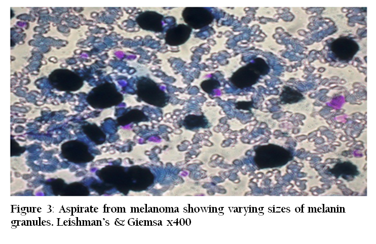
Figure 3: Aspirate from melanoma showing varying sizes of melanin granules. Leishman’s & Giemsa x400
Histopathologically, all the tumour masses were characterized by moderate degree of orthokeratosis, hyper pigmentation of epidermal cell layer. The dermis revealed aggregates of melanin pigments interspersed between the dermal collagen. The collagens were severely hyperplastic and were arranged as wavy bundles without any adnexal structures (Figure 4).

Based on these features, the case was diagnosed as multiple cutaneous melanomas. The cases of melanoma in dogs have been reported by many earlier worker (Schultheiss, 2006; Bidur et al., 2007) but the unusual occurrence of spontaneous multiple cutaneous melanomas in a one year old dog as in the present case appears to have not been reported earlier.
AKNOWLEDGEMENT
The authors are thankful to the Dean and faculty of Veterinary Surgery and Radiology division, Rajiv Gandhi College of Veterinary and Animal Sciences, Puducherry for extending necessary help.
REFERENCES
Bidur P; Min-Soo K; Il-Hong B; Mi-Sun P; Hyang J; Mi-Hyeon Y; Jae-Hoon K; Byung-Il Y; Yang-Kyu C and Dae-Yong K (2007). Retrospective study of canine cutaneous tumours in Korea. J. Vet. Sci. 8: 229–236.
http://dx.doi.org/10.4142/jvs.2007.8.3.229
PMCid:PMC2868128
Goldschmidt MH and Schofer FS (1992). Skin Tumours in Dogs and Cats. Buterworth Heinemann, Oxford, UK. J. Vet. Diagn. Invest. 17: 403–411.
Luna LG (1968). Manual of Histologic Staining Methods of the Armed Forces Institute of Pathology. Mc Graw Hill, New York, USA.
Moulton JE (1990). Tumours of the skin and soft tissue. In: Tumours in Domestic Animals. 3rd ed., Moulton JE. Berkely, University of California press, USA.
Schultheiss PC (2006). Histologic features and clinical outcomes of melanomas of lip, haired region, and nail bed locations of dogs. J. Vet Diagn Invest. 18: 422-425.
http://dx.doi.org/10.1177/104063870601800422
PMid:16921890
Scott DW; Miller WH and Griffin CE (2001). Muller and Kirk's Small Animal Dermatology. 6th ed. W.B. Saunders Company, Philadelphia, USA.


