Advances in Animal and Veterinary Sciences
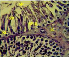
Section in the testis of control animal showing normal arrangement of seminiferous tubules, Leydig cells (LC) in the interstitial tissue, sertoli cells (SC), spermatogona A and spermatogona B (SP-B), spermatocytes (SP-A), spermatozoa (SP) in their lumen. (H and E stain 40X).
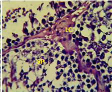
Section in the testis of animal treated with atenolol showing edema in the innterstitial tissue with atrophy of leydig cells (LC), incomplete spermatogenesis (SP) separated of epithelial cells from basement membrane with large spaces between few epithelial cells. (H and E stain 40X).
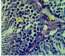
Section in the testis of animal treated with Atenolol and vitamin E showing edema with proliferation of leydig cells in the interstitial tissue (LC) and complete spermatogenes process and sperm in the lumen of seminiferous tubules (SP) (H andE stain 40X).
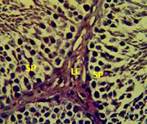
Section in the testis of animal treated with vitamin E showing proliferation of leydig cells (LC) with active complete spermatogenesis (SP). (H andE stain 40X).
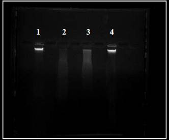
Digital printout of an agarose gel electrophoresis Showing lane (1): DNA from control sperm, lane (2) sperm DNA from atenolol treated group, lane (3) sperm DNA from atenolol plus vitamin E group, lane (4) sperm DNA from vitamin E treated group.


