Advances in Animal and Veterinary Sciences
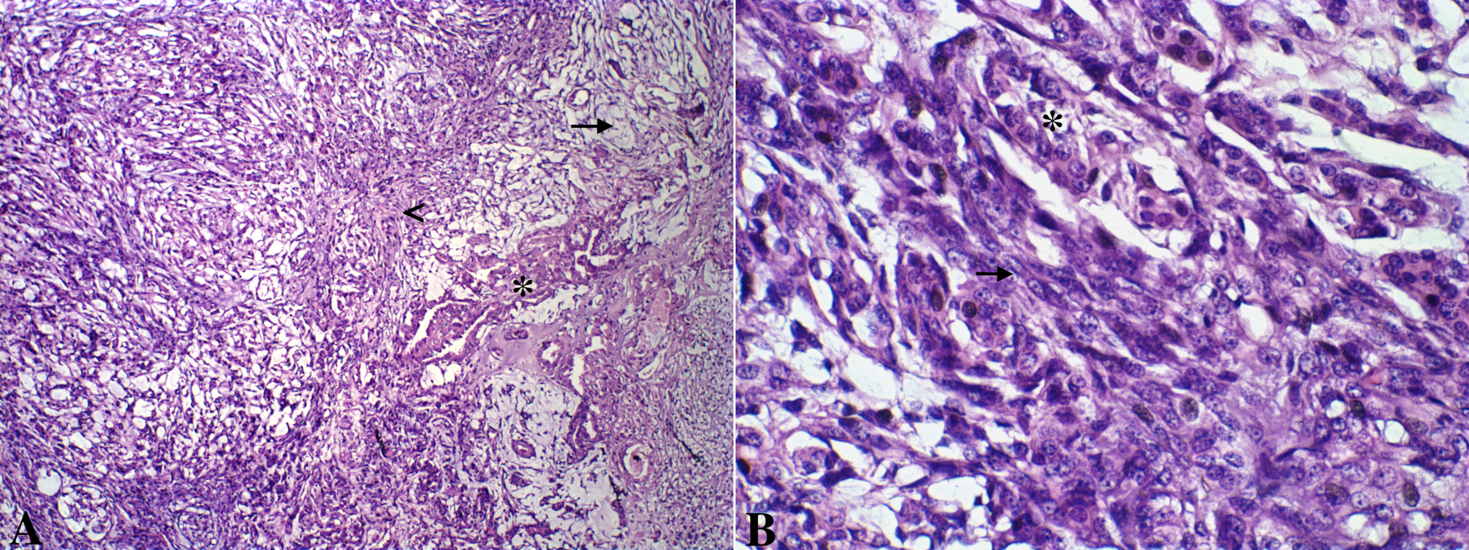
A. Canine mammary gland carcinosarcoma presenting a neoplastic epithelial component (asterisk) and a mesenchymal component with spindle cells(arrow head) and myxoid matrix (arrow). Hematoxylin and eosin, x100. B. Greater details of epithelial (asterisk) and mesenchymal spindle cells (arrow) shown. Hematoxylin and eosin, x600
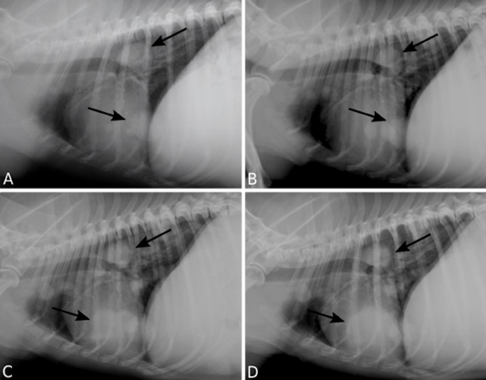
Pulmonary metastasis of a canine mammary gland carcinosarcoma. Right lateral recumbency thoracic radiographs (TR) presenting multiple nodules (arrows). TR performed 12 (A), 18 (B), 24 (C), 30 (D) months following surgical excision of the primary neoplasm.
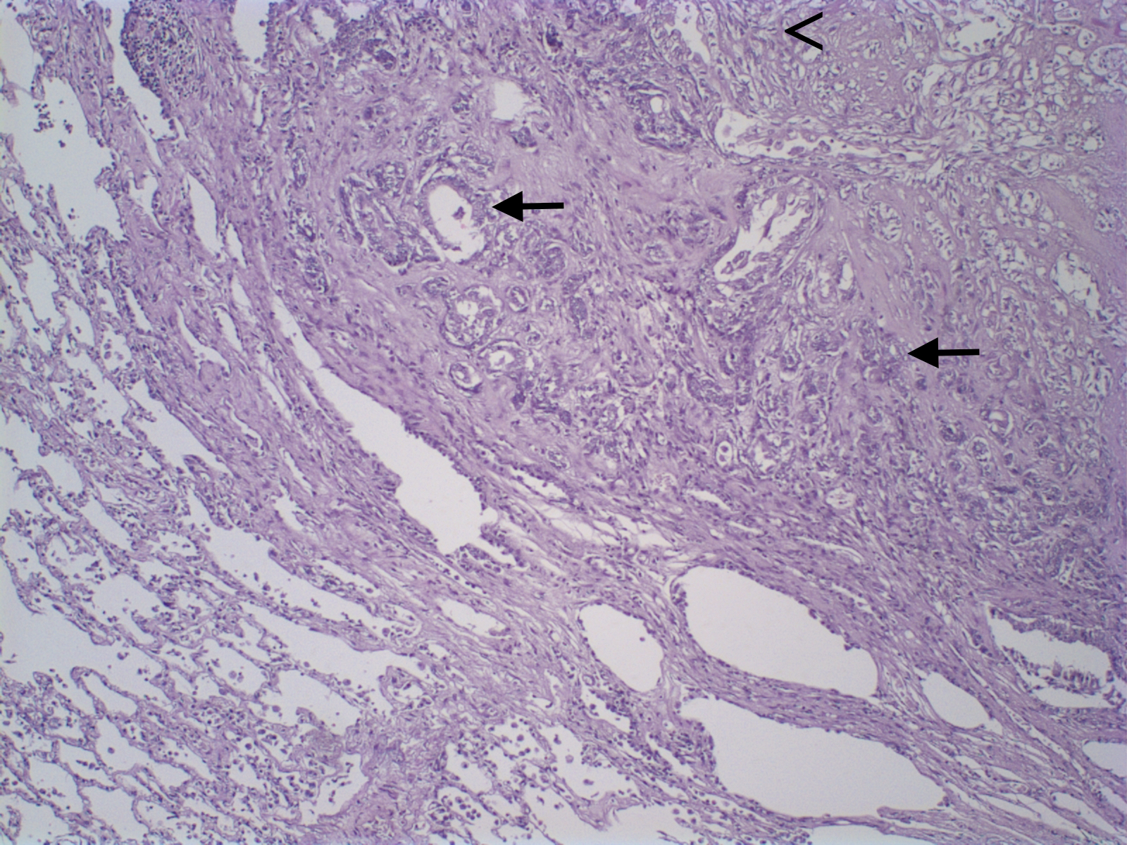
Lung tissue presenting a malignant epithelial (arrows) and spindle cells (arrowhead). Hematoxylin and eosin, x200.
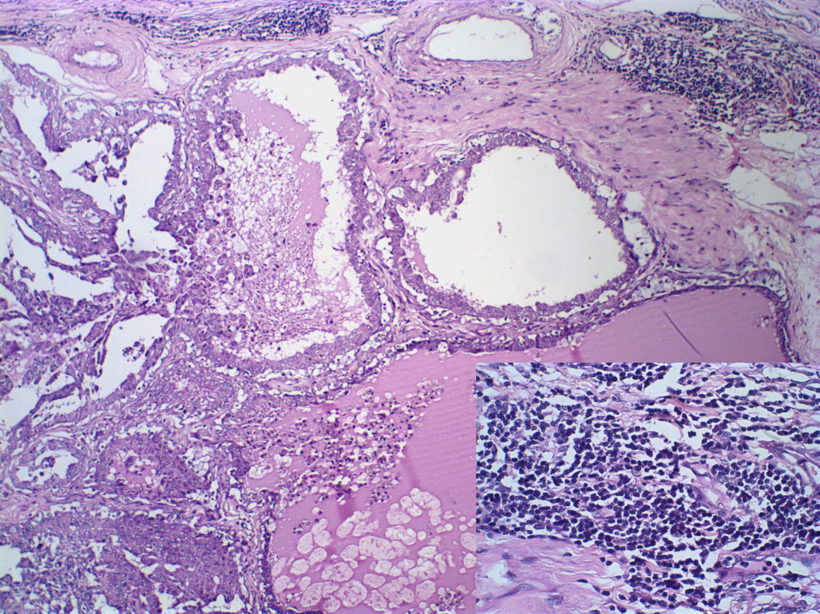
Canine mammary gland carcinosarcoma associated with a multifocal and intense inflammatory infiltrate, presenting an intense lymphocytic component (insert, Hematoxylin and eosin, x1000). Hematoxylin and eosin, x600.
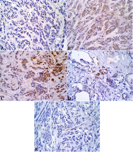
A. Canine mammary gland presenting neoplastic Ki-67-immunoreactive epithelial cells stained in brown (nucleous). DAB immunohistochemistry, counterstained with Harris’s hematoxylin, x600. B. Canine mammary gland presenting neoplastic ER-immunoreactive epithelial cells stained in brown (nucleous). DAB immunohistochemistry, counterstained with Harris’s hematoxylin, x600. C. Canine mammary gland presenting neoplastic PR-immunoreactive epithelial cells stained in brown (nucleous). DAB immunohistochemistry, counterstained with Harris’s hematoxylin, x600. D. Canine mammary gland presenting neoplastic Cox-2-immunoreactive epithelial cells stained in brown (cytoplasm). DAB immunohistochemistry, counterstained with Harris’s hematoxylin, x600. E. Canine mammary gland presenting neoplastic CD31-immunoreactive endothelial cells stained in brown (cytoplasm). DAB immunohistochemistry, counterstained with Harris’s hematoxylin, x600.


