Advances in Animal and Veterinary Sciences
Research Article
Advances in Animal and Veterinary Sciences 1 (4): 107 – 110Screening of Foods of Animal Origin for Coxiella burnetii in India
SatyaVeer Singh Malik1, Durga Prasad Das*, DeepakBhiwa Rawool1, Ashok Kumar, 1, Rahul D. Suryawanshi1, Mamta Negi1, Jess Vergis1, Swapnil Doijad2, Sukhdeo Baliram Barbuddhe2
- Division of Veterinary Public Health, Indian Veterinary Research Institute, Izatnagar, U.P., India–243 122
- ICAR Research Complex for Goa, Ela, Old Goa, India–403402
*Corresponding author:drdurga.prasad.das@gmail.com
ARTICLE CITATION:
Malik SVS, Das DP, Rawool DB, Kumar A, Suryawanshi RD, Negi M, Vergis J, Doijad S, and Barbuddhe S.B (2013). Screening of foods of animal origin for Coxiella burnetii in India. Adv. Anim. Vet. Sci. 1 (4): 107 – 110.
Received: 2013–08–19, Revised: 2013–08–27, Accepted: 2013–08–28
The electronic version of this article is the complete one and can be found online at
(
http://www.nexusacademicpublishers.com/table_contents_detail/4/79/html
)
which permits unrestricted use, distribution, and reproduction in any medium, provided the original work is properly cited
ABSTRACT
The epidemiology of Q–fever with regard to the role of foods of animal origin in transmission of Coxiella burnetii and their risk assessment remains largely unknown in most parts of the world including India. In the present study, a total of 591 food samples of different animal origin including milk (518), meat (60) and shell eggs (13) were screened for the presence of the pathogen by trans–PCR assay targeting transposon like element IS1111 of C. burnetii. . The analysis revealed an overall positivity in 4.06% (24/591) food samples, all of which were of milk. The milk samples showed an overall positivity of 4.63% (24/518), with the detection of C. burnetii in 5.55% (23/414) bovine and 1.42% (1/70) ovine milk samples. However, all the samples of camel milk (n = 34), meat (n = 60) and shell eggs (n = 13) turned out to be negative for the pathogen. The detection of C. burnetii in the bovine and ovine milk indicates a potential health risk for domestic livestock as well as human beings, especially those who consume raw or unpasteurized milk. The study of screening foods of animal origin for occurrence of C. burnetii in India by highly sensitive and specific molecular diagnostic tools is largely lacking, and in this context, the present study appears to be the first of its kind in the country. It is recommended that milk should be consumed only after pasteurization and, dairy as well as other foods of animal origin should be included in surveillance and monitoring programmes for food–borne pathogens for the risk assessment of C. burnetii infection.
INTRODUCTION
Q fever (query fever), erstwhile classified as a rickettsial zoonosis, is now better known as an OIE notifiable bacterial zoonotic disease caused by the most contagious, Gram–negative, obligate, intracellular bacterium – Coxiella burnetii (OIE, 2010; ILRI, 2012). The disease has a worldwide distribution with the notable exception of New Zealand (Hiblink et al., 1993; Norlander, 2000; Angelakis and Raoult, 2010). It remains endemic in many parts of the world and its prevalence has been confirmed in at least 51 countries including India (Acha and Szyfres, 2003; Marrie, 2003). Of late, Q fever is emerging or re–emerging in several countries worldwide (Arricau–Bouvery and Rodolakis, 2005; Gwida et al., 2012; Natale et al., 2012). It is noteworthy that the prevalence of Q fever has increased many–fold in recent times not only in the human beings and animals (Gwida et al., 2012), but also in foods of animal origin (Kim et al., 2005, Rahimi and Doosti, 2012) In humans, Q fever manifestations range from asymptomatic or mild flu–like illness to chronic fatal endocarditis (Angelakis and Raoult, 2010).
Major reservoirs of C. burnetii include farm animals and pets (Agerholm, 2013), and the infection gets transmitted to humans mainly through inhalation of contaminated aerosols of infected parturition materials or dusts containing C. burnetii shed by infected animals (Tissot–Dupont et al., 1999; Angelakis and Raoult, 2010; Agerholm, 2013). Moreover, continual shedding of the bacteria in the milk of infected animals (Maurin and Raoult, 1999; Kim et al., 2005), predominantly of cattle, for prolonged time periods ranges from 13 months (Arricau–Bouvery and Rodolakis, 2005) to 32 months (Angelakis and Raoult, 2010).
The pathogen has also been detected in foods of animal origin like milk (Ongor et al., 2004; Kim et al., 2005; Fretz et al., 2007), meat (Schaal, 1977), eggs and their products (Tatsumi et al., 2006; Rahimi and Doosti, 2012). The prevalence of pathogen in milk has been reported to range from a high of 94% in bovine milk bulk tank samples in USA (Kim et al., 2005) to 6.2% in bovine and 1.8% caprine bulk milk samples in Iran (Rahimi et al., 2009); 4.7% of bovine milk samples in Switzerland (Fretz et al., 2007), and 3.5% of ovine milk samples in Turkey (Ongor et al., 2004). The screening of eggs and their products for C. burnetii by PCR revealed positivity in 4.2% egg samples and 17.6% of the mayonnaise specimens in Japan (Tatsumi et al., 2006) and 1.5% of hen eggs and 7.7% duck eggs in Iran (Rahimi and Doosti, 2012).
Molecular method like PCR has become a useful tool with wide applicability in direct detection of C. burnetii DNA in different samples for diagnosis of Q fever because of its high specificity and sensitivity (Levy et al., 1991), as it can detect specific DNA sequences that may be present even in very low numbers in the fresh or frozen samples including milk, vaginal swabs, feces (Berri et al., 2000, 2001). Of late, a Trans–PCR assay, targeting the transposon– based IS1111 insertion sequence of C. burnetii has been widely used for detection of the pathogen in various clinical samples (Willems et al., 1994; Berri et al., 2000; Rolain and Raoult, 2005; Vaidya et al., 2008, 2010; Das, 2010).
In India, the association of C. burnetii with clinical conditions in animals and humans has been reported based on the recent and reliable molecular and serological tools (Vaidya et al., 2008, 2010; Das, 2010; Das et al., 2013), however, studies on the prevalence of this important food borne pathogen in different foods of animal origin by sensitive and reliable diagnostic methods are largely lacking. In this context, the present study was envisaged to analyze the occurrence of C. burnetii in different foods of animal origin by PCR, a highly sensitive and specific molecular test for the pathogen detection in milk (Kim et al., 2005; Fretz et al., 2007) and eggs (Tatsumi et al., 2006; Rahimi and Doosti, 2012).
MATERIALS AND METHODS
Collection of Sample
In the present study, a total of 591 food samples comprising of milk (n = 518), meat (n = 60) and eggs (n = 13) were collected. Milk samples were randomly collected from 105 cows, 309 buffaloes, 60 ewes and 34 camels belonging to organized or unorganized farms in Goa, Maharashtra, Odisha, Rajasthan, and Uttar Pradesh states of India. Meat samples from female goats (n = 40) with reproductive disorders and indigenous hens (n = 20) were collected from slaughter house and retail market of Bareilly, U.P., respectively. Some eggs (n = 13) of backyard poultry were collected from retail markets of Bareilly. The samples were collected aseptically in screw–capped sterile vials (15 mL) with 10 mL transport medium (sterile Bovarnick’s buffer) or straightly in sterile vials or polybags; and then transported under refrigerated conditions to the laboratory and stored at –200C until used for PCR testing.
Standard C. burnetii DNA procurement
The DNA of standard C. burnetii Nine Mile strain was obtained from Dr. Eric Ghigo, URMITE–IRD, Faculté de Médecine, France.
DNA Extraction from food samples
Samples of different foods of animal origin were processed for extraction of C. burnetii DNA by using DNeasy Blood and Tissue kit 50 (Qiagen, USA) as per the basic protocol given by the manufacturer.
Milk
Meat
Eggs
The sample of milk (1 mL) was centrifuged at 13,000 x g for 60 min at room temperature, and the layers of cream and milk were removed as recommended by Berri et al. (2000). Buffer ATL (180 mL) was added to the pellet, reacted overnight with proteinase K and then processed for DNA extraction employing DNeasy Blood and Tissue kit 50 (Qiagen, USA).
The meat samples (25 mg) were cut into small pieces, put in a 1.5 mL micro centrifuge tube, added with 180 mL Buffer ATL, and processed for DNA extraction.
Firstly, the C. burnetii was extracted from egg samples as per the method described by Fretz et al. (2007) and the resultant pellet was further processed for extraction of DNA as described with milk samples following the protocols of DNeasy Blood and Tissue kit 50 (Qiagen, USA).
Trans–PCR Assay
The reported primers, targeting the transposon–like repetitive element of C. burnetii, namely trans–3 (5’–GTA ACG ATG CGC AGG CGAT–3’) and trans–4 (5’–CCA CCG CTT CGC TCG CTA–3’), amplifying a 243 bp gene fragment (Lorenz et al., 1998) were synthesized by Sigma Genosys, USA and used in our study. The trans–PCR method of Willems et al. (1994) was employed with some modifications for C. burnetii detection in samples. Briefly, the PCR cycling reactions included an initial denaturation (95°C for 2 min), followed by 35 cycles, each consisting of 94°C for 30s, 61°C for 30s and 72°C for 1 min; and a final extension reaction of 72°C for 10 min. The PCR reaction mixture (25 μL) comprised of 10 X PCR buffer (2.5 μL), 10 mM dNTP mix (2.5 μL ), 25 mM MgCl2 (3.0 μL), 10 pM of primers (1.0 μL each), Taq DNA polymerase (0.5 μL ), DNA extract (5 μL) and sterilized nuclease free water for making the full reaction volume. For positive control, the standard DNA of C. burnetii RSA 493 Nine Mile 1 strain was used. The PCR amplification was carried out in a Master Cycler Gradient Thermocycler (Eppendorf, Germany), and the amplified products were subjected to agarose gel (1.5%) electrophoresis with ethidium bromide staining, and visualized under a UV transilluminator (UVP Gel Seq Software, UK).
RESULTS
Trans–PCR Assay
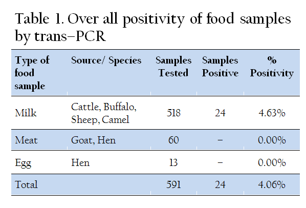
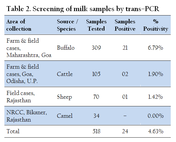
The trans–PCR assay, using trans–3 and trans–4 primers of C. burnetii, specifically generated a 243 bp amplified products (Figure 1). Sensitivity testing of the trans–PCR assay using standard DNA of C. burnetii Nine Mile I strain of known concentration (70 ng/μL) as well as DNA extracted from the spiked samples, diluted 10–fold, revealed C. burnetii DNA detection in dilutions up to 7 x 10⁻³ ng/μL. Of the 591 food samples screened, the trans–PCR assay revealed 24 (4.06%) samples to be positive for C. burnetii, and all of them were of milk samples (Table 1). Out of the total 518 milk samples tested by trans–PCR, 24 (4.63%) samples detected as positive were comprised of 23 (5.55%) from bovines (414) including 21 (6.79%) of buffaloes (309) and 02 (1.90%) of cattle (105), and 01 (1.42%) from sheep (70) (Table 2). However, all the camel milk samples tested were found negative. None of the meat and egg samples screened by the PCR assay showed positivity for C. burnetii.

DISCUSSION
Of late,the detection of C. burnetii in bulk tank milk samples from dairy herds in USA (year 2002 to 2004) with over 94% positivity by Real time PCR has suggested that the pathogen might persist in the dairy animals, with minor variations as regards to temporal or regional environment (Kim et al., 2005). However, only few of the reported studies till the year 2006 have analyzed foodstuffs for the presence of C. burnetii employing the molecular tool of PCR (Fretz et al., 2007). Similarly, the study of screening foods of animal origin for occurrence of C. burnetii in India, especially by highly sensitive and specific molecular diagnostic tools is largely lacking, and in this context, our study appears to be the first of its kind in India.
An overall positivity of 4.63% milk samples (5.55% from bovines and 1.42% from ovines) for C. burnetii by trans–PCR observed in our study is comparable to earlier reported studies with a positivity of 4.7% of bovine milk samples in Switzerland by nested PCR (Fretz et al., 2007); 3.5% of 400 sheep milk samples by IMS–PCR in Turkey (Ongor et al., 2004); and 6.2% of 210 bovine milk samples by nested PCR assay in Iran (Rahimi et al., 2009). The relatively high positivity of bovine milk samples and low positivity of ovine milk samples for the pathogen observed in our study is in agreement with the reported high (over 94%) detection rate of pathogen in bovine bulk milk tank in USA (Kim et al., 2005) but a complete absence of C. burnetii in ovine bulk milk samples tested in Iran (Rahimi et al., 2009) and Switzerland (Fretz et al., 2007). These reports indicate that the bovine milk, even from apparently healthy animals may serve as significant sources of C. burnetii infection. Moreover, the relatively high positivity (5.55%) of bovine milk in India observed in the present study also corroborates with our previous findings where the prevalence of Q fever among bovines with a history of reproductive disorders was observed to be higher (12.78% in cattle, 16.66% in buffaloes) compared to small ruminants (11.04% in sheep, 6.13% in goats) (Vaidya et al., 2010). Moreover, the excretion of the pathogen in bovine milk has been reported to last as long as 13–32 months (Arricau–Bouvery and Rodolakis, 2005; Angelakis and Raoult, 2010). The present study indicate that the occurrence of C. burnetii in bovine milk in India might be common, and therefore, a large scale screening is needed for evaluation and implementation of necessary legal regulations with regard to coxiellosis in dairy herds.
Non–detection of C. burnetii in the meat samples observed in our study might be due to the absence of the pathogen in the animal population screened. The published reports on occurrence of C. burnetii in meat are largely lacking. Moreover, the detection of C. burnetii in meat and offals, even in the infected animals, seems to be low as the screening of meat, offal and animal byproducts destined for human consumption in Germany has revealed the pathogen in only 8% of musculature (meat) of Q fever infected cattle whereas other animal parts used for meat purposes were found less infected (Schaal, 1977).
Mass screening of eggs and their products in Toyama prefecture in Japan revealed 4.2% of eggs and 17.6% of mayonnaise specimens to be positive for C. burnetii by PCR (Tatsumi et al., 2006). In a recent study from Iran, PCR revealed positivity for C. burnetii in 1.5% of hen eggs and 7.7% duck eggs (Rahimi and Doosti, 2012). However, small number of eggs (13) screened by PCR in our study were found free from C. burnetii infection, which is similar to a reported Swiss study, wherein all the 504 shell eggs analyzed with nested PCR were found negative for C. burnetii (Fretz et al., 2007). However, the lack of association of egg samples with C. burnetii infection observed in our study on limited samples should not be considered as a measure of prevalence of C. burnetii in these widely consumed foods of animal origin, especially in the light of high prevalence of the pathogen in eggs from other parts of the world (Tatsumi et al., 2006; Rahimi and Doosti, 2012).
In conclusion, the Q fever infection in countries like India remains grossly under diagnosed and the exact burden of the disease largely remains unknown. The detection of C. burnetii DNA in the bovine and ovine milk indicate that dairy animals in India may serve as the potential source of the infection through their milk and represent a risk for other domestic livestock animals, especially in suckling calves, as well as human beings, especially in breast fed babies (Kumar et al., 1981) and those who consume raw or unpasteurized milk as the diet or sacred offerings in the temples, which has been reported to be the vehicle of zoonotic pathogens like Listeria monocytogenes (Sheela Mary and Shrinithivihahshini, 2013). In the light of these observations, it is recommended that milk from dairy animals should be consumed only after pasteurization and it would be better to include dairy as well as other food animals in surveillance and monitoring programmes. Besides this, the risk assessment studies addressing the type of food animal involved, areas or regions affected are of prime importance to ascertain the exact burden of C. burnetii infection in foods of animal origin.
ACKNOWLEDGEMENT
The authors gratefully acknowledge the generous help and support received from Dr. Eric Ghigo, URMITE–IRD, Faculté de Médecine, France, for providing DNA of standard C. burnetii Nine Mile I strain (RSA 493); Director, Indian Veterinary Research Institute (I.V.R.I.), Izatnagar, India for providing the necessary financial support; Director, NRCC, Bikaner, Rajasthan as well as Dr. Shirish D. Narnaware, Scientist, NRCC, Bikaner, Rajasthan, for providing the camel milk samples; and Mr. K. K. Bhat, Technical officer, V.P.H. Division, I.V.R.I. for his technical support, for this study.
REFERENCES
Acha PN and Szyfres B (2003). Chlamydiosis, Rickettsiosis and Virosis. In: Zoonoses and Communicable diseases common to man. 3rd ed. Pan Amer. Hlth. Org. : 16–27.
Agerholm JS (2013). Coxiella burnetii associated reproductive disorders in domestic animals–a critical review. Acta Vet. Scand. 55: 13.
http://dx.doi.org/10.1111/rda.12117
PMid:23106815
Angelakis E and Raoult D (2010). Q fever. Vet. Microbiol. 140: 297–309.
http://dx.doi.org/10.1016/j.vetmic.2009.07.016
PMid:19875249
Arricau–Bouvery N and Rodolakis A (2005). Is Q fever an emerging or re–emerging zoonosis? Vet. Res. 36: 327–349.
http://dx.doi.org/10.1051/vetres:2005010
PMid:15845229
Berri M, Laroucau K and Rodolakis A (2000). The detection of Coxiella burnetii from ovine genital swabs, milk and fecal samples by the use of a single touchdown polymerase chain reaction. Vet. Microbiol. 72: 285–293.
http://dx.doi.org/10.1016/S0378-1135(99)00178-9
Berri M, Souriau A, Crosby M, Crochet D, Lechopier P and Rodolakis A (2001). Relationship between the shedding of Coxiella burnetii, clinical signs and serological responses of 34 sheep. Vet. Rec. 48: 502–505.
http://dx.doi.org/10.1136/vr.148.16.502
Das DP (2010). Detection of Coxiella burnetii infection in man, animals and ticks by different diagnostic tests employed for Q fever. M.V.Sc. Thesis. Indian Veterinary Research Institute, Izatnagar, India.
Das DP, Malik SVS, Mohan V, Rawool DB and Barbuddhe SB (2013). Screening of fecal droppings of wild birds for coxiellosis by a duplex PCR targeting Com1 and IS1111 genes of Coxiella burnetii. J. Foodborne Zoonotic Dis. (Accepted).
Fretz R, Schaeren W, Tanner M and Baumgartner A (2007). Screening of various foodstuffs for occurrence of Coxiella burnetii in Switzerland. Int. J. Food Microbiol. 116: 414–418.
http://dx.doi.org/10.1016/j.ijfoodmicro.2007.03.001
PMid:17434220
Gwida M, El–Ashker M and Khan I (2012). Q fever: A re–emerging disease? J. Vet. Sci. Technol. 3: 120 doi:10.4172/2157–7579.1000120)
http://dx.doi.org/10.4172/2157-7579.1000120
Hiblink F, Penrose M, Kovacova E and Kazar J (1993). Q fever is absent from New Zealand. Int. J. Epidemiol. 22: 945–949.
http://dx.doi.org/10.1093/ije/22.5.945
ILRI (2012). Mapping of poverty and likely zoonoses hotspots. Zoonoses Project 4, Report to Department for International Development, UK, International Livestock Research Institute, 119.
Kim SG, Kim EH, Lafferty CJ and Dubovi E (2005). Coxiella burnetii in bulk tank milk samples, United States. Emerg. Infect. Dis. 11: 619–621.
http://dx.doi.org/10.3201/eid1104.041036
PMid:15829205 PMCid:PMC3320343
Kumar A, Yadav MP and Kakkar S (1981). Human milk as a source of Q fever infection in breast–fed babies. Indian J. Med. Res. 73: 510–512.
PMid:7262921
Levy PY, Drancourt M, Etienne J, Auvergnat C, Beytout J, Sainty M, Goldstein F and Raoult D (1991). Comparison of different antibiotic regimens for therapy of 32 cases of Q fever endocarditis. Antimicrob. Agents Chemother. 35: 533–537.
http://dx.doi.org/10.1128/AAC.35.3.533
PMid:2039204 PMCid:PMC245045
Lorenz H, Jager C, Willems H and Balger G (1998). PCR detection of Coxiella burnetii from different clinical specimens, especially bovine milk on the basis of DNA preparation with silica matrix. Appl. Environ. Microbiol. 64: 4234–4237.
PMid:9797270 PMCid:PMC106632
Marrie TJ (2003). Coxiella burnetii pneumonia. Eur. Respir. J. 21: 713–719.
http://dx.doi.org/10.1183/09031936.03.00099703
PMid:12762362
Maurin M and Raoult D (1999). Q fever. Clin. Microbiol. Rev. 12: 518–553.
PMid:10515901 PMCid:PMC88923
Natale A, Buccia G, Capelloa K, Barberiob A, Tavellac A, Nardellia S, Marangona S and Cegliea L (2012). Old and new diagnostic approaches for Q fever diagnosis: Correlation among serological (CFT, ELISA) and molecular analyses. Comp. Immunol. Microbiol. Infect. Dis. 35: 375–379.
http://dx.doi.org/10.1016/j.cimid.2012.03.002
PMid:22463984
Norlander L (2000). Q fever epidemiology and pathogenesis. Microb. Infect. 2: 417–424.
http://dx.doi.org/10.1016/S1286-4579(00)00325-7
OIE (2010). Q fever. In: OIE Terrestrial Manual, Version adopted by the World Assembly of Delegates of the OIE in May 2010, Chapter 2.1.12. Office International des Epizootics Paris, France, 1–13.
Ongor H, Cetinkaya B, Karahan M, Acik MN, Bulut H and Muz A (2004). Detection of Coxiella burnetii by immunomagnetic separation–PCR in the milk of sheep in Turkey. Vet. Rec. 154: 570–572.
http://dx.doi.org/10.1136/vr.154.18.570
PMid:15144006
Rahimi E and Doosti A (2012). Detection of Coxiella burnetii in poultry egg samples in Iran using Nested PCR assay. Asian J. Anim. Vet. Adv. 7(3): 273–276.
http://dx.doi.org/10.3923/ajava.2012.273.276
Rahimi E, Doosti A, Ameri M, Kabiri E and Sharifian B (2009). Detection of Coxiella burnetii by Nested PCR in bulk milk samples from dairy bovine, ovine, and caprine herds in Iran. Zoonoses Publ. Hlth. 57(7–8): e38–41.
Rolain, JM and Raoult D (2005). Molecular detection of Coxiella burnetii in blood and sera during Q fever. Q. J. Med. 98: 615–621.
http://dx.doi.org/10.1093/qjmed/hci099
PMid:16027172
Schaal E (1977). Occurrence of Coxiella burnetii in foodstuffs in animal origin. Berl Munch Tierarztl Wochenschr. 90(19): 376–379.
PMid:911292
Sheela Mary M and Shrinithivihahshini ND (2013). Prevalence of Listeria monocytogenes in temple milks offered to the devotees as sacred liquid in Tiruchirappalli, Tamilnadu, India. Food Publ. Hlth. 3(2): 97–99.
Tatsumi N, Baumgartner A, Qiao Y, Yamamoto I and Yamaguchi K (2006). Detection of Coxiella burnetii in market chicken eggs and mayonnaise In: Hechemy, K.E., Oteo, J.O., Raoult, D.A., Silverman, D.J., Blanco, J.R. (Eds.), Century of Rickettsiology – Emerging, Reemerging Rickettsioses, Molecular Diagnostics, and Emerging Veterinary Rickettsioses. Ann. N. Y. Acad. Sci. 1078: 502–505.
Tissot–Dupont H, Torres S, Nezri M and Raoult D (1999). A hyper endemic focus of Q fever related to sheep and wind. Am. J. Epidemiol. 150: 67–74.
http://dx.doi.org/10.1093/oxfordjournals.aje.a009920
PMid:10400556
Vaidya VM, Malik SVS, Kaur S, Kumar S and Barbuddhe SB. (2008). Comparison of PCR, immunofluorescence assay, and pathogen isolation for diagnosis of Q fever in humans with spontaneous abortions. J. Clin. Microbiol. 46(6): 2038–2044.
http://dx.doi.org/10.1128/JCM.01874-07
PMid:18448698 PMCid:PMC2446837
Vaidya VM, Malik SVS, Bhilegaonkar KN, Rathore RS, Kaur S and Barbuddhe SB (2010). Prevalence of Q fever in domestic animals with reproductive disorders. Comp. Immunol. Microbiol. Infect. Dis. 33(4): 307–321.
http://dx.doi.org/10.1016/j.cimid.2008.10.006
PMid:19101035
Willems H, Thiele D, Frolech–Retter R and Krauss H (1994). Detection of Coxiella burnetii in cow's milk using the polymerase chain reaction. J. Vet. Med. B 41: 580–587
http://dx.doi.org/10.1111/j.1439-0450.1994.tb00267.x


