Advances in Animal and Veterinary Sciences
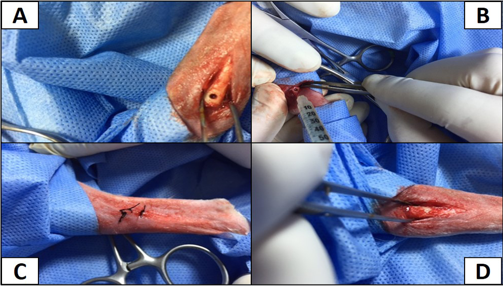
A) The site of operation shows the hole in right tibia; B) Inoculation of MRSA intramedullary; C) The hole filled with bone wax; D) The site of operation sutured with silk.

Plaques of bacteria lysis by Phage after incubation 24 hr at 37°C.
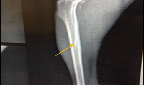
Site of operation, shows mediolateral view of the right tibia in non-infected group 21 days post-operatively, the hole had disappeared completely (healed, ML view)
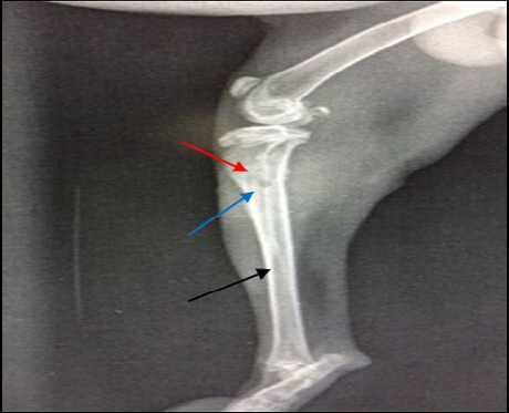
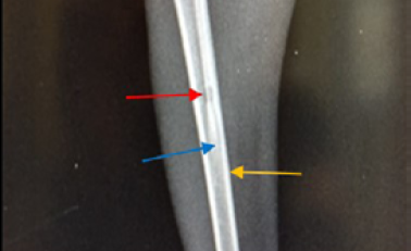
14 days post infection with MRSA, Area of bone lysis (Radiolucent spot) (→) surrounded by bone production (Radiopaque area) (→).Thickened cortex and severe periosteal reaction (→) (ML view)
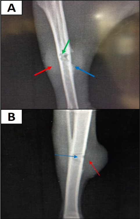
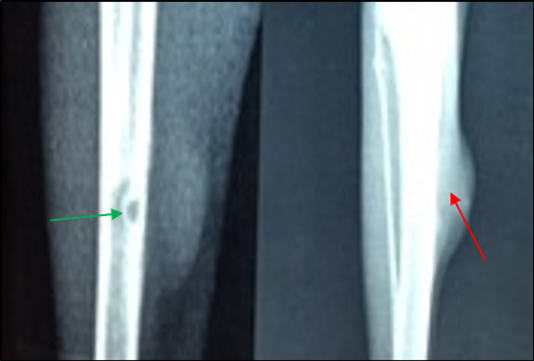
A) 21 days post infection with MRSA shows Sever bone lysis (→), Thickened of cortex and sever periosteal reaction (→), Soft tissue suppuration (→) (ML view); B) Seqeustrum appeared (→) and abscesses (→) (AP view)
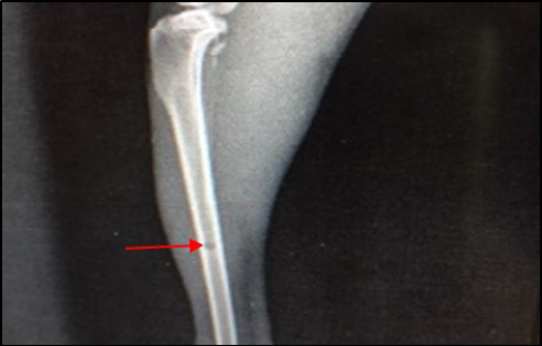
Site of operation, 7 days post treatment with Staph specific – bacteriophage given intramuscularly (Group C), all features of osteomyelitis disappeared, almost clear medullary canal (→), uniformed bone density and cortex returned to normal thickness (→), but the hole still exists (→) (ML View)
Site of operation, 7 days post treatment with Ceftaroline given intramuscularly (group D): A) Chronic Osteomyelitis (Seqeustrum still exists) (→) (ML View); B) Radiopaque area in soft tissue present represent accumulated pus (→) (AP View)
Site of operation, 14 days post treatment with Staph specific –bacteriophage given intramuscularly (Group C). Signs of osteomyelitis had disappeared. (Medullary canal is clear), bone cortex uniformed, normal bone density, the hole still present (ML View)
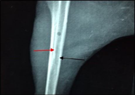
Site of operation, 14 days post treatment with Ceftaroline given intramuscularly (Group D). Still the medullary canal is not clear. Thickened cortex (→) and Endosteal reaction appeared clearly (→) (ML View)


