Advances in Animal and Veterinary Sciences
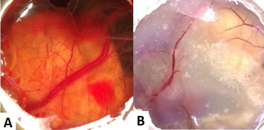
(A) CAM of control eggs (Group I) with well arbourized vascular system; (B) CAM of copper chloride treated eggs (Group II) showing thin blood vessels with less number of branch points
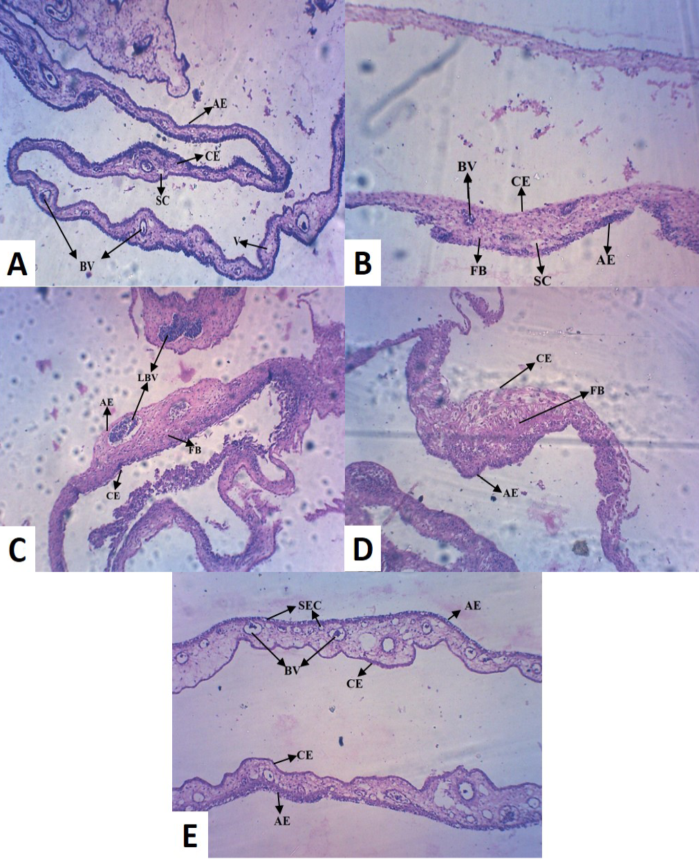
Histopathological analysis of CAM (100X) treated with different Cu salts (A) Control CAM showing mesodermal LBV with SC and AE and CE of equal thickness (B) Copper chloride treated CAM showing reduced thickness in CE (C) Copper acetate treated CAM showing hypodermic LBV and FB cells (D) Copper tartrate treated CAM showed thin AE, but no blood vessel and densely accumulated FB cells (E) Copper carbonate treated CAM showed numerous BV with SEC, indicative of pro-angiogenic activity [LBV (Large Blood Vessels); BV (Blood Vessels); V (Vein); AE (Allantoic Epithelium); CE (Chorionic Epithelium); FB (Fibroblast); SEC (Sub Epidermal Capillary plexus); SC (Small Capillary)]
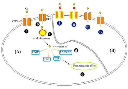
Schematic representation of [A] Mode of action of Cu traffic; (a) ATP7A/ B are Cu transporter proteins regulating intracellular Cu concentration (b) CTR1 is Cu uptake protein (c) Atx1, a Cu uptake protein which is responsible for intracellular Cu binding and trafficking and release of Cu activates (d) VEGF, FGF, TNF and PD-ECGF growth factors (e) resulting in pro-angiogenic effect. [B] Illustration of possible mechanism by which different Cu salts altered angiogenesis and tissue patho-physiology (i) CuCl2, (acidic) and (ii) C4H4CuO6 (basic), possibly inactivated copper transporters by altering the net charge on them and showed inflammatory response evident by accumulation of fibroblast cells (Figure 2B and D) (iii) Cu(CH3COO)2 resulted in Cu transport mediated anti-angiogenesis, but caused inflammation too (Figure 2C) (iv) CuCO3 allowed entry of Cu and acted as pro-angiogenic material


