Advances in Animal and Veterinary Sciences
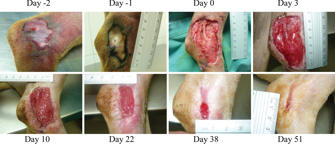
Necrotic compression lesion on the lateral right hock of a dog. Days were assigned in relation to the application of the antioxidant dressing. Bright red granulation tissue is evident at Day 3. Also, very healthy epithelization borders can be observed quickly after surgery (arrowheads day 3 and 10). These epithelization borders remain healthy and active until closure of the wound
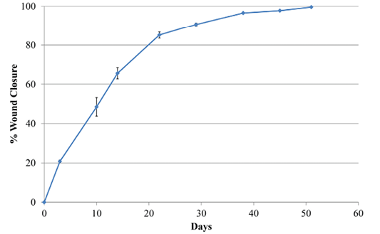
Graphical representation of the rate of closure of the wound in Case 1. Wound area estimates were calculated with the program Image J (NIH v1.50i) (Rasband, 1997) from different pictures. All pictures had a ruler placed near the lesion. Data were then expressed in relation to the size of the wound at day 0 after surgery and before treatment with the antioxidant wound dressing HR006
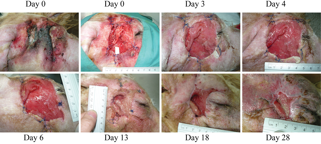
Necrosis due to foreign body in the right cheek of a dog. At the first wound dressing change, some areas with purulent exudate can still be observed, however, wound progression is evident. Granulation tissue starts quickly filling in the lesion. Dressing change at day 13 was due to removal of the penrose drain and dressing by the patient


