Advances in Animal and Veterinary Sciences
Research Article
Adv. Anim. Vet. Sci. 4 (5): 218 - 229
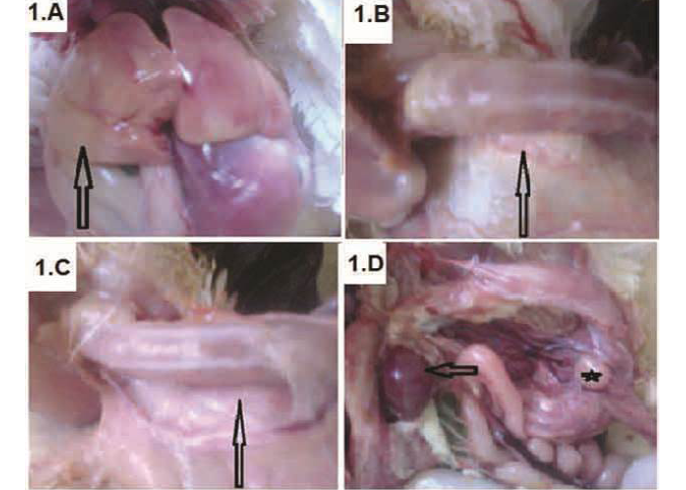
Figure 1
Broiler chickens infected with CAV showed pale liver (A-arrow); Atrophied thymus (B-arrow) sometimes resulting in an almost complete absence of thymic lobes (C-arrow), Enlarged spleen (D-arrow) and atrophied bursa of Fabricius (D-star)
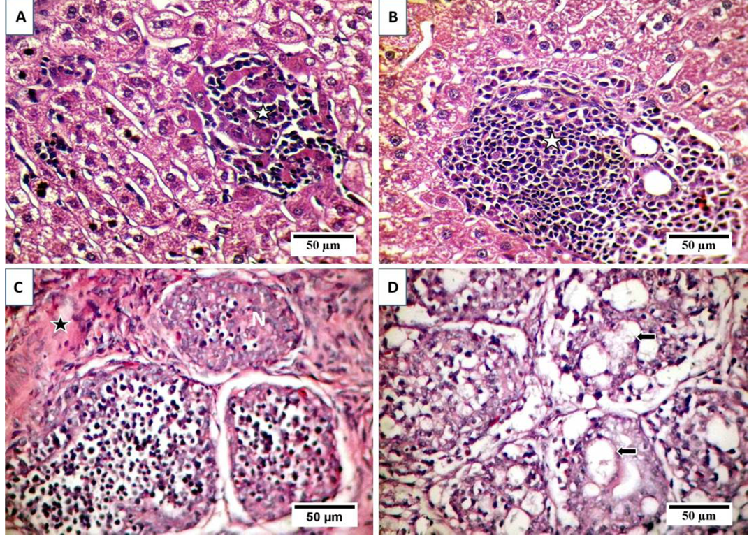
Figure 2
Effect of CIAV on liver (A and B) and bursa (C and D); stained with HE
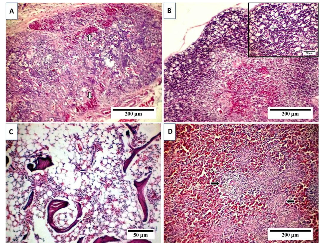
Figure 3
Effect of CIAV on thymus (A, B), bone marrow (C), and spleen (D), stained with HE
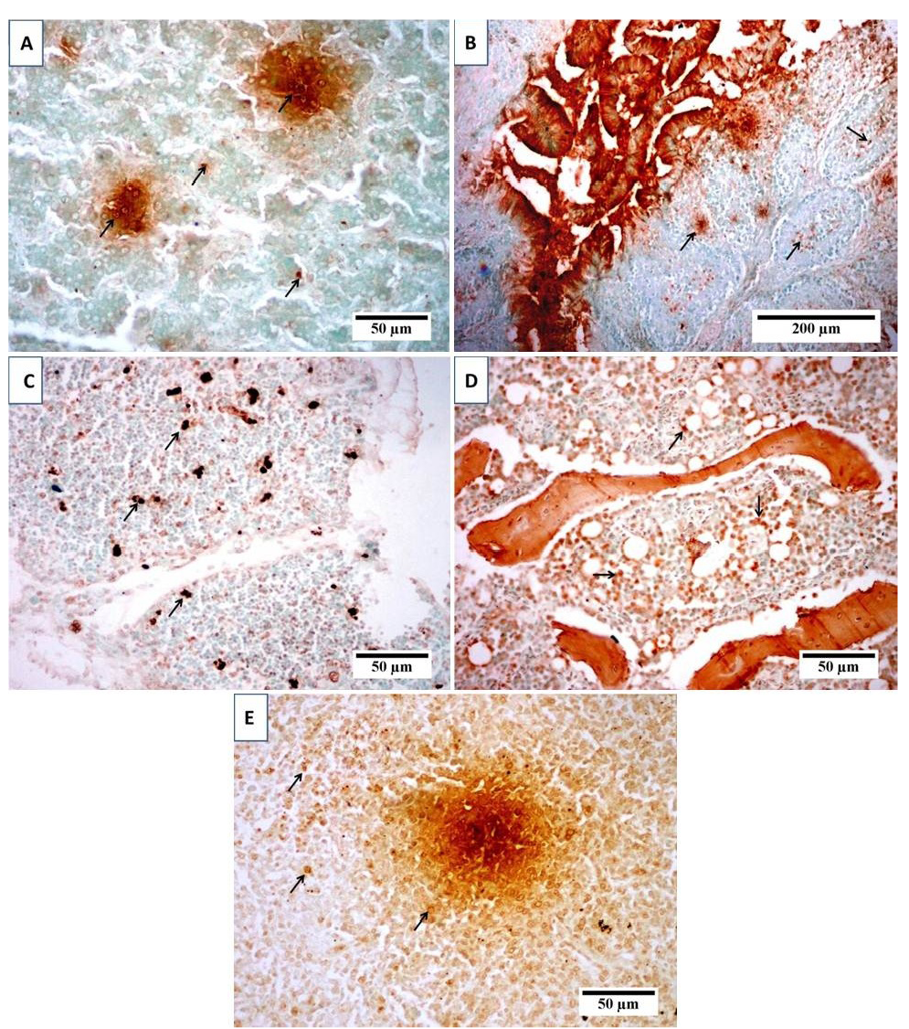
Figure 4
Terminal deoxynucleotidyl transferase-mediated dUTP nick end labeling
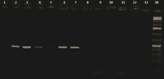
Figure 5
PCR products (418 bp) of amplified CAV-DNA extracted from tissues of diseased chicks
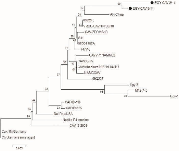
Figure 6
Phylogenetic tree for the 2 Egyptian CAV and other related CAV strains based on the partial VP1 gene sequence. The viruses used in this study were indicated by black dots


