Advances in Animal and Veterinary Sciences
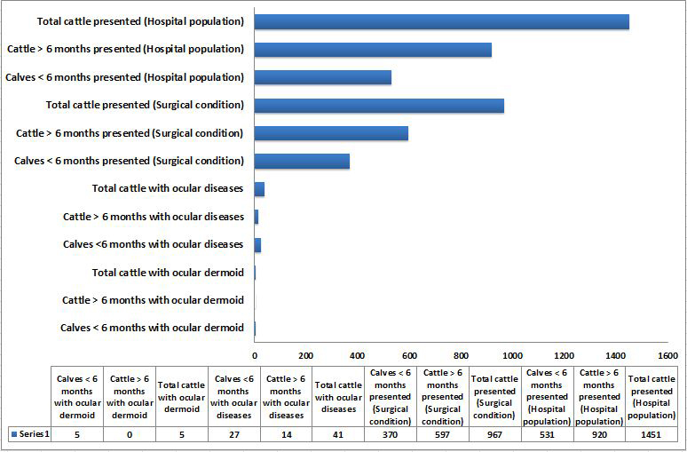
Graphical representation of hospital data on number of calves less than 6 months of age and cattle above 6 months of age presented with ocular dermoids, ocular diseases and other conditions
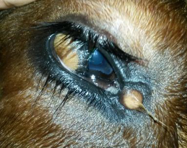
The ventrolateral corneoconjunctival dermoid on the right eye of a calf with haired corneal tissue merging with the hairless tissue of conjunctiva
A minute dermoid was noticed in the medial canthus also. The dermoid was bilateral and symmetrical
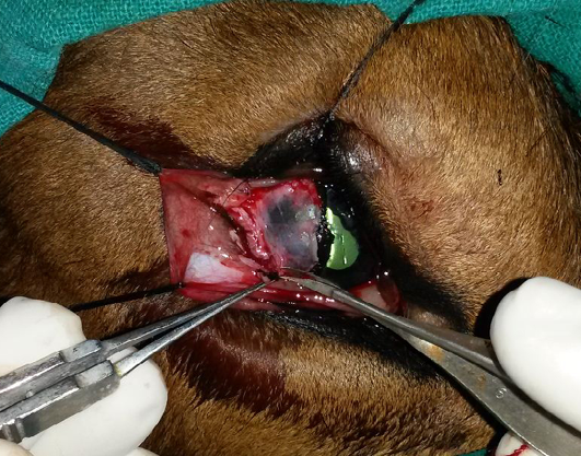
Excision of the conjunctival dermoid and superficial kerectotomy performed for the removal of the corneoconjunctival dermoid
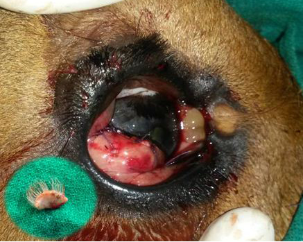
Postoperative photograph of the conjunctival flap applied on to the cornea for augmenting healing of cornea following superficial keratectomy
The minute dermoid on the medial canthus was left unoperated. The inset shows the excised corneoconjunctival dermoid.
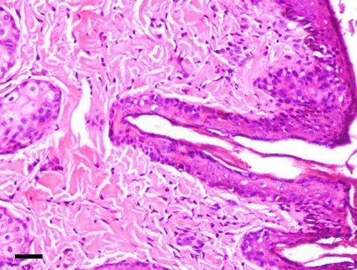
Microphotograph showing histopathology of corneoconjunctival dermoid
The dermoid showed stratified squamous keratinized epithelium above a thick collagenous stroma with hair follicles and bulbs in addition to apocrine gland [H&E; bar = 100 μm]


