Advances in Animal and Veterinary Sciences
Case Report
Adv. Anim. Vet. Sci. 4 (1): 1 - 4
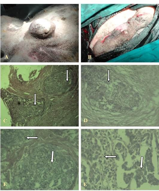
Figure 1
Gross and histopathological changes in invasive ductal carcinoma
A, B: Gross cutaneous projection in post umbilical region with involvement of neighbouring glands; C, (4X), D (10X), E, F (40X): Showing pleomorphic cells with increased mitotic figures, without involvement of basal cells (in the arrows)
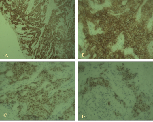
Figure 2
Immunohistochemical findings
A: High-grade ductal carcinoma in situ associated with invasive mammary carcinoma positive for estrogen receptor (40x); B: High- grade ductal carcinoma in situ positive for HER2 (100x); C, D: Weak staining with Ki67 indicating luminal A type invasive ductal carcinoma (40X).


