Advances in Animal and Veterinary Sciences
Short Communication
Adv. Anim. Vet. Sci. 3 (10): 522 - 526
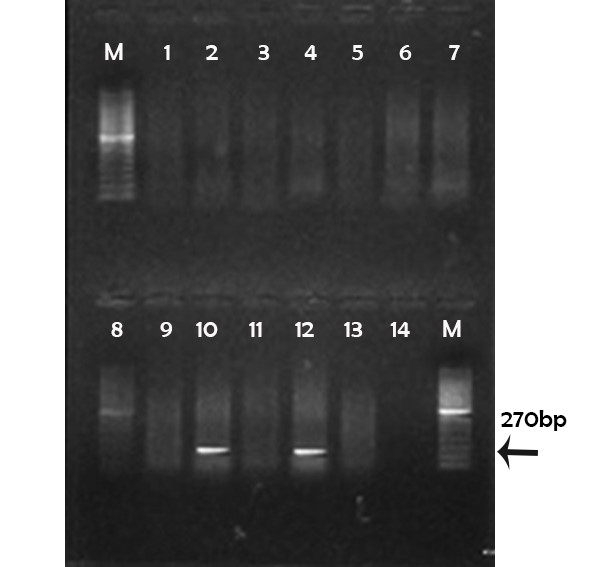
Figure 1
Agarose gel electrophoresis showing amplification of 270 pb fragment using tox A primer
(M) marker 100pb; Lane 1, 2, 3, 4, 5, 6, 7, 8, 9, 11 and 13: negative; Lane 10, 12: positive; Lane 14: controle negative
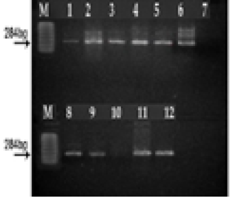
Figure 2
Agarose gel electrophoresis showing amplification of 284 pb fragment using las B primer
(M) marker 100 pb. Lane 1, 2, 3, 4, 5, 6, 7, 8, 9, 11 and12: positive; Lane 10: negative; Lane 7: controle negative
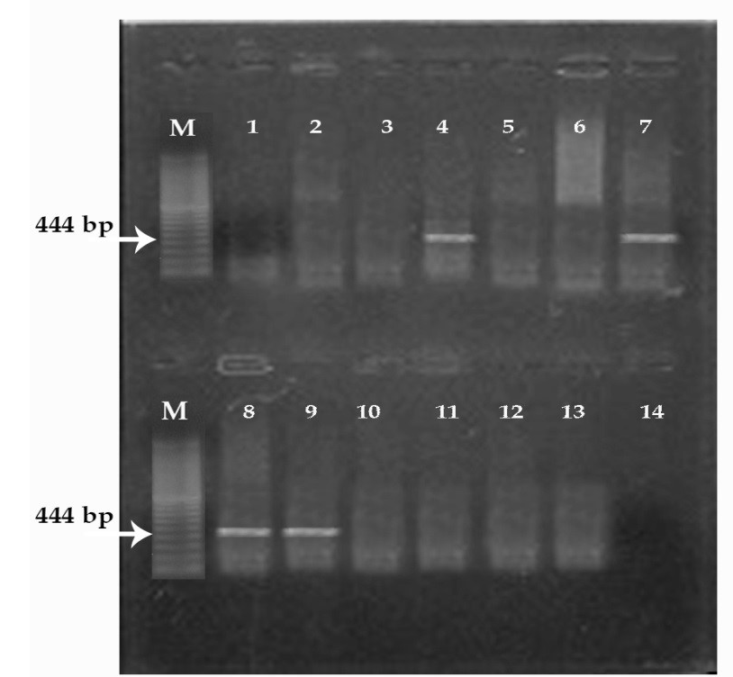
Figure 3
Agarose gel electrophoresis showing amplification of 444pb fragment using exo S primer
(M) marker 100 pb. Lane 1, 2, 3, 5, 6, 10, 11 and 12: negative; Lane 4, 7, 8, 9: positive; Lane 14: controle negative


