Advances in Animal and Veterinary Sciences
Review Article
Adv. Anim. Vet. Sci. 3 (4S): 9 - 22. Special Issue-4 (Advances in animal disease diagnosis, vaccines and therapeutics)
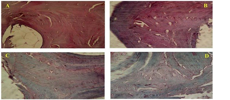
Figure 1
Photomicrograph showing (A-Native bone) intense GAG staining, (B-SDS treated bone) preserved GAG, (C-Acetone/Ethanol treated) less staining for GAG and (D-Freeze and Thawing group) less GAG content as compared to native bone. (Safranin-O; x40)
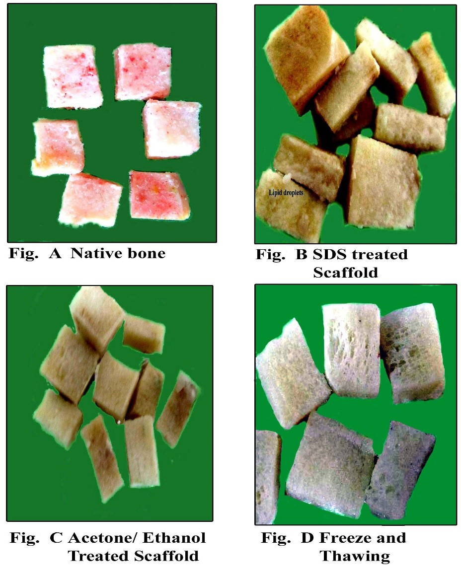
Figure 2
Gross appearance of the scaffolds of Native bone and decellularized bone
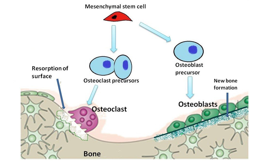
Figure 3
Diagram is showing evolution of osteoblasts and osteoclasts in the formation and resorption of bone
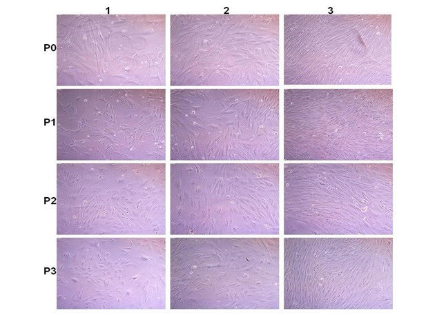
Figure 4
Rabbit fatal Osteoblast cell culture: P0; P1; P2 and P3 are passaged. 1; 2 and 3 images after 2; 6 and 8 days after passages. Cells become confluence (80-90% growth) within 6-8 days after passage
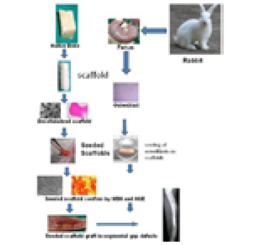
Figure 5
The process of graft preparation and in vitro and in vivo evaluation


