Advances in Animal and Veterinary Sciences

Download PDF Download ePUB
AAVS_Nexus 618


Review Article


Zoonotic Pathogens Transmitted from Equines: Diagnosis and Control


Sandip Kumar Khurana1*, Kuldeep Dhama2, Minakshi Prasad3, Kumaragurubaran Karthik4, Ruchi Tiwari5

1National Research Centre on Equines, Hisar, 125 001, Haryana, India; 2Division of Pathology, 4Divison of Bacteriology and Mycology, Indian Veterinary Research Institute, Izatnagar, Bareilly, 243 122, Uttar Pradesh, India; 3Department of Biotechnology, College of Veterinary Sciences, LUVAS, Hisar, 125 004, Haryana, India; 5Department of Veterinary Microbiology, College of Veterinary Science and Animal Husbandry, Uttar Pradesh Pandit Deen Dayal Upadhayay Pashu Chikitsa Vigyan Vishwa Vidyalaya Evam Go-Anusandhan Sansthan (DUVASU), Mathura (UP) – 281001.


Abstract | The diseases of equines and other animal species that are shared between animals and humans come under the category of zoonotic diseases, and always put threat to veterinarians, animal handlers, animal health personnel and general public. These pose a greater threat to pregnant women, infants, children, immunocompromised and old persons, individuals with stress of antibiotic therapy, and other susceptible humans. Equines also play an important role in transmitting several zoonotic diseases causing human illnesses such as those caused by encephalitic alphaviruses, hendravirus, West Nile virus and equine rabies, salmonellosis, glanders, anthrax, methicillin resistant Staphylococcus aureus (MRSA) infection, brucellosis and Rhodococcus equi infections, therefore acts as substantial global health threat. Among these zoonotic threat agents, anthrax and glanders are also potential biological weapons and at times have been used as bio-terroristic agents also. The emergence and re-emergence of equine zoonotic pathogens have been observed from time to time. Antibiotic resistance is a hot topic around the globe at present and certain strains like MRSA, extended spectrum beta lactamase producing Enterobacteriacea can also be transmitted between horses and humans. These drug resistant strains pose greater threat to human beings. Rapid detection of the causative agents of zoonosis, close attention to personal hygiene, identification of potential fomites and vectors, and the use of protective clothing, newer therapeutics and vaccines may contribute to reduce the risk of zoonoses. Equines are used for antivenom and antitoxin production against various antigens, therefore the serum being obtained from equines should be properly screened for various pathogens either by serological methods or molecular assays which are specific for detection of zoonotic agents. The present review discusses several important aspects of zoonotic diseases of equines with special focus on the recent advances in their diagnosis and control.


Keywords | Equine zoonotic diseases, Diagnosis, Control


Editor | Ruchi Tiwari, College of Veterinary Sciences, Department of Veterinary Microbiology and Immunology Uttar Pradesh Pandit Deen Dayal Upadhayay Pashu Chikitsa, Vigyan Vishvidhyalaya Evum Go-Anusandhan Sansthan (DUVASU), Mathura (U.P.) – 281001, India.

Special Issue | 2 (2015) “Reviews on Trends and Advances in Safeguarding Terrestrial /Aquatic Animal Health and Production”

Received | January 20, 2015; Revised | February 10, 2015; Accepted | February 12, 2015; Published | February 16, 2015

*Correspondence | Sandip Kumar Khurana, National Research Centre on Equines, Hisar, Haryana, India; Email: sandipkk2003@yahoo.co.in

Citation | Khurana SK, Dhama K, Prasad M, Karthik K, Tiwari R (2015). Zoonotic pathogens transmitted from equines: diagnosis and control. Adv. Anim. Vet. Sci. 3(2s): 32-53.

DOI | http://dx.doi.org/10.14737/journal.aavs/2015/3.2s.32.53

ISSN (Online) | 2307-8316; ISSN (Print) | 2309-3331

Copyright © 2015 Khurana et al. This is an open access article distributed under the Creative Commons Attribution License, which permits unrestricted use, distribution, and reproduction in any medium, provided the original work is properly cited.


INTRODUCTION


Zoonotic diseases, naturally transmitted between vertebrate animals and man, are highly variable on the basis of their severity and transmissibility. The “one medicine concept” involving a convergence of animal, human and environmental science professionals for prevention, control and eradication of cross-species disease transmission is gaining momentum, where zoonoses has assumed central position (Dhama et al., 2013a; Mukarim et al., 2015; Plowright et al., 2015). The emergence and re-emergence of zoonotic diseases poses a greater threat to pregnant women, infants and children, immunocompromised and old persons, persons under antibiotic therapy stress, veterinarians, animal handlers and animal health personnel (Stull et al., 2012).


Zoonoses constitute nearly 60% of all known human infections and over 75% of all emerging pathogens. They are caused by a diverse group of microorganisms and infectious syndromes caused by zoonotic pathogens are equally diverse. Classification of zoonoses is broadly based on the nature of the pathogen, animal host, severity of disease and mode of transmission between animals to humans. Zoonoses have major implications on economics, labour and health productivity globally. Preventing and controlling zoonoses is even more critical today in the context of globalization of international trade, changes in agricultural practices and global warming (Dhama et al., 2013b). They are responsible for affecting productivity of both humans and animals severely, thus contributing to aggravation of poverty.


Equines are used for various purposes like riding, racing, sports, draught, transport, ceremonies, antitoxin/antibody production, etc., throughout the world (Burnouf et al., 2004). There remains a contact between the human and these equines at various stages which pave way for the spread of various diseases of equines to human and hence human beings may acquire equine zoonotic bacterial, viral or other infectious diseases either directly or by indirect means. The number of diseases affecting horses and other members of the Equidae family carries zoonotic potential including viral, bacterial, rickettsial, anaplasma associated, fungal and parasitic infections, which will be elaborated through this review. Zoonotic diseases may occur as mild transient infection to severe lethal or highly contagious infection and thus can be a suitable candidate of bioterrorism also under certain circumstances. Veterinarians have a key role in revealing various emerging zoonotic infections due to their close involvement with both animals and owners. Growing possibilities of exploring zoonotic pathogens as bioterrorism agents is another worry which reflects the main functioning of veterinarians in early detection of bioterrorism-associated outbreaks of zoonotic diseases. Antivenom are commonly produced from equine serum, so critical attention should be given during this time as this can be a major source for the transmission of dangerous pathogens to human (Lalloo and Theakston, 2003; Theakston et al., 2003). Inference accredited to equine zoonoses depends on prevalence of particular disease along with its case-fatality rate in human population. Encephalitic alphaviruses, hendravirus, equine rabies, salmonellosis, glanders, anthrax, MRSA infection, brucellosis and Rhodococcus equi are among the very important zoonoses. Weese (2002) has reviewed the risk of zoonotic diseases to veterinary practitioners with emphasis on occupational aspects. The present review covers several important diseases which may be transmitted through equines to man or vice-versa with emphasis on their epidemiology, diagnosis and control calling for emergent need of collaborative approach of medical doctors, horse breeders, veterinarians, other governmental and non-governmental agencies for early preparedness; however there could be several others which have not been described here.


ZOONOTIC PATHOGENS TRANSMITTED FROM EQUINES VIRAL PATHOGENS


Encephalitic Alphaviruses

Western equine encephalitis virus (WEEV), eastern equine encephalitis virus (EEEV) and Venezuelan equine encephalitis virus (VEEE) are common zoonotic encephalitic alphaviruses. These have been isolated from horses, humans, mosquitoes, birds, rodents and some other animals also. These are vector borne and transmitted through blood sucking arthropods, thus considered as arboviral infection and condition is referred as Arboviral Encephalitis including EEE, WEE, VEE and WNV as a cause of encephalitis in horse (Strauss et al., 1995; Smith et al., 1997; Kapoor et al., 2010). Ordinarily arboviruses are not directly transmitted from horses to humans under usual circumstances, though aerosolization could be one possible risk factor. As per the recommendations of the Centers for Disease Control and Prevention (CDC), suspected clinical specimens such as cerebrospinal fluid and serum must be handled under level 2 biocontainment facility equipped laboratories with limited entrance. Lundstrom (2014) has reviewed progress in alphavirus vector development and vaccine technology which enabled clinical trials in humans.


Eastern Equine Encephalitis Virus (EEEV) Infection

EEEV was first isolated from horses in 1933 (Giltner and Shahan, 1933; TenBroeck and Merrill, 1933). The virus is propagated in nature between birds and mosquitos. The virus is considered to be most virulent among the alphaviruses causing encephalitis with case-fatality rate of up to 70% in human beings (Zacks and Paessler, 2010). EEEV infections has greater significance American continent where it was responsible for more than 180 cases in human (Aguilar et al., 2007). Mosquito vectors have a role in the transmission of the virus form horses from human. The incubation period is 4-10 days, fever, headache, vomiting, respiratory distress, seizures and coma may occur in human encephalitis cases. The mortality rate in horses is higher than in WEEV infection. Diagnosis is done by ELISA, haemagglutination-inhibition, neutralization assay and virus isolation. Formalin inactivated vaccine is used in horses as double vaccine with WEEV. Honnold et al. (2014) demonstrated that chimeric live-attenuated EEEV vaccine candidates protest mice against a lethal aerosol challenge. Trobaugh et al. (2014) have reviewed recent advances in alphavirus virulence mechanisms that could be used to design live-attenuated vaccine against EEEV/ alphaviruses.


Western Equine Encephalitis Virus (WEEV) Infection

WEEV was first isolated from brain of a horse (Meyer et al., 1931). First human WEEV was confirmed in 1938. WEEV is naturally propagated in enzootic cycle between passerine birds and specific mosquito vector. Some rodents and lagomorphs also act as reservoir hosts (Pfeffer and Dobler, 2010). Horses and humans are dead end hosts (Go et al., 2014). WEEV infections generally have an incubation period of 2-7 days with early non-specific symptoms like fever, anorexia, headache, nausea and vomiting. Diagnosis of WEEV infection is done by ELISA, haemagglutination-inhibition and neutralization assay (Martin et al., 2000). RT-PCR has also been developed for its diagnosis (Linssen et al., 2000; Lambert et al., 2003). Formalin inactivated vaccine is used in horses as double vaccine with EEEV.


Venezuelan Equine Encephalitis Virus (VEEV) Infection

VEEV is propagated in nature in enzootic cycle between rodents and mosquito vector (Taylor and Paessler, 2013; Carossino et al., 2014; Go et al., 2014). Epidemics occur when mosquitoes transmit the virus to humans and equids (Tigrett and Downs, 1962; Walton et al., 1973). Horses, donkeys and mules have sufficient viremia and are able to transmit the infection through mosquitoes (Young, 1972; Mackenzie et al., 1976). Some reports suggest that humans are also able to transmit the infection especially in urban settings (Watts et al., 1997, 1998; Morrison et al., 2008). Madsen et al. (2014) demonstrated that inhibition of Ago2, an important component of RNA-induced silencing complex (RISC), resulted in decreased replication of encephalitic alphaviruses and thus may be a future therapeutic. A live attenuated vaccine TC83 is thought to available option for humans as well as horses (Engler et al., 1992). However, formalin inactivated whole viral vaccines are also available (Taylor and Paessler, 2013). DNA based vaccines are also available which can be produced rapidly and are cost effective (Dupuy et al., 2011; Tretyakova et al., 2013; Carossino et al., 2014). Encouraging results have also been obtained with chimeric vaccines (Paessler et al., 2003; Paessler and Weaver, 2009; Carossino et al., 2014).


Japanese Encephalitis (JE)

Japanese encephalitis (JE) is very important and common mosquito borne flavivirus causing encephalitis and is regarded a major public health problem in Asian countries (van-den Hurk et al., 2008, 2009; Pawaiya et al., 2010a). The disease affects primarily human beings, horses and pigs. The infection in horses is usually subclinical with signs of pyrexia, depression, tremors and ataxia. Abortions and stillbirths are common manifestations in pigs.


JE virus was first isolated from a case of fatal human encephalitis in Japan in 1935 (Lewis et al., 1947). In human beings incubation period is 5 to 15 days with majority of cases being asymptomatic. Encephalitis is reported in about 0.04% cases only. Severe rigors, pyrexia and malaise are non-specific symptoms lasting one to six days. Signs of encephalitis are neck rigidity, cachexia, hemiperesis, convulsions and pyrexia. There is only one serotype of JEV and four genotypes (Chen et al., 1990, 1992; Tsarev et al., 2000). Urbanization, population spurt in tropical areas, increased transportation and global warming are responsible for spread of infections to newer areas (Go et al., 2014).


JEV is transmitted by Culex tritaeniorhynchus, C annulus, C annulirostris and Aedis mosquitoes (Rosen, 1986). Pigs and aquatic birds are amplifying hosts that have high titre viremia which acts as a source of infection for mosquitoes (Rosen, 1986). Humans and horse do not have sufficient viremia to transmit the infection and thus are dead end hosts. Yeh et al. (2010) developed a duplex reverse transcriptase PCR for rapid differential detection of west nile and japanese encephalitis viruses which is rapid, sensitive and specific and is useful both in humans and horses. Yeh et al. (2012) developed a diagnostic algorithm to serologically differentiate west nile virus from japanese encephalitis virus infection and its validation in field surveillance of horses.


Earlier inactivated vaccines were used. A purified vaccine from vero-cell adapted SA 14-14-2 strain has been developed (Tauber et al., 2007). Another chimeric vaccine containing pr M and E proteins of JEV has also been developed and found to show high level of immunogenicity (Guy et al., 2010; Halstead and Thomas, 2011). Singh et al. (2015) demonstrated JENVAC, a Vero-cell derived vaccine with a long lasting, broadly protective immunity.


West Nile Virus (WNV) Infection

West Nile virus (WNV), a member of the Flavivirus genus of the Flaviviridae family, is one of the most widely distributed mosquito–transmitted arbovirus having potential global public health concerns (Gulati et al., 2014). WNV was first isolated in Uganda in 1937 (Smithburn et al., 1940). There have been outbreaks in Africa, the Middle East, Asia, and Australia before spread to USA and Canada (Weaver and Barrett, 2004). The infection is maintained in nature between Culex mosquitoes and birds, whereas horses, humans and other mammals are dead end hosts (Blitvich, 2008; Dhama et al., 2010a; Beck et al., 2013; Go et al., 2014). Birds like crow are also affected by this viral infection (Mishra et al., 2012). Molaei et al. (2010) have studied the vector and host interactions governing the epidemiology of WNV in Southern California. Most of the WNV infections are subclinical and only less than 1% humans develop neurologic disease (Mostashari et al., 2001; Hayes et al., 2005; Hayes and Gubler, 2006; Porter et al., 2011).


Yeh et al. (2010) developed a duplex reverse transcriptase PCR for rapid differential detection of West Nile and Japanese encephalitis viruses which is useful both in humans and horses. Yeh et al. (2012) could serologically differentiate West Nile from Japanese encephalitis by a diagnostic algorithm. Currently, there is no suitable therapy for WNV infection (Paterson et al., 2011). A number of vaccines have been explored for horses including inactivated whole West Nile virus (West Nile–Innovator®, Vetera® WNV vaccine) and chimeric recombinant canarypoxvirus - Recombitek® Equine WNV Vaccine (De Filette et al., 2012; Gulati et al., 2014). DNA vaccine for WNV has been licensed in USA, and with the use of prime boost approaches it may protect WNV (Kumaragurubarn and Kaliaperumal, 2013), but such vaccine (West Nile–Innovator® DNA) has recently been discontinued by Pfizer (Brandler and Tangy, 2013; Gulati et al., 2014).


Hendra Virus (HeV) Infection

Hendra virus (HeV) was first isolated in 1994 from an outbreak among humans and horses (Selvey et al., 1995; Murray, 1996). This is one of deadliest human and veterinary pathogen causing respiratory and encephalitic illness in humans with high mortality rate which may exceed 70% (Dhama et al., 2010b; Croser and Marsh, 2013). Hendra virus (HeV) is a zoonotic paramyxovirus in the genus Henipavirus. There had been 48 outbreaks with increasing number of outbreaks with each passing year since it was first reported (Aljofan, 2013). Horses acquire infection from flying foxes. Clinical signs in horses include fever, anorexia followed by respiratory signs that include frothy nasal discharge. Human beings get infection through direct contact with secretions from infected horses. No evidence of human to human, human to horse and flying fox to human has been reported (Selvey et al., 1996), however, Williamson et al. (1998) have shown the role of fruit bats, horses and cats in transmission of Hendra virus. Flying fox bats acts as reservoirs and horses may encounter the infection from contaminated secretions or excretions through environment. Veterinarian get the disease while physical examination of oral cavity of horses. Human have flu like symptoms with mainly respiratory signs. The diagnosis of this infection is based on ELISA (IgG and IgM), RT-PCR and isolation of virus. There are no effective therapeutics against HeV infections and protective measures while examining horses are the only way to minimize the risk of its spread (Mahalingam et al., 2012). Human monoclonal antibody against HeV glycoprotein (G) protein is considered to be most promising passive immunotherapy (Bossart et al., 2011).
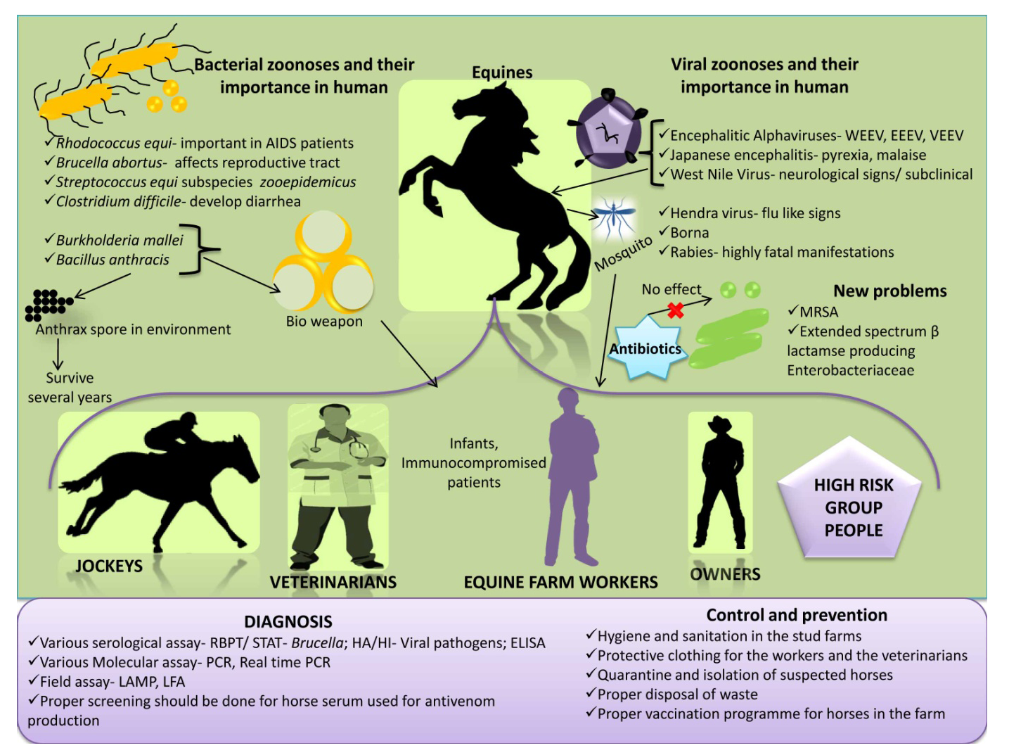


Equine Rabies

Rabies is a well-documented zoonotic disease that causes huge fatalities throughout the world. Rabies is a fatal neurological disease of mammals which is not an exception in equines, affects horses, mules and donkeys (Pawaiya et al., 2010b). It is caused by Lyssavirus of Rhabdoviridae family. The major animal involved is the dog and affected dogs can transmit the virus through their saliva. Inspite of low incidences in horses, literature reveal documented reports of equine rabies in past years as 82 reports of rabies in 1998, 65 cases in 1999 and 52 reports of equine rabies in 2000 have been from United States. Rabies in horse’s do not have the typical symptomps as in dog but the animal seems alert, lack of muscular in-coordination and seizures are common (Hudson et al., 1996). Though as compared to small animal practice probability of acquiring rabies from equines in veterinarian is low but possibility cannot completely be ignored due to severity of infection and variation in expression of symptoms. In equines, among furious and dumb form, paralytic or dumb form is more reported with the signs of rubbing of site of wound, gradual lameness and colic (Krebs et al., 2000, 2001; Weese, 2002). Animal handlers, veterinarians and horse owners are at highest risk for the transmission of rabies virus to them. Humans experience various symptoms like ataxia, loss of awareness, paralysis of muscles, etc. Vaccines are available for animals and humans that can prevent rabies, however history of vaccination does not necessarily exclude the risk of rabies, as one report described that even among 21 vaccinated horse, 5 animals could acquire the rabies (Green et al., 1999). Initial diagnosis can be made by differentially diagnosing all undifferentiated neurological diseases or encephalitis from Rabies. Animals may die due to cardiac arrest within 2-7 days or 14 days after appearance of clinical symptoms as nerves are affected in this disease. In infected animals, central nervous system components, saliva and salivary glands all are rich source of rabies virus hence any contact between infected saliva and breached skin or mucous membrane of horse or handler, jockey, owner must be avoided by taking biosecurity precautions. If any case is observed or suspected, higher authorities must be notified.

An overview on equine zoonosis is presented in Figure 1.


BACTERIAL PATHOGENS


Rhodococcus equi Infection

Rhodococcus equi is a gram positive, aerobic soil actinomycete responsible primarily for severe respiratory disease of young foals with high mortality rate (Prescott, 1991; Yager et al., 1991; Khurana et al., 2009; Giguere et al., 2011a, b; Khurana, 2014; Khurana et al., 2014; Khurana, 2015). R. equi also causes extra-pulmonary complications in equines including enteritis, arthritis and abscesses in abdomen (Giguere and Prescott, 1997). R. equi was first recovered from lung of a foal as Corynebacterium equi (Magnusson, 1923) and was reclassified as R. equi (Goodfellow and Alderson, 1977). This organism is emerging as an important pathogen in AIDS patients (Weinstock and Brown, 2002), drug therapy (Mizuno et al., 2005) and some other immunosuppressive conditions (Napoleao et al., 2005). The most common manifestation of human R. equi infections is pneumonia, others include fever, diarrhoea, abscesses in various internal organs and arthritis.


Virulence associated protein A (Vap A), a cell surface lipoprotein is essentially required for virulence in foals whereas virulence associated protein B (Vap B) is often associated with disease in human beings and pigs. Intracellular localization of R. equi is responsible for its prolonged and difficult therapeutic management. No suitable serodiagnostic test or vaccination is available for R. equi infection of equines as well as humans till date (Khurana, 2015).


Human beings acquire infection mainly through inhalation of dust harboring bacteria, from domestic animals including equines and wound, however man to man transmission is thought to be rare (Weinstock and Brown, 2002).


Agar gel diffusion test developed by Nakazawa et al. (1987) and ELISA by Giguere et al. (2003) have not been found to be of much promise for diagnosis of R. equi serologically. In India, diagnosis through post-mortem examination (Garg et al., 1985; Saxena and Narwal, 2009) and isolation of R. equi from clinical samples (Khurana et al., 2009). Various PCR assays have been developed (Sellon et al., 2001; Arriaga et al., 2002; Ladron et al., 2003; Oldfield et al., 2004; Ocampo-Sosa et al., 2007; Pusterla et al., 2007; Letek et al., 2008; Monego et al., 2009). The most valuable diagnostic procedures are combination of cultural methods along with PCR assay (Khurana, 2015).


Rifampicin along with macrolides is the drug of choice for treatment of R. equi infection. Rifampicin resistance have been reported which is posing a challenge in therapy (Asoh et al., 2013; Burton et al., 2013; Goldstein, 2014; Liu et al., 2014). Proper management and sanitation at farms is very important for control of disease at equine farms. Hygiene is important in immunocompromised human beings.


Anthrax

Anthrax is caused by Bacillus anthracis, an extremely resistant spore forming bacteria. Horses generally get infection by grazing in areas contaminated by anthrax. The typical incubation period is 3 to 7 days. Bacteria multiply and disseminate throughout the body through blood and lymphatic system, and release lethal toxin causing cell death and breakdown of tissues.


Symptoms appear very rapidly which include high fever, agitation, chills, colic, anorexia, dullness, laboured breathing and seizures. Bloody diarrhoea, swelling around the neck may also be observed. Chest, abdomen and genitals may also get swollen. Death may occur in 2 to 3 days after occurrence of first symptoms. This is controlled by vaccination in endemic areas, early diagnosis and treatment, burning and burial of dead animals.


Horses are usually less susceptible than ruminants, but reports of outbreaks of anthrax are available in horses from Minnesota and North Dakota. Horses may have prolonged course of disease. Affected horses exhibit symptoms of marked pyrexia, colic, dyspnea, and subcutaneous edema and death may occur abruptly.


Human beings contract infection from contaminated horses and their accessories and are often associated with three forms cutaneous, gastrointestinal and inhalation (Hicks et al., 2012). One more form of injectional anthrax has also been reported (Lalitha et al., 1988; Beaumont, 2010; Booth et al., 2011; Jallali et al., 2011). Spores of anthrax bacilli are so sturdy that they can survive for years in the environment and that can cause major problem for human population.


Control of anthrax is done through vaccination, early detection and reporting, quarantine, antibiotic therapy of exposed animals, burning or burial of dead animals that had suspected or confirmed anthrax infection. Vaccination of horses is done only in endemic farms and areas.


Glanders

Glanders is a contagious and fatal disease of horse, mules and donkey with zoonotic potential (Malik et al., 2010; Varga et al., 2012; Verma et al., 2014a). This is a disease known since ancient times and was identified in 4th century BC by Hippocrates (Colahan et al., 1999). The disease is caused by a non-spore forming gram negative bacillus called Burkholderia mallei. Most common mode of transmission of this organism is inhalation and ingestion of contaminated feed and water. The disease occurs in chronic form in horses, where bacteria are found in nasal discharges and skin lesions (OIE 2004, 2008), whereas in mules and donkeys the disease occurs in acute form (Hunting, 1913; Gulati and Gautam, 1962). The acute form of glanders involves pulmonary, cutaneous and nasal sites (Jubb et al., 1993), and is characterized by pyrexia, cough, discharges from nostrils, ulcers on nasal mucosa and nodules on the skin finally leading to death.


Chronic form in horse also shows all three clinical manifestations which include pulmonary, nasal and cutaneous forms. The human glanders is not very common, but it is having very high mortality rate of 90-95% in untreated septicaemic infection and 50% in treated humans. Human outbreaks have not been reported.


This organism is very important from biological warfare angle due to high rate of mortality and ability of small number of organisms to establish the infection. The diagnosis of glanders is done by isolation and allergic test (Mallein test) which is prescribed test for international trade. The test is not very specific, so it should be used along with complement fixation test which is also prescribed for international trade. The test is not very specific, so it should be used along with complement fixation test (CFT), which is also prescribed for international trade. Various serological tests for diagnosis of glanders include complement fixation test (CFT), indirect haemagglutination assay (IHA) and ELISA. Singha et al. (2014) have developed an indirect ELISA using truncated TssB protein for serodiagnosis of glanders.


Various PCR assays have been developed (Grishkina and Samygi, 2010; Zhang et al., 2012) which have been found to be rapid, sensitive and specific. Janse et al. (2013) developed a multiplex qPCR for detection and differentiation of B. mallei and B. pseudomallei. Diagnosis in human beings is generally done by CFT and imaging studies.


No vaccine is available for prevention of glanders for animals or humans (Burtnick et al., 2012). In case of death suspected due to glanders, the carcass should not be opened and must be buried deep or incinerated.


Brucellosis

Brucellosis is one of the most important disease problems of animals and human beings. This is caused by Brucella abortus in equines, and is manifested by fistulous withers, poll evil, lameness due to joint infection and rarely late abortions in mares. Horizontal transfer of Brucella spp. to horses from cattle and pigs has been documented (Forbes, 1990). Brucellosis is considered to be an occupational disease from public health point of view that mainly affects slaughter house workers, butchers and veterinarians. This causes undulant fever in human beings.


Ehizibolo et al. (2011) have reported a sero-prevalence of 14.7% of equine brucellosis in Nigeria. Tahamtan et al. (2010) have reported a sero-prevalence of 2.5% for brucellosis in horses in Iran. A sero-prevalence of 5.88%, 12.89% and 5.78% has been reported from Egypt (Montasser et al., 1999), India (Sharma et al., 1979) and Pakistan (Ahmed and Munir, 1995a, b), respectively.


The incubation period in humans varies from 1-3 weeks. The symptoms may include irregular fever, headache, weakness, malaise, profuse sweating especially during night. Coughing, chest pain, irritation, insomnia, depression are occasionally encountered. In many patients, the symptoms last for 2 to 4 weeks and are followed by spontaneous recovery. Others develop recurrent bouts at 2-14 day intervals. Most people with this undulant form recover completely in 3 to 12 months. A few patients become chronically ill, with symptoms of chronic fatigue, depressive episodes and arthritis. Relapses can be seen months after the initial symptoms, even in successfully treated cases. Occasional complications include arthritis, endocarditis, granulomatous hepatitis, meningitis, uveitis, orchitis, cholecystitis, osteomyelitis, and rare cases of encephalitis. Asymptomatic infections are also common in humans.


Diagnosis relies on the detection of circulating antibodies followed by the bacteriological isolation and serum agglutination tests [Rose Bengal plate agglutination test (RBPT) and standard tube agglutination test (STAT)]. Molecular diagnostic assays can be used for detection instead of serological tests because serological assays like RBPT, STAT have the disadvantage of false positive reaction against other gram negative bacteria (Karthik et al., 2014a). Various molecular assays like polymerase chain reaction (PCR), Real time-PCR, etc., can be employed for detection of the pathogen. Recent field assays like loop mediated isothermal amplification assay and lateral flow assay can be employed (Karthik et al., 2014a, 2014b).


Brucellosis being intracellular requires the need for antibiotics that can target intracellular pathogen. Treatment in humans can be done with combination of various antibiotics like doxycyline, streptomycin and rifampicin. Combinational therapy is followed to reduce the infection quickly and also to reduce the toxicity (MacMillan et al., 1982; Yousefi-Nooraie et al., 2012).


Salmonellosis

Salmonellosis is an enteric disease with clinical manifestation of chronic diarrhea, fever which may turn into acute toxic enterocolitis or septicaemia in various vertebrate hosts including horses and human beings. Etiological agent is Gram negative bacteria of Salmonella genus with multiple serotypes. Horses of all age groups are susceptible. Multidrug resistant S. Typhimurium DT104 (Salmonella enterica subspecies enterica Serotype Typhimurium Definitive Type 104) has been recovered from many horses in Ontario and has significant zoonotic potential due to its high lethality rate in human beings as well. Initially S. Typhimurium DT104 was isolated from cattle in 1988 in England and Wales but consequently was reported from sheep, pigs, poultry and horse. Fecal-oral route with high inoculums size containing more number of bacteria is the most common way for zoonotic spread of salmonellosis in human population, though in immunocompromised persons low inoculums may also produce the disease. Higher mortality rate and resistance to commonly preferred antibiotics further aids to the potential of this pathogen for zoonotic transmission. Suspected cases should be monitored and treated separately. Strict follow up of personal hygienic measures and proper disinfection of stables, contaminated equipments and utensils will help in reducing the menace of zoonotic transmission (Fone and Barker, 1994; Weese et al., 2001a; Weese, 2002).


Streptococcus equi Subspecies zooepidemicus Infection

Streptococcus equi subspecies zooepidemicus is considered opportunistic pathogen in horse, but causes infection in cattle, sheep, goat, pig and dog also. S. equi subspecies zooepidemicus has more than 98% DNA sequence homology with S. equi subspecies equi. Pelkonen et al. (2013) have shown that S. equi subspecies zooepidemicus is transmitted from horse to human beings and they found that human and equine isolates were identical or closely related. Lindahl et al. (2013) reported an outbreak of repiratory disease due to S. equi subspecies equi. Downar et al. (2001) have documented infection of Streptococcal meningitis from close contact with an infected horse (Downar et al. (2001)).


S. equi subspecies zooepidemicus is β-haemolytic streptococci in Lancefield group C (Lancefield, 1933). The equine Lancefield group C streptococci are differentiated biochemically by their ability to ferment sorbitol, lactose and trehalose (Quinn et al., 1994). In addition, PCR can be used to genetically identify different species and subspecies (Alber et al., 2004; Baverud et al., 2007; Preziuso et al., 2010).


Clostridium difficile Infection

Clostridium difficile (C. difficile) is an anaerobic causative agent for colitis in horses and human (Jones et al., 1987; George et al., 1978; Weese et al., 2001b). C. difficile associated diarrhoea can be mild, self-limiting or peracute leading to fatal outcomes in horses. It affects all ages of horses ranging from neonatal to adults. In acute cases of diarrhea rapid diagnosis is based on detection of bacterial toxins in fecal samples. C. difficile is a well-recognized pathogen of human also. The infection in man varies from mild to severe pseudomembranous colitis leading to intestinal perforation and death. Transmission between animals and humans has been poorly studied. Equines suffering with C. difficile diarrhoea should be considered infectious, particularly to people undergoing antimicrobial or chemotherapeutic treatment. Proper precautions (gloves, gowns and boots) and close attention to personal hygiene may prevent the chance of zoonotic transmission. Sporicidal disinfectant (5–10% bleach solution) should be used to clean the contaminated equipment and the areas under use. The veterinarians who develop acute diarrhoea following contact with suspected or confirmed case of infection in a horse should take medical help to confirm the infection.


Methicillin Resistant Staphylococcus spp.

In the era of antibiotic resistance, methicillin resistant Staphylococcus aureus (MRSA) is one of the prime concerns. Different Staphylococcus spp. can harbour horses of which S. aureus and its variant MRSA are the important species as they can be transmitted to humans and can cause major infections. In the early phase of MRSA, concern was restricted to humans and community associated MRSA which were increasing throughout the world. Livestock associated MRSA has spread recently among animals (Hartmann et al., 1997; McCarthy et al., 2012). Though the report of MRSA in horses was late but there are arrays of documentation after its first report in later part of nineteenth century (Stull et al., 2012). Literature reveal that approximately 10% of healthy vigorous horses cart MRSA in their nasal passages, intestinal tracts and on their skin, thereafter these colonized horses act as reservoirs of MRSA in the community which are efficiently capable of transmitting MRSA in the human population in and across the globe due to frequent international movement and trading of horses (Weese et al., 2005, 2006). Most of the methicillin resistant Staphylococcus organisms affect soft tissues and joints in horses but these are resistant to treatment with antibiotics. Some instances pneumonia, metritis and sinusitis are also documented in horses (Smiet et al., 2012). There is always danger of transfer of these drug resistance bacteria from animals to humans, and these methicillin resistant organisms can jump both ways from human to animal and also from animal to human (Weese and Lefebvre, 2007). Veterinarians top the list of humans suffering from this methicillin resistant Staphylococcus spp. transmission form horses (van Duijkeren et al., 2011). Bacteriophage therapy has yielded good response against MRSA (Karthik et al., 2014c). Similarly, broad spectrum beta lactamase producing Enterobacteriacea can also be transmitted from horses to human (Boyen et al., 2013).


Tuberculosis

Tuberculosis (TB) affects different mammals and many variants including extremely drug resistant TB has emerged which poses serious threat to human beings (Karthik, 2012; Karthik et al., 2013). Incidences of TB are sparse in horses mainly due to established control programmes, however few workers have reported the disease as confirmed by the presence of causative agent (Pavlik et al., 2004). TB is a zoonotic disease caused by Mycobacterium tuberculosis, M. bovis, and members of M. tuberculosis complex in several mammalian hosts including horses though they are considered comparatively resistant. Horses residing in close proximity with infected cattle acquire the infection as evidenced from the earlier reports showing presence of M. tuberculosis and M. bovis as well and advocate a possibility of interspecies transmission and zoonotic prospective of M. tuberculosis. Horses infected with pulmonary TB illustrate lesions of multiple tuberculoid granulomas in lung with manifestation of granulomatous lymphadenitis in mediastinal and tracheobronchial lymph nodes, which can be further confirmed by laboratory culture techniques and quantitative real-time PCR (Keck et al., 2010; Blahutkova et al., 2011; Konstantin et al., 2012).


PARASITIC PATHOGENS


Cryptosporidiosis

Equine cryptosporidiosis is caused by a protozoal pathogen, Cryptosporidium parvum, causing enteric disease in several species including humans and is most commonly associated with foals and immunodeficient animals. Infected horses shed oocysts with the shedding rate of 0–21% which have infective potential, horses develop asymptomatic cryptosporidiosis. High prevalence of disease with 71% infection rate is documented in foals and zoonotic transmission of Cryptosporidium from foal to handling veterinarians is also reported (Coleman et al., 1989; Cole et al., 1998). Humans express symptoms of copious watery diarrhea which may lead to critical grave condition. In immunocompromised persons, the disease exists in a self-limiting form. Zoonotic implications occur due to high shedding rates, hence precautions must be taken while dealing with diarrhoic animals to minimize the risk of zoonosis (Snyder et al., 1978; Konkle et al., 1997; Majewska et al., 1999; McKenzie and Diffay, 2000).


Giardiasis

Fecal shedding of Giardia oocysts from horses is an indication of zoonotic risk. Giardiasis caused by Giardia intestinalis is the most common intestinal parasitic disease characterized by mild or severe diarrhea. Zoonotic transmission of Giardia is supported by feco-oral route. Asymptomatic shedding of Giardia in 25% of adult horses and 71% cumulative infection rate in foals further suggest possibility of zoonotic potential of this pathogen in horses besides other species (Xiao and Herd, 1994). Fecal cyst detection and ELISA are available for diagnosis for giardiasis (Rishniw et al., 2010).


Other Zoonotic Diseases of Equines

Several important zoonoses shared between man and equines have been described above, however all the possible zoonotic threats related to equines are not detailed and still there are many others like Borna virus, Nipah virus infection as geographically limited zoonotic, dourine, crytosporidisis (Sellon, 2007; Xiao, 2008), giardiasis, leptospirosis, dermatophytosis, Halicephalobus gingivalis (Micronema deletrix) under certain specific conditions, are also among the several other infections capable of being transmitted from equines to human beings thus adding to the disease burden. Besides viral and bacterial pathogens, parasitic, protozoan and fungal agents are also involved in equine zoonoses. Leptospirosis is another important disease of public health concern that can be transmitted from various animals. Leptospirosis is a zoonosis of global concern caused by spirochetes of genus Leptospira (Ebani et al., 2012). Leptospira spp. is endemic in various parts of the world. Various serovars like Pomana, Icterohaemorrhagiae and Bratislava are recorded in horses but risk of zoonotic transmission of leptospirosis from horses is not immense. Hall and Bryan (1952) and other workers have reported leptospirosis in horse, which harbor the infection as accidental host (Hall and Bryan, 1952; Hogg, 1974; Barwick et al., 1998). Symptoms in horse include anorexia, fever, lethargyness, renal dysfunction, jaundice; abortion and still birth can also occur in pregnant mares (Divers et al., 1992; Timoney et al., 2011). Equine recurrent uveitis or moon blindness occurs after weeks or months after the onset of systemic leptospirosis in horses. Symptoms in human include jaundice, fever, muscular pain, vomiting, uveitis etc. (Hartskeerl et al., 2004; Verma et al., 2013). Microscopic agglutination test is the gold standard test for diagnosis of leptospirosis but several disadvantages makes it difficult to perform. Various recombinant proteins are used in ELISA and latex agglutination assay formats for diagnosis of leptospirosis which are effective in diagnosis (Deneke et al., 2014). Dermatophytosis (ringworm) is a fungal dermatologic disease of zoonotic concern affecting variety of animals including horses, caused by species of Microsporum or Trichophyton. Horses are mainly affected by T. equinum with the clinical presentation of mild or subclinical form of disease to severe lesions imitating to pemphigus foliaceus. Dermatophytosis can be transmitted from horses to persons in contact through direct and indirect routes (Pascoe, 1976; Pier and Zancanella, 1993; Huovinen et al., 1998).


Preparedness and strategic planning to counter equine zoonosis

To protect the public and animal health of equine sector from devastating effects of zoonoses, equine emergency preparedness plans are required to be implemented by collaboration of local, regional, state officials, academic institutions, tribal and other allied government agencies to safeguard the equine industry and personnel involved by providing public education, following integrated surveillance plans, epidemiological disease investigations and prevention strategies on zoonotic threats of already mentioned emerging equine zoonotic diseases. As swiftly as possible any suspected incidence should be detected and controlled, and containment of the incidence should be arranged to protect the health and environment for stabilizing the economy. Local veterinary surgeon and state personnel should assure educating equine veterinarians, owners, trainers and farm managers about equine industry organizations and effective disease management practices.


In the current era of increasing global population, globalization trends, tourism expansion, ecosystem and biodiversity changes like global warming, emerging drug resistance, need for effective therapeutics and vaccines, immune stresses, we need to strengthen research and development programmes, implement strategic and planned veterinary and medical approaches, the one world health one medicine concept, multi-disciplinary and international level surveillance /networking employing geographical information system (GIS), early warning systems and to tackle emerging/re-emerging infectious diseases of animals and their increasing zoonotic and pandemic risk (Slingenbergh et al., 2004; Kahn et al., 2007; Jones et al., 2008; Bergquist, 2011; Dhama et al., 2013a, 2013b, 2013c, 2014a; Tiwari et al., 2013; Verma et al., 2014b). Important vectors and reservoirs of infectious zoonotic pathogens need to be brought under control to prevent the spread of infectious agents and disease risks and threats to equines and related public health concerns (Daszak et al., 2000; Bengis et al., 2004; Zinsstag et al., 2007; Dhama et al., 2013d). Recent advances in diagnostics and molecular detection tools for delivering rapid and confirmatory diagnosis of zoonotic infectious pathogens of animals and affecting humans need to be explored to their full potential, including PCR, real-time PCR, multiplex PCR, LAMP, recombinant protein based diagnostics, biosensors, biochips, microarrays, gene sequencing, phylogenetic analysis and nanodiagnostics (Schmitt and Henderson, 2005; Belak, 2007; Belak et al., 2009; Bollo, 2007; Ratcliff et al., 2007; Balamurugan et al., 2010; Bergquist, 2011; Deb and Chakraborty, 2012; Dhama et al., 2012, 2014b; Ayyar and Arora, 2013). Apart from conventional killed and live vaccines, due priority need to be given for developing effective and safer new generation prophylactics comprising of DNA vaccines, plant based (edible) vaccines, reverse genetics vaccines, vector vaccines, protein/peptide vaccines, gene deleted mutant vaccines, reassortant vaccines, chimeric vaccines, virus like particles (VLP), vaccine cocktails, and vaccine delivery systems (oral, spray administration) (Meeusen et al., 2007; Dhama et al., 2008, 2013e; Koff et al., 2013). Adaptation of regular and judicious vaccination strategies, DIVA strategy, prime boost regimens and giving booster vaccinations must be implemented appropriately. Emphasis should be given in utilizing potential of novel and alternative/complementary immunomodulatory and treatment regimens comprising of immunotherapy, cytokine therapy, si-RNAs, avian egg antibodies, toll like receptors, phages, enzybiotics, probiotics, nutritional immunomodulation, herbs and nanomedicines for devising appropriate prevention and control programmes to counter infectious pathogens including zoonosis (Mahima et al., 2012; Dhama et al., 2013f, 2014a; Malik et al., 2013; Tiwari et al., 2014). RNA interference has been used successfully in various parasitic infections which can be adopted for other diseases (Sudhakar et al., 2013). Implementation of good management practices, strict biosecurity rules, proper hygiene and sanitation procedures, follow up of isolation and quarantine, and trade restrictions need to be taken care of very timely for checking and controlling the transmission and spread of zoonotic pathogens. This holistic approach would pave road to lower the disease incidences / outbreaks as well as in controlling the zoonotic pathogens of equines and their public health concerns.


CHALLENGES AHEAD AND FUTURE OUTLOOK


Lack of awareness, inadequate communication between veterinarians and public health organizations, and weak surveillance systems for zoonoses are the major problem areas. Infections that affect animals and humans fall in ‘no man’s land’. In most human-animal episodes, neither the human nor the veterinary health systems have the capacity to deal with the outbreaks. Strengthening of risk assessments and early warning systems, laboratory capacity for diagnosis, monitoring and treatment are the much needed priorities. Monitoring and testing of all antivenom or antitoxin produced from horse serum against various pathogens like Brucella, Streptococcus, Burkholderia mallei and other viral pathogens using different specific diagnostic assays can prevent spread of zoonotic diseases. Zoonotic illnesses can infect humans by entering the body in a variety of ways: animal bites, insect bites, by ingestion, by inhalation, through cuts/scratches and through the eyes or contact with other mucous membranes. A combination of precautions including breaking the transmission cycle especially in arboviral infections is effective in preventing zoonotic infections. Appropriate follow up of early diagnosis; maintain high personal hygiene and precautions while handling animals, fomites, tissues and various specimens effectively reduces the probability of disease transmission to a great extent. Physicians, veterinarians and public health professionals must work together to recognize and control zoonotic diseases. Approaches to the control of zoonoses differ according to the type of zoonoses, because majority of direct and cyclozoonoses and some saprozoonoses are most effectively controlled by techniques involving the animal host, and methods used to combat these diseases are almost entirely the responsibility of veterinary medicine. The control of metazoonoses may be directed at the infected vertebrate host, at the infected invertebrate vector or both. National and international agencies with mandates to control zoonotic diseases should coordinate their activities and share resources to accomplish prevention and control of zoonotic diseases. The key success to prevent and control equine zoonotic pathogens lies in optimum utilization of recent developments in advances in diagnostics, surveillance /networking, vaccines, therapeutics and good management practices. Proper hygienic and sanitary practice while handling horses or any other animals can prevent spread of infectious agents not only from animals but also vice versa. Veterinarians who are at most risk should wear protective clothings as gloves, mask, boots etc., while examining animals to prevent spread of zoonotic infection.


REFERENCES


  • Aguilar PV, Robich RM, Turell MJ, O’Guinn ML, Klein TA, Huaman A, Guevara C, Rios Z, Tesh RB, Watts DM, Olson J, Weaver SC (2007). Endemic eastern equine encephalitis in the amazon region of Peru. Am. J. Trop. Med. Hyg. 76(2): 293–298.
  • Ahmed R, Munir M (1995a). Epidemiological investigations of brucellosis in horses, dogs, cats and poultry. Pak. Vet. J. 15(2): 85-88.
  • Ahmed R, Munir M (1995b). Epidemiological investigations of brucellosis in Pakistan. Pak. Vet. J. 15(4): 169-172.
  • Alber J, El Sayed A, Lammler C, Hassan AA, Weiss R, Zschock M (2004). Multiplex polymerase chain reaction for identification and differentiation of Streptococcus equi subsp. zooepidemicus and Streptococcus equi subsp. Equi. J. Vet. Med. B Infect. Dis. Vet. Public Health. 51(10): 455-458. http://dx.doi.org/10.1111/j.1439-0450.2004.00799.x
  • Aljofan M (2013). Hendra and nipah infection: emerging paramyxoviruses. Virus Res. 177(2): 119-126. http://dx.doi.org/10.1016/j.virusres.2013.08.002
  • Arriaga JM, Cohen ND, Derr JN, Chaffin MK, Martens RJ (2002). Detection of Rhodococcus equi by polymerase chain reaction using species-specific nonproprietary primers. J. Vet. Diagn. Invest. 14(4): 347-353. http://dx.doi.org/10.1177/104063870201400416
  • Asoh N, Watanabe H, Fines-Guyon M, Watanabe K, Oishi K, Kositsakulchai W, Sanchai T, Kunsuikmengrai K, Kahintapong S, Khanawa B, Tharavichitkul P, Sirisanthana T, Nagatake T (2013). Emergence of rifampin-resistant Rhodococcus equi with several types of mutations in rpoB gene among AIDS patients in northern Thailand. J. Clin. Microbiol. 41(6): 2337-2340. http://dx.doi.org/10.1128/JCM.41.6.2337-2340.2003
  • Ayyar BV, Arora S (2013). Antibody-based biosensors for detection of Veterinary viral pathogens. Adv. Anim. Vet. Sci. 1(4S): 37-44.
  • Balamurugan V, Venkatesan G, Sen A, Annamalai L, Bhanuprakash V, Singh RK (2010). Recombinant protein-based viral disease diagnostics in veterinary medicine. Expert Rev. Mol. Diagn. 10(6): 731-753. http://dx.doi.org/10.1586/erm.10.61
  • Barwick RS, Mohammed HO, McDonough PL, White ME (1998). Epidemiological features of equine Leptospira interrogans of human significance. Prev. Vet. Med. 36: 153–165. http://dx.doi.org/10.1016/S0167-5877(98)00069-5
  • Baverud V, Johansson SK, Aspan A (2007). Real-time PCR for detection and differentiation of Streptococcus equi subsp. equi and Streptococcus equi subsp. zooepidemicus. Vet. Microbiol. 124(3-4): 219-229. http://dx.doi.org/10.1016/j.vetmic.2007.04.020
  • Beamont G (2010). Anthrax in a Scottish intravenous drug user. J. Forensic Leg. Med. 17(8): 443-445. http://dx.doi.org/10.1016/j.jflm.2010.09.008
  • Beck C, Jimenez-Clavero MA, Leblond A, Durand B, Nowotny N, Laparc-Goffart I, Zientara S, Jourdain E, Lecollinet S (2013). Flaviviruses in Europe: Complex circulation patterns and their consequences for diagnosis and control of West Nile disease. Int. J. Environ. Res. Public health. 10: 6049-6083. http://dx.doi.org/10.3390/ijerph10116049
  • Belak S (2007). Molecular diagnosis of viral diseases, present trends and future aspects: A view from the OIE collaborating centre for the application of polymerase chain reaction methods for diagnosis of viral diseases. Veterinary Medocone. Vaccine. 25(30): 5444-5452. http://dx.doi.org/10.1016/j.vaccine.2006.11.068
  • Belak S, Thoren P, Le Blanc N, Viljoen G (2009). Advances in viral disease diagnostic and molecular epidemiological technologies. Expert Rev. Mol. Diagn. 9(4): 367-381. http://dx.doi.org/10.1586/erm.09.19
  • Bengis RG, Leighton FA, Fischer JR, Artois M, Mörner T, Tate CM (2004). The role of wildlife in emerging and re-emerging zoonoses. Revue Scientifique et Technique de l Office International des Epizooties. 23(2): 497-511.
  • Bergquist R (2011). New tools for epidemiology: a space odyssey. Memórias do Instituto Oswaldo Cruz. 106(7): 892-900. http://dx.doi.org/10.1590/S0074-02762011000700016
  • Blahutkova M, Fictum P, Skoric M (2011). Mycobacteruim avium subsp. Hominissuis infection in two sibling Fjord horses diagnosed using quantitative real time PCR: a case report. Veterinarni Medicina. 56: 294-301.
  • Blitvich BJ (2008). Transmission dynamics and changing epidemiology of West Nile virus. Anim. Health Res. Rev. 9(1): 71-86. http://dx.doi.org/10.1017/S1466252307001430
  • Bollo E (2007). Nanotechnologies applied to veterinary diagnostics. Vet. Res. Commun. 1: 145-147. http://dx.doi.org/10.1007/s11259-007-0080-x
  • Booth MG, Hood J, Brooks TJ, Hart A (2011). Health protection Scotland anthrax clinical network. Anthrax infection in drug users. Lancet. 375: 1345-1346. http://dx.doi.org/10.1016/S0140-6736(10)60573-9
  • Bossart KN, Geisbert TW, Feldmann H, Zhu ZY, Feldmann F, Geisbert JB, Yang LY, Feng YR, Brining D, Scott D, Wang YP, Dimitrov AS, Callison J, Chan YP, Hickey AC, Dimitrov DS, Broder CC, Rockx B (2011). A neutralizing human monoclonal antibody protects African green monkeys from hendra virus challenge. Sci. Transl. Med. 3(105): 105ra103.
  • Bowen GS, Calisher CH (1976). Virological and serological studies of Venezuelan equine encephalomyelitis in humans. J. Clin. Microbiol. 4(1): 22-27.
  • Boyen F, Smet A, Hermans K, Butaye P, Martens A, Martel A, Haesebrouck F (2013). Methicillin resistant staphylococci and broad-spectrum b-lactamase producing Enterobacteriaceae in horses. Vet. Microbiol. 167(1-2): 67–77. http://dx.doi.org/10.1016/j.vetmic.2013.05.001
  • Brandler S, Tangy F (2013). Vaccines in development against West Nile Virus. Viruses. 5(10): 2384–2409. http://dx.doi.org/10.3390/v5102384
  • Burnouf T, Griffiths E, Padilla A, Seddik S, Stephanoe MA, Gutie´rrez JM (2004). Assessment of the viral safety of antivenoms fractionated from equine plasma. Biologicals. 32(3): 115-128. http://dx.doi.org/10.1016/j.biologicals.2004.07.001
  • Burtnick MN, Heiss C, Roberts RA, Schweizer HP, Azadi P, Brett PJ (2012). Development of capsular polysaccharide-based glycoconjugates for immunization against melioidosis and glanders. Front Cell. Infect. Microbiol. 2: 108. http://dx.doi.org/10.3389/fcimb.2012.00108 http://dx.doi.org/10.3389/fcimb.2012.00148
  • Burton AJ, Giguère S, Sturgill TL , Berghaus LJ, Slovis NM, Whitman JL, Levering C, Kuskie KR, Cohen ND (2013). Macrolide-and rifampin-resistant Rhodococcus equi on a horse breeding farm, Kentucky, USA. Emerg. Infect. Dis. 19(2): 282-285. http://dx.doi.org/10.3201/eid1902.121210
  • Carossino M, Thiry E, Grandiere A, Barrandeguy ME (2014). Novel vaccination approaches against equine alphavirus encephalitides. Vaccine. 32(3): 311-319. http://dx.doi.org/10.1016/j.vaccine.2013.11.071
  • Chen W, Tesh RB, Rico-Hesse R (1990). Genetic variation of Japanese encephalitis virus in nature. J. Gen. Virol. 71(Pt 12): 2915-2922. http://dx.doi.org/10.1099/0022-1317-71-12-2915
  • Chen W, Rico-Hesse R, Tesh RB (1992). A new genotype of Japanese encephalitis virus from Indonesia. Am. J. Trop. Med. Hyg. 47(1): 61-69.
  • Colahan PT, Mayhew IG, Merritt AM, Moore JN (1999). Equine Medicine and Surgery. Vol. 1-2, 5th Edn. Mosby Inc, St. Louis. Pp. 536-537.
  • Cole DJ, Cohen ND, Snowden K, Smith R (1998). Prevalence of and risk factors for fecal shedding of Cryptosporidium parvum oocysts in horses. J. Am. Vet. Med. Assoc. 213(9): 1296-1302.
  • Coleman SU, Klei TR, French DD, Chapman MR, Corstvet RE (1989). Prevalence of Cryptosporidium sp. in equids in Louisiana. Am. J. Vet. Res. 50(4): 575-577.
  • Croser EL, Marsh GA (2013). The changing face of henipaviruses. Vet. Microbiol. 167(1-2): 151-158. http://dx.doi.org/10.1016/j.vetmic.2013.08.002
  • Daszak P, Cunningham AA, Hyatt AD (2000). Emerging infectious diseases of wildlife: threats to biodiversity and human health. Science. 287(5452): 443-449. http://dx.doi.org/10.1126/science.287.5452.443
  • De Filette M, Ulbert S, Diamond M, Sanders NN (2012). Recent progress in West Nile virus diagnosis and vaccination. Vet. Res. 43: 16. http://dx.doi.org/10.1186/1297-9716-43-16
  • Deb R, Chakraborty S (2012). Trends in Veterinary diagnostics. J. Vet. Sci. Technol. 3: 1.
  • Deneke YSabarinath TGogia NLalsiamthara JViswas KNChaudhuri P (2014). Evaluation of recombinant LigB antigen-based indirect ELISA and latex agglutination test for the serodiagnosis of bovine leptospirosis in India. Mol. Cell Probes. 28(4): 141-146. http://dx.doi.org/10.1016/j.mcp.2014.01.001
  • Dhama K, Mahendran M, Gupta PK, Rai A (2008). DNA vaccines and their applications in veterinary practice: current perspectives. Vet. Res. Commun. 32(5): 341-356. http://dx.doi.org/10.1007/s11259-008-9040-3
  • Dhama K, Pawaiya RVS, Kapoor S, Mathew T (2010a). West Nile virus infection in horses. In: Advances in Medical and Veterinary Virology, Immunology, and Epidemiology - Vol. 7 : Tropical Viral Diseases of Large Domestic Animals- Part 1, Editor : Thankam Mathew, Thajema Publishers, 31 Glenview Dr. West Orange NJ 07052-1010, USA / Xlibris Corporation, United Kingdom, ISBN 978-1-4415-8160-0. Pp. 372-392.
  • Dhama K, Pawaiya RVS, Kapoor S (2010b). Hendra virus infection in horses. In: Advances in Medical and Veterinary Virology, Immunology, and Epidemiology - Vol. 7 : Tropical Viral Diseases of Large Domestic Animals- Part 1, Editor : Thankam Mathew, Thajema Publishers, 31 Glenview Dr., West Orange NJ 07052-1010, USA / Xlibris Corporation, United Kingdom, ISBN 978-1-4415-8160-0. Pp. 292-308.
  • Dhama K, Wani MY, Tiwari R, Kumar D (2012). Molecular diagnosis of animal diseases: the current trends and perspectives. Livest. Sphere. 1(5): 6-10.
  • Dhama K, Chakraborty S, Kapoor S, Tiwari R, Kumar A, Deb R, Rajagunalan S, Singh R, Vora K, Natesan S (2013a). One world, one health - veterinary perspectives. Adv. Anim. Vet. Sci. 1(1): 5-13.
  • Dhama K, Tiwari R, Chakraborty S, Kumar A, Karikalan M, Singh R, Rai RB (2013b). Global warming and emerging infectious diseases of animals and humans: Current scenario, challenges, solutions and future perspectives – A review. Int. J. Curr. Res. 5(7): 1942-1958.
  • Dhama K, Verma AK, Tiwari R, Chakraborty S, Vora K, Kapoor S, Deb R, Karthik K, Singh R, Munir M, Natesan S (2013c). A perspective on application of Geographical Information System (GIS); and advanced tracking tool for disease surveillance and monitoring in Veterinary epidemiology. Adv. Anim. Vet. Sci. 1(1): 14-24.
  • Dhama K, Karthik K, Chakraborty S, Tiwari R, Kapoor S. (2013d). Wildlife: a hidden warehouse of zoonosis – A review. Int. J. Curr. Res. 5(7): 1866-1879.
  • Dhama K, Wani MY, Deb R, Karthik K, Tiwari R, Barathidasan R, Kumar A, Mahima, Verma AK, Singh SD (2013e). Plant based oral vaccines for human and animal pathogens-a new era of prophylaxis: current and future prospective. J. Exp. Biol. Agri. Sci. 1(1): 1-12.
  • Dhama K, Chakraborty S, Mahima Wani MY, Verma AK, Deb R, Tiwari R, Kapoor S (2013f). Novel and emerging therapies safeguarding health of humans and their companion animals: A review. Pak. J. Biol. Sci. 16(3): 101-111. http://dx.doi.org/10.3923/pjbs.2013.101.111
  • Dhama K, Chakraborty S, Tiwari R, Verma AK, Saminathan M, Amarpal, Malik YS, Nikousefat Z, Javdani M, Khan, RU (2014a). A concept paper on novel technologies boosting production and safeguarding health of humans and animals. Res. Opin. Anim. Vet. Sci. 4(7): 353-370.
  • Dhama K, Karthik K, Chakraborty S, Tiwari R, Kapoor S, Kumar A, Thomas P (2014b). Loop-mediated isothermal amplification of DNA (LAMP) – a new diagnostic tool lights the world of diagnosis of Animal and Human Pathogens: A review. Pak. J. Biol. Sci. 17(2): 151-166. http://dx.doi.org/10.3923/pjbs.2014.151.166
  • Divers TJ, Byars TD, Shin SJ (1992). Renal dysfunction associated with infection of Leptospira interrogans in a horse. J. Am. Vet. Med. Assoc. 201(9): 1391–1392.
  • Downar J, Willey BM, Sutherland JW, Mathew K, Low DE (2001). Streptococcal meningitis resulting from contact with an infected horse. J. Clin. Microbiol. 39(6): 2358–2359. http://dx.doi.org/10.1128/JCM.39.6.2358-2359.2001
  • Dupuy LC, Richards MJ, Ellefsen B, Chau L, Luxembourg A, Hannaman D, Livingstone BD, Schmaljohn CS (2011). A DNA vaccine for Venezuelan equine encephalitis virus delivered by intramuscular electroporation elicits high levels of neutralizing antibodies in multiple animal models and provides protective immunity to mice and nonhuman primates. Clin. Vaccine Immunol. 18(5): 707-716. http://dx.doi.org/10.1128/CVI.00030-11
  • Ebani VV, Bertelloni F, Pinzauti P, Cerri D (2012). Seroprevalence of Leptospira spp. and Borrelia burgdorferi sensu lato in Italian horses. Ann. Agric. Environ. Med. 19(2): 237–240.
  • Ehizibolo DO, Gusi AM, Ehizibolo PO, Mbuk EU, Ocholi RA (2011). Serologic prevance of brucellosis in horse stables in two nortern states of Nigeria. J. Equine Sci. 22(1): 17-19. http://dx.doi.org/10.1294/jes.22.17
  • Engler RJ, Mangiafico JA, Jahrling P, Ksiazek TG, Pedrotti-krueger M, Peters CJ (1992). Venezuelan equine encephalitis-specific immunoglobulin responses: live attenuated TC-83 versus inactivated C-84 vaccine. J. Med. Microbiol. 38(4): 305-310.
  • Fone D, Barker R (1994). Associations between human and farm animal infections with Salmonella typhimurium DT104 in Herefordshire. Comun. Dis. Rep. CDR Rev. 4: R136–R140.
  • Forbes LB (1990). Brucella abortus infection in 14 farm dogs. J. Am. Vet. Med. Assoc. 196: 911–916.
  • Garg DN, Manchanda VP, Chandramani NK (1985). Etiology of post-natal foal mortality. Ind. J. Comp. Microbiol. Immunol. Infect. Dis. 6(1): 29-35.
  • George RH, Symonds JM, Dimock F, Brown JD, Arabi Y, Shinagawa N (1978). Identification of Clostridium difficile as a cause of pseudomembranous colitis. Br. Med. J. 1: 695.
  • Giguere S, Hernandez J, Baskin J, Prescott JF, Takai S, Miller C (2003). Performance of five serological assays for diagnosis of Rhodococcus equi pneumonia in foals. Clin. Diagn. Lab. Immunol. 10(2): 241–245.
  • Gigue`re S, Prescott JF (1997). Clinical manifestations, diagnosis, treatment, and prevention of Rhodococcus equi infections in foals. Vet. Microbiol. 56: 313-334.
  • Giguère S, Cohen ND, Keith Chaffin M, Hines SA, Hondalus MK, Prescott JF, Slovis NM (2011a). Rhodococcus equi: Clinical manifestations, virulence, and immunity. J. Vet. Intern. Med. 25(6): 1221-1230. http://dx.doi.org/10.1111/j.1939-1676.2011.00804.x
  • Giguère S, Cohen ND, Keith Chaffin M, Slovis NM, Hondalus MK, Hines SA, Prescott JF (2011b). Diagnosis, treatment, control, and prevention of infections caused by Rhodococcus equi in foals. J. Vet. Intern. Med. 25(6): 1209-1220. http://dx.doi.org/10.1111/j.1939-1676.2011.00835.x
  • Giltner LT, Shahan MS (1933). The 1933 outbreak of infectious equine encephalomyelitis in the eastern states. North Am. Vet. 14(11): 25-27.
  • Go YY, Balasuriya UBR, Lee C (2014). Zoonotic encephalitides caused by arboviruses: transmission and epidemiology of alphaviruses and flaviviruses. Clin. Exp. Vaccine Res. 3(1): 58-77. http://dx.doi.org/10.7774/cevr.2014.3.1.58
  • Goldstein BP (2014). Resistance to rifampicin: a review. J. Antibiot. 67(9): 625-630. http://dx.doi.org/10.1038/ja.2014.107
  • Goodfellow M, Alderson G (1977). The actinomycete-genus Rhodococcus: A home for “rhodochrous complex”. J. Gen. Microbiol. 100(1): 99-122. http://dx.doi.org/10.1099/00221287-100-1-99
  • Green SL, Smith L, Vernau W, Beacock SM (1999). Rabies in horses: 21 cases (1970–1990). J. Am. Vet. Med. Assoc. 200: 1133– 1137.
  • Grishkina TA, Samygin VM (2010). Molecular methods of detection and identification of pathogenic Burkholderia. Zh Mikrobiol. Epidemiol. Immunobiol. 5: 98-105.
  • Gulati, RL, Gautam OP (1962). Glanders in mules. Indian Vet. J. 39: 588-593.
  • Gulati BR, Gupta AK, Kadian SK (2014). West Nile virus infection among animals and humans in India. Adv. Anim. Vet. Sci. 2 (4S): 17 – 23. http://dx.doi.org/10.14737/journal.aavs/2014/2.4s.17.23
  • Guy B, Guirakhoo F, Barban V, Higgs S, Monath TP, Lang J (2010). Preclinical and clinical development of YFV 17D-based chimeric vaccines against dengue, west Nile and Japanese encephalitis viruses. Vaccine. 28(3): 632-649. http://dx.doi.org/10.1016/j.vaccine.2009.09.098
  • Hall CE, Bryan JT (1952). A case of leptospirosis in a horse. Cornell Vet. 44: 345–348.
  • Halstead SB, Thomas SJ (2011). New Japanese encephalitis vaccines: alternatives to production in mouse brain. Expert Rev. Vaccines. 10(3): 355-364. http://dx.doi.org/10.1586/erv.11.7
  • Hartmann FA, Trostle SS, Klohnen AAO (1997). Isolation of methicillin- resistant Staphyloccus aureus from a postoperative wound infection in a horse. J. Am. Vet. Med. Assoc. 211(5): 590–592.
  • Hartskeerl RA, Goris MG, Brem S, Meyer P, Kopp H, Gerhards H, Wollanke B (2004). Classification of leptospira from the eyes of horses suffering from recurrent uveitis. J. Vet. Med. B Infect. Dis. Vet. Public Health. 51(3): 110-115. http://dx.doi.org/10.1111/j.1439-0450.2004.00740.x
  • Hayes EB, Komar N, Nasci RS, Montogomery SP, O’Leary DR, Campbell GL (2005). Epidemiology and transmission dynamics of West Nile virus disease. Emerg. Infect. Dis. 11(8): 1167-1173. http://dx.doi.org/10.3201/eid1108.050289a
  • Hayes EB, Gubler DJ (2006). West Nile virus: epidemiology and clinical features of an emerging epidemic in the United States. Annu. Rev. Med. 57: 181-197. http://dx.doi.org/10.1146/annurev.med.57.121304.131418
  • Hicks CW, Sweeney DA, Cui X, Li Y, Eichacker PQ (2012). An overview of anthrax infection including recently identified form of disease in injection drug users. Intensive Care Med. 38(7): 1092-1104. http://dx.doi.org/10.1007/s00134-012-2541-0
  • Hogg GG (1974). The isolation of Leptospira pomona from a sick foal. Aust. Vet. J. 50: 326. http://dx.doi.org/10.1111/j.1751-0813.1974.tb05336.x
  • Honnold SP, Baken RR, Fisher D, Lind CM, Cohen JW, Eccleston LT, Spurgers KB, Maheshwari RK, Glass PJ (2014) Second generation inactivated eatern equine encephalitis virus vaccine candidates protect mice against lethal aerosol challenge. PLoS One. 9(8): e104708. http://dx.doi.org/10.1371/journal.pone.0104708
  • Howitt B (1938). Recovery of virus of equine encephalomyelitis from the brain of a child. Science. 88(2289): 455-456. http://dx.doi.org/10.1126/science.88.2289.455
  • Hudson LC, Weinstock D, Jordan T, Bold-Fletcher NO (1996). Clinical presentation of experimentally induced rabies in horses. Zentralbl Veterinarmed B. 43(5):277-85.
  • Hunting W (1913). Glanders. In: A System of Veterinary Medicine, Hoarse, E.W. (Ed.). Balliere Tindall and Cox, London.
  • Huovinen S, Tunnela E, Kuijpers AF, Suhonen R, Huovinen P (1998). Human onychomycosis caused by Trichophyton equinum transmitted from a racehorse. Br. J. Dermatol. 138(6): 1082–1084. http://dx.doi.org/10.1046/j.1365-2133.1998.02286.x
  • Jallali N, Hettianatchy S, Gordon AC, Jain A (2011). The surgical management of injectional anthrax. J. Plast. Recontr. Aesthet. Surg. 64(2): 276-277. http://dx.doi.org/10.1016/j.bjps.2010.06.003
  • Janse I, Hamidjaja RA, Hendriks AC, von Rotterdam BJ (2013). Multiplex qPCR for reliable detection and differentiation of Burkholderia mallei and Burkholderia pseudomallei. BMC Infect. Dis. 13: 86. http://dx.doi.org/10.1186/1471-2334-13-86
  • Jones KE, Patel NG, Levy MA, Storeygard A, Balk D (2008). Global trends in emerging infectious diseases. Nature. 451: 990-993. http://dx.doi.org/10.3201/eid1409.080585
  • Jones RL, Adney WS, Shideler RK (1987). Isolation of Clostridium difficile and detection of cytotoxin in the feces of diarrheic foals in the absence of antimicrobial treatment. J. Clin. Microbiol. 25(7): 1225–1227.
  • Jubb KVF, Kennedy PC, Palmer N (1993). Pathology of Domestic Animals. Vol. 2, 4th Edn. Academic Press, San Diego. Pp. 253-255. http://dx.doi.org/10.1016/B978-0-08-057133-1.50008-4
  • Kahn LH, Kaplan B, Steele JH (2007). Confronting zoonoses through closer collaboration between medicine and veterinary medicine (as ‘one medicine’). Veterinaria Italiana. 43(1): 5-19.
  • Kapoor S, Dhama K, Pawaiya, RVS, Mahendran M, Mathew T (2010). Eastern, Western and Venezuelan equine encephalomyelitis. In: Advances in Medical and Veterinary Virology, Immunology, and Epidemiology - Vol. 7 : Tropical Viral Diseases of Large Domestic Animals- Part 1, Editor : Thankam Mathew, Thajema Publishers, 31 Glenview Dr., West Orange NJ 07052-1010, USA / Xlibris Corporation, United Kingdom, ISBN 978-1-4415-8160-0. Pp. 90-109.
  • Karthik, K (2012). Tuberculosis Goes Wild: Emphasis on Elephants. J. Vet. Adv. 2(11): 534-538.
  • Karthik K, Kesavan M, Tamilmahan P, Saravanan M and Dashprakash M (2013). Neutrophils in tuberculosis: will code be unlocked? Vet. World. 6(2): 118-121. http://dx.doi.org/10.5455/vetworld.2013.118-121
  • Karthik K, Rathore R, Thomas P, Arun TR, Viswas KN, Agarwal RK, Manjunathachar HV, Dhama K (2014a). Loop-mediated isothermal amplification (LAMP) test for specific and rapid detection of Brucella abortus in cattle. Vet. Q. http://dx.doi.org/10.1080/01652176.2014.966172
  • Karthik K, Rathore R, Thomas P, Arun TR, Viswas KN, Dhama K, Agarwal RK (2014b). New closed tube loop mediated isothermal amplification assay for prevention of product cross contamination. MethodsX. 1: e137-e143. http://dx.doi.org/10.1016/j.mex.2014.08.009
  • Karthik K, Muneeswaran NS, Manjunathachar HV, Gopi M, Elamurugan A, Kalaiyarasu S. (2014c). Bacteriophages: Effective Alternative to Antibiotics. Adv. Anim. Vet. Sci. 2(3S): 1 – 7. http://dx.doi.org/10.14737/journal.aavs/2014/2.3s.1.7
  • Keck N, Dutruel H, Smyej F, Nodet M, Boschiroli ML (2010). Tuberculosis due to Mycobacterium bovis in a Camargue horse. Vet. Rec. 166(16): 499-500. http://dx.doi.org/10.1136/vr.b4785
  • Khurana SK, Malik P, Virmani N, Singh BR (2009). Prevalence of Rhodococcus equi infection in foals. Ind. J. Vet. Res. 18(1): 20-22.
  • Khurana SK (2014). Rhodococcus equi infection. In: SR Garg, ed. Zoonoses: bacterial Diseases. Daya Publishing House. New Delhi. India. Pp. 390-401.
  • Khurana SK, Kanu Priya, Singh N, Singha H, Punia S (2014). Comparative analysis of whole cell proteins of Rhodococcus equi isolates using SDS-PAGE. Int. J. Bioassays. 3(3): 1803-1805.
  • Khurana SK (2015). Current understanding of Rhodococcus equi infection and its zoonotic implications. Adv. Anim. Vet. Sci. 3(1): 1-10. http://dx.doi.org/10.14737/journal.aavs/2015/3.1.1.10
  • Koff WC, Burton DR, Johnson PR, Walker BD, King CR, Nabel GJ, Ahmed R, Bhan MK, Plotkin SA (2013). Accelerating next generation vaccine development for global disease prevention. Science. 340 (6136): 1232910. http://dx.doi.org/10.1126/science.1232910
  • Konkle DM, Nelson KM, Lunn DP (1997). Nosocomial transmission of Cryptosporidium in a veterinary hospital. J. Vet. Int. Med. 11(6): 340–343. http://dx.doi.org/10.1111/j.1939-1676.1997.tb00477.x
  • Konstantin PL, Rena G, Javan E, Alexis L, Ray Waters W, Horst P, Bodmer T,  Janssens JP,  Aloisio F, Graubner C, Grosclaude E, Piersigilli A, Schiller I (2012). Pulmonary Disease due to Mycobacterium tuberculosis in a Horse: Zoonotic Concerns and Limitations of Antemortem Testing. Vet. Med. Int. http://dx.doi.org/10.1155/2012/642145
  • Krebs JW, Rupprecht CE, Childs JE (2000). Rabies surveillance in the United States during 1999. J. Am. Vet. Med. Assoc. 217(12): 1799-1811. http://dx.doi.org/10.2460/javma.2000.217.1799
  • Krebs JW, Mondul AM, Rupprecht CE, Childs, JE (2001). Rabies surveillance in the United States during 2000. J. Am. Vet. Med. Assoc. 219: 1687–1699. http://dx.doi.org/10.2460/javma.2001.219.1687
  • Kumaragurubaran K, Kaliaperumal K (2013). DNA Vaccine: the miniature miracle. Vet. World. 6(4): 228-232. http://dx.doi.org/10.5455/vetworld.2013.228-232
  • Ladro´n N, Ferna´ndez M, Agu¨ero J, Zo¨rn BG, Va´zquez-Boland JA, Navas J (2003). Rapid identification of Rhodococcus equi by a PCR assay targeting the choE gene. J. Clin. Microbiol. 41(7): 3241–3245. http://dx.doi.org/10.1128/JCM.41.7.3241-3245.2003
  • Lalitha MK, Anandi V, Walter N, DevaduttaJO, Pulimood BM (1988). Primary anthrax presenting as an injection abscess. Ind. J. Pathol. Microbiol. 31: 254-256.
  • Lalloo DG, Theakston RD (2003). Snake antivenoms. J. Toxicol. Clin. Toxicol. 41: 277-90. http://dx.doi.org/10.1081/CLT-120021113
  • Lambert AJ, Martin DA, Lanciotti RS (2003). Detection of North American and western equine encephalitis viruses by nucleic acid amplification assays. J. Clin. Microbiol. 41(1): 379-385. http://dx.doi.org/10.1128/JCM.41.1.379-385.2003
  • Lancefield RC (1993). A serological differentiation of human and other groups of hemolytic streptococci. J. Exp. Med. 57(4): 571-595.
  • Letek M, Ocampo-Sosa AA, Sanders M, Fogarty U, Buckley T, Leadon DP, Gonza´lez P, Scortti M, Meijer WG, Parkhill J, Bentley S, Va´zquez-Boland JA (2008). Evolution of the Rhodococcus equi vap pathogenicity island seen through comparison of host-associated vapA and vapB virulence plasmids. J. Bacteriol. 190: 5797-5805. http://dx.doi.org/10.1128/JB.00468-08
  • Lewis L, Taylor HG, Sorem MB, Norcross JW, Kindsvatter VH (1947). Japanese B encephalitis: Clinical observation in an outbreak on Okinawa Shima. Arch. Neurol. Psychiatry. 57(4): 430-463. http://dx.doi.org/10.1001/archneurpsyc.1947.02300270048004
  • Lindahl S, Aspán A, Båverud V, Paillot R, Pringle J, Rash NL, Söderlund R, Waller AS (2013). Outbreak of respiratory disease in horses caused by Streptococcus equi subsp. Zooepidemicus ST-24.Vet. Microbiol. 166(1-2), 281-285. http://dx.doi.org/10.1016/j.vetmic.2013.05.006
  • Linssen B, Kinney RM, Aguilar P, Russel KL, Watts DM, Kaaden OR, Pfeffer M (2000). Development of reverse transcription-PCR assays specific for detection of equine encephalitis viruses. J. Clin. Microbiol. 38(4): 1527-1535.
  • Liu H, Wang Y, Yan J, Wang C, He H (2014). Appearance of multidrug-resistant virulent Rhodococcus equi clinical isolates obtained in China. J. Clin. Microbiol. 52(2): 703. http://dx.doi.org/10.1128/JCM.02925-13
  • Lundstrom K (2014). Alphavirus-based vaccines. Viruses. 6(6): 2392-2415. http://dx.doi.org/10.3390/v6062392
  • Mackenzie RM, De SJ, Parra D (1976). Venezuelan equine encephalitis virus: comparison of infectivity and virulence of strains V-38 and P676 in donkeys. Am. J. Trop. Med. Hyg. 25(3): 494-499.
  • MacMillan AP, Baskerville A, Hambleton P, Corbel MJ (1982). Experimental Brucella abortus infection in the horse: observations during the three months following inoculation. Res. Vet. Sci. 33(3): 351–359.
  • Madsen C, Hooper I, Lundberg L, Shafagati N, Johnson A, Senina S, de la Feunte C, Hoover LI, Fredricksen BL, Dinman J, Jacobs JL, Kuhn-Hall K (2014). Small molecule inhibitors of Ago2 decrease Venezuelan equine encephalitis virus replication. Antiviral Res. 112: 26-37. http://dx.doi.org/10.1016/j.antiviral.2014.10.002
  • Magnusson H (1923). Spezifische infektiose Pneumonie beim Fohlen. Ein neuer Eiterreger beim Pferd. Arch. Wiss. Prakt. Tierhelkd. 50: 22-38.
  • Mahalingam S, Herrero LJ, Playford EG, Spann K, Herring B, Rolph MS, Middleton D, McCall B, Field H, Wang LF (2012). Hendra virus: an emerging paramyxovirus in Australia. Lancet. Infect. Dis. 12(10): 799-807. http://dx.doi.org/10.1016/S1473-3099(12)70158-5
  • Mahima Rahal A, Deb R, Latheef SK, Samad HA, Tiwari R, Verma AK, Kumar A, Dhama K (2012). Immunomodulatory and therapeutic potentials of herbal, traditional / indigenous and ethnoveterinary medicines. Pak. J. Biol. Sci. 15(16): 754-774. http://dx.doi.org/10.3923/pjbs.2012.754.774
  • Majewska AC, Werner A, Sulima P, Luty T (1999). Survey on equine cryptosporidiosis in Poland and the possibility of zoonotic transmission. Ann. Agri. Environ. Med. 6: 161–165.
  • Malik P, Khurana SK, Dwivedi SK (2010). Re-emergence of glanders in India- Report of Maharashtra state. Ind. J. Microbiol. 50(3): 345-348. http://dx.doi.org/10.1007/s12088-010-0027-8
  • Malik YS, Sharma K, Jeena LM, Kumar N, Sircar S, Rajak KK, Dhama K (2013). Toll-like receptors: the innate immune receptors with ingenious anti-viral paradigm. South Asian J. Exp. Biol. 3(5): 207-213.
  • Martin DA, Muth DA, Brown T, Johnson AJ, Karabatsos N, Roehrig JT (2000). Standardization of immunoglobulin M capture enzyme-linked immunosorbent assays for routine diagnosis of arboviral infections. J. Clinical Microbiol. 38(5): 1823–1826.
  • McKenzie DM, Diffay BC (2000). Diarrhoea associated with cryptosporidal oocyst shedding in a quarterhorse stallion. Aust. Vet. J. 78: 27–28. http://dx.doi.org/10.1111/j.1751-0813.2000.tb10351.x
  • McCarthy AJ, Breathnach AS, Lindsay JA (2012). Detection of mobile-genetic element variation between colonizing and infecting hospital-associated methicillin-resistant Staphylococcus aureus isolates. J. Clin. Microbiol. 50(3): 1073–1075.
  • Meeusen EN, Walker J, Peter A, Pastorate PP, Jungersen G (2007). Current status of veterinary vaccines. Clin. Microbiol. Rev. 20(3): 489-510. http://dx.doi.org/10.1128/CMR.00005-07
  • Meyer KF, Haring CM, Howitt B (1931). The etiology of epizootic encephalomyelitis in horses in San Joachin Valley, 1930. Science. 74(1913): 227-228.
  • Mishra NKalaiyarasu SNagarajan SRao MVGeorge ASridevi RBehera SPDubey SCMcCracken TNewman SH (2012). Serological evidence of West Nile virus infection in wild migratory and resident water birds in Eastern and Northern India. Comp. Immunol. Microbiol. Infect. Dis. 35(6): 591-8. http://dx.doi.org/10.1016/j.cimid.2012.08.002
  • Mizuno Y, Sato F, Sakamoto M, Yoshikawa K, Yoshida M, Shiba K, Onodera S, Matsuura R, Takai SJ (2005). VapB-positive Rhodococcus equi infection in an HIV-infected patient in Japan. J. Infect. Chemother. 11(1): 37-40. http://dx.doi.org/10.1007/s10156-004-0355-X
  • Molaei G, Cummings RF, Su T, Armstrong PM, Williams GA, Cheng M, Webb JP, Andreadis TG (2010). Vector-host interactions governing epidemiology of West Nile Virus in Southern California. Am. J. Trop. Med. Hyg. 83(6): 1269-1282. http://dx.doi.org/10.4269/ajtmh.2010.10-0392
  • Monego F, Maboni F, Krewer C, Vargas A, Costa M, Loreto E (2009). Molecular characterization of Rhodococcus equi from horse-breeding farms by means of multiplex PCR for the vap gene family. Curr. Microbiol. 58(4): 399-403. http://dx.doi.org/10.1007/s00284-009-9370-6
  • Montasser AM, Saleh S, Ibrahim SI, Gilaby SE (1999). Recent studies on brucellosis in domestic animals in Egypt. In: 5th Science Congres Egyptian Society for Cattle Diseases. Assiut, Egypt.
  • Morrison AC, Forshey BM, Notyce D, Astete H, Lopez V, Rocha C, Carrion R, Carey C, Eza D, Mongomery JM, Kochel TJ (2008). Venezuelan equine encephalitis virus in Iquitos, Peru: urban transmission of sylvatic strain. PLoS. Negl. Trop. Dis. 2: e349. http://dx.doi.org/10.1371/journal.pntd.0000349
  • Mostashari F, Bunning ML, Kitsutani PT (2001). Epidemic West Nile encephalitis, New York, 1999: results of a household-based seroepidemiological survey. Lancet. 358(9278): 261-264. http://dx.doi.org/10.1016/S0140-6736(01)05480-0
  • Mukarim A, Dechassa T, Mahendra P (2015). Equine Bacterial and Viral Zoonosis: A Systematic Review. Austin. J. Trop. Med. Hyg. 1(1): 1001-1006.
  • Murray PK (1996). The evolving story of the equine morbillivirus. Aust. Vet. J. 74(3): 214. http://dx.doi.org/10.1111/j.1751-0813.1996.tb15406.x
  • Nakazawa M, Isayama Y, Kashiwazaki M, Yasui T (1987). Diagnosis of Rhodococcus equi in foals by agar gel diffusion test with protein antigen. Vet. Microbiol. 41(7): 3241-3245.
  • Napoleão F, Damasco PV, Camello TC, do Vale MD, de Andrade AF, Hirata RJ, de Mattos-Guaraldi AL (2005). Pyogenic liver abscess due to Rhodococcus equi in an immunocompetent host. J. Clin. Microbiol. 43(2):1002-1004. http://dx.doi.org/10.1128/JCM.43.2.1002-1004.2005
  • Ocampo-Sosa AA, Lewis DA, Navas J, Quigley F, Callejo R, Scortti M, Leadon DP, Fogarty U, Va´zquez-Boland JA (2007). Molecular epidemiology of Rhodococcus equi based on traA, vapA, and vapB virulence plasmid markers. J. Infect. Dis. 196(5): 763-769. http://dx.doi.org/10.1086/519688
  • OIE (2004). Glanders. In: Manual of Diagnostic Tests and Vaccines for Terrestrial Animals, OIE (Ed.). 5th Edn. OIE, Paris.
  • OIE (2008). Glanders. In: OIE Terrestrial Manual, OIE (Ed.). Organisation for Animal Health, New York.
  • Oldfield C, Bonella H, Renwick L, Dodson HI, Alderson G, Goodfellow M (2004). Rapid determination of vapA/vapB genotype in Rhodococcus equi using a differential polymerase chain reaction method. Antonie Van Leeuwenhoek. 85(4):317-326. http://dx.doi.org/10.1023/B:ANTO.0000020383.66622.4d
  • Paessler S, Fayzulin RZ, Anishchenko M, Greene IP, Weaver SC, Frolov I (2003). Recombinant sindbis/Venezuelan equine encephalitis virus is highly attenuated and immunogenic. J. Virol. 77(17): 9278-9286. http://dx.doi.org/10.1128/JVI.77.17.9278-9286.2003
  • Paessler S, Weaver SC (2009). Vaccines for Venezuelan equine encephalitis. Vaccine. 27 (Suppl. 4): D80 - D85. http://dx.doi.org/10.1016/j.vaccine.2009.07.095
  • Pascoe RR (1976). Studies on the prevalence of ringworm among horses in racing and breeding stables. Aust. Vet. J. 52(9): 419–421. http://dx.doi.org/10.1111/j.1751-0813.1976.tb09515.x
  • Paterson BJ, Mackenzie JS, Durrheim DN, Smith D (2011). A review of epidemiology and surveillance of viral zoonotic encephalitis and the impact on human health in Australia. NSW Public Health Bull. 22(5-6): 99-104. http://dx.doi.org/10.1071/NB10076
  • Pavlik I, Jahn P, Dvorska L, M Bartos, L Novotny, R Halouzka (2004). Mycobacterial infections in horses: a review of the literature. Veterinarni Medicina. 49: 427-440.
  • Pawaiya RVS, Dhama K, Kapoor S, Mahendran M, Mathew T (2010a). Japanese encephalitis in horses. In: Advances in Medical and Veterinary Virology, Immunology, and Epidemiology - Vol. 7 : Tropical Viral Diseases of Large Domestic Animals- Part 1, Editor : Thankam Mathew, Thajema Publishers, 31 Glenview Dr., West Orange NJ 07052-1010, USA / Xlibris Corporation, United Kingdom, ISBN 978-1-4415-8160-0. 322-344.
  • Pawaiya, RVS, Dhama K, Kapoor S, Mahendran M (2010b). Equine rabies. In: Advances in Medical and Veterinary Virology, Immunology, and Epidemiology - Vol. 7 : Tropical Viral Diseases of Large Domestic Animals- Part 1, Editor : Thankam Mathew, Thajema Publishers, 31 Glenview Dr., West Orange NJ 07052-1010, USA / Xlibris Corporation, United Kingdom, ISBN 978-1-4415-8160-0. Pp. 200-223
  • Pelkonen S, Lindahl SB, Suomala P, Karhukorpi J, Vuorinen S, Koivula I, Vaisanen T, Pentikainen J, Autio T, Tuuminen T. (2013). Transmission of Streptococcus equi subspecies zooepidemicus infection from horses to humans. Emerg. Infect. Dis. 19(7): 1041-1048. http://dx.doi.org/10.3201/eid1907.121365
  • Pfeffer M, Dobler G (2010). Emergence of zoonotic arboviruses by animal trade and migration. Parasit. Vectors. 3(1): 35. http://dx.doi.org/10.1186/1756-3305-3-35
  • Pier AC, Zancanella PJ (1993). Immunization of horses against dermaytophytosis caused by Trichophyton equinum. Equine Pract. 15: 23–27.
  • Plowright RK, Eby P, Hudson PJ, Smith IL, Westcott D, Bryden WL, Middleton D, Reid PA, McFarlane RA, Martin G, Tabor GM, Skerratt LF, Anderson DL, Crameri G, Quammen D, Jordan D, Freeman P, Wang LF, Epstein JH, Marsh GA, Kung NY, McCallum H (2015). Ecological dynamics of emerging bat virus spillover. Proc. R. Soc. B 282: 20142124. http://dx.doi.org/10.1098/rspb.2014.2124
  • Porter RS, Leblond A, Lecolilinet S, Tritz P, Cantile C, Kutasi O, Zientara S, Pradier S, van Galen G, Speybroek N, Saegerman C (2011). Clinical diagnosis of West Nile fever in equids by classification and regression tree (CART) analysis and comparative study of clinical appearance in three European countries. Transbound. Emerg. Dis. 58: 197-205. http://dx.doi.org/10.1111/j.1865-1682.2010.01196.x
  • Prescott JF (1991). Rhodococcus equi an animal and human pathogen. Clin. Microbiol. Rev. 4(1): 20-34.
  • Preziuso, S., Laus, F., Tejeda, A.R., Valente, C. & Cuteri, V (2010). Detection of Streptococcus dysgalactiae subsp. equisimilis in equine nasopharyngeal swabs by PCR. J. Vet. Sci. 11(1). 67-72. http://dx.doi.org/10.4142/jvs.2010.11.1.67
  • Pusterla N, Wilson WD, Mapes S, Leutenegger CM (2007). Diagnostic evaluation of real-time PCR in the detection of Rhodococcus equi in faeces and nasopharyngeal swabs from foals with pneumonia. Vet. Rec. 161(8): 272-275. http://dx.doi.org/10.1136/vr.161.8.272
  • Quinn PJ, Carter ME, Markey B, Carter GR (1994). The streptococci and related cocci. In: Clinical Veterinary Microbiology. pp. 127-136. London: Wolfe Publishing. Mosby-Year Book Europe Limited.
  • Ratcliff RM, Chang G, Kok T, Sloots TP (2007). Molecular diagnosis of medical viruses. Curr. Issues Mol. Biol. 9(2): 87-102.
  • Rishniw MLiotta JBellosa MBowman D, Simpson KW (2010). Comparison of 4 Giardia diagnostic tests in diagnosis of naturally acquired canine chronic subclinical giardiasis. J. Vet. Intern. Med. 24(2):293-297. http://dx.doi.org/10.1111/j.1939-1676.2010.0475.x
  • Rosen L. (1986). The natural history of Japanese encephalitis virus. Annu. Rev. Microbiol. 40: 395-414. http://dx.doi.org/10.1146/annurev.mi.40.100186.002143
  • Saxena V, Narwal PS (2009). Rhodococcus equi infection in foals. J. Remount Vet. Corps. 48: 27-31.
  • Schmitt B, Henderson L (2005). Diagnostic tools for animal diseases. Revue scientifique et technique de l office international des epizooties. 24(1): 243-250.
  • Sellon DC, Besser TE, Vivrette SL, McConnico RS (2001). Comparison of nucleic acid amplification, serology, and microbiologic culture for diagnosis of Rhodococcus equi pneumonia in foals. J. Clin. Microbiol. 39(4): 1289-1293. http://dx.doi.org/10.1128/JCM.39.4.1289-1293.2001
  • Sellon DC (2007). Miscellaneous parasitic diseases. Ed: DC Sellon, MT Long, WB Saunders, St Louis. Pp. 473-480. 
  • Selvey LA, Taylor R, Arklay A, Gerrard J (1996). Screening of bat carers for antibodies to morbillivirus. Commun. Dis. Intell. 20(22): 477-478.
  • Selvey LA, Wells RM, McCormack JG, Ansford AJ, Murray K, Rogers RJ Lavercombe PS, Selleck P, Sheridan JW (1995). Infection of humans and horses by a newly described morbillivirus. Med. J. Aust. 162(12): 642-645.
  • Sharma VD, Sethi MS, Yadav MP, Dube DC (1979). Sero-epidemiologic investigations on brucellosis in the states of Uttar Pradesh (U.P.) and Delhi (India). Int. J. Zoonoses. 6(2): 75-81.
  • Singh A, Mitra M, Sampath G, Venugopal P, Rao JV, Krishnamurthy B, Gupta MK, Krishna SS, Sudhakar B, Rao NB, Kaushik Y, Gopinathan K, Hegde NR, Gore MM, Mohan YK, Ella KM (2015). A japanese encephalitis vaccine from India induces durable and cross-protective immunity against temporally and spatially wide-ranging global field strains. J. Infect. Dis. pii: jiv023. http://dx.doi.org/10.1093/infdis/jiv023
  • Singha H, Malik P, Goyal SK, Khurana SK, Mukhopadyaya C, Eshwara VK, Singh RK (2014). Optimization and validation of indirect ELISA using truncated TssB protein for serodiagnosis of glanders amongst equines. Scientific World J. 2014: 469407. http://dx.doi.org/10.1155/2014/469407
  • Slingenbergh J, Gilbert M, de Balog K, Wint W (2004). Ecological sources of zoonotic diseases. Revue scientifique et technique de l office international des epizooties. 23(2): 467-484.
  • Smiet E, Grinwis GCM, van den Top JGB, Sloet van Oldruitenborgh-Oosterbaan MM (2012). Equine mammary gland disease with a focus on botryomycosis: a review and case study. Equine Vet. Edu. 24: 357–366. http://dx.doi.org/10.1111/j.2042-3292.2011.00352.x
  • Smith JF, Davis K, Hart MK, Luwig GV, McClain DJ, Parker MD, Pratt WD (1997). Medical aspects of chemical and biological warfare Washington, D.C.: Office of The Surgeon General, Borden Institute, Walter Reed Army Medical Centre, Office of The Surgeon General United States Army. Chapter 28: Viral Encephalitides: Pp. 561-569.
  • Smithburn KC, Hughes TP, Burke AW, Paul JH (1940). A neurotropic virus isolated from the blood of a native of Uganda. Am. J. Trop. Med. 20(4): 471-492.
  • Snyder SP, England JJ, McChesney AE (1978). Cryptosporidiosis in immunodeficient Arabian foals. Vet. Pathol. 15: 12–17. http://dx.doi.org/10.1177/030098587801500102
  • Strauss JH, Calisher CH, Dalgarno L, Dalrymple, JM, Frey TK, Petterson RF, Rice CM, Spaan WJM (1995). In: Murphy FA, Fauquet CM, Bishop DHL, Ghabrial SA, Jarwis, AW, Martelli GP, Mayo MA, Summers MD, editors. Virus Taxonomy. Sixth Report of the International Committee on Taxonomy of Viruses. New York: Springer-Verlag. p. 428-433.
  • Stull JW, Slavic´ D, Rousseau J, Weese JS (2012). Staphylococcus delphini and methicillin-resistant Staphylococcus pseudintermedius in horses at a veterinary teaching hospital. J. Equine Vet. Sci. 32: S5–S6. http://dx.doi.org/10.1016/j.jevs.2012.08.015
  • Sudhakar NR, Manjunathachar HV, Karthik K, Sahu S, Gopi M, Shantaveer SB, Madhu DN, Maurya PS, Nagaraja KH, Shinde S, Tamilmahan P (2013). RNA interference in parasites; prospects and pitfalls. Adv. Anim. Vet. Sci. 1(2S): 1 – 6.
  • Tahamtan Y, Namavari MM, Mohammadi G, Jula GM (2010). Prevalence of brucellosis in horse north-east of Iran. J. Equine Vet. Sci. 30(7): 376-378. http://dx.doi.org/10.1016/j.jevs.2010.05.007
  • Tauber E, Kolluritsch H, Korinek M, Rendi-Wagner P, Jilma B, Firbas C, Schranz S, Jong E, Klingler A, Dewasthaly S, Klade CS (2007). Safety and immunogenicity of a vero-cell-derived inactivated japanese encephalitis vaccine: a non-inferiority, phase III, randomized controlled trial. Lancet. 370(9602): 1847-1853. http://dx.doi.org/10.1016/S0140-6736(07)61780-2
  • Taylor KG, Paessler (2013). Pathogenesis of Venezuelan equine encephalitis. Vet. Microbiol. 167(1-2): 145-150. http://dx.doi.org/10.1016/j.vetmic.2013.07.012
  • TenBroeck C, Merrill MH (1933). A serological difference between eastern and western equine encephalomyelitis virus. Proc. Soc. Exp. Biol. Med. 31: 217-220. http://dx.doi.org/10.3181/00379727-31-7066C
  • Theakston RD, Warrell DA, Griffiths E (2003). Report of a WHO workshop on the standardization and control of antivenoms. Toxicon. 41(5): 541-557. http://dx.doi.org/10.1016/S0041-0101(02)00393-8
  • Tigrett WD, Downs, WG (1962). Studies on the virus of Venezuelan equine encephalomyelitis in Trinidad, W.I.I. The 1943-1944 epizootic. Am. J. Trop. Med. Hyg. 11: 822-834.
  • Timoney JF, Kalimuthusamy N, Velineni S, Donahue JM, Artiushin SC, Fettinger M (2011). A unique genotype of Leptospira interrogans serovar Pomona type kennewicki is associated with equine abortion. Vet. Microbiol. 150(3-4): 349–353. http://dx.doi.org/10.1016/j.vetmic.2011.02.049
  • Tiwari R, Chakraborty S, Dhama K, Rajagunalan S, Singh SV (2013). Antibiotic resistance- an emerging health problem: causes, worries, challenges and solutions- a review. Int. J. Curr. Res. 5(7): 1880-1892
  • Tiwari R, Chakraborty S, Dhama K, Wani MY, Kumar A, Kapoor S (2014). Wonder world of phages: potential biocontrol agents safeguarding biosphere and health of animals and humans - current scenario and perspectives. Pak. J. Biol. Sci. 17(3): 316-328. http://dx.doi.org/10.3923/pjbs.2014.316.328
  • Tretyakova I, Lukashevich IS, Glass P, Wang E, Weaver S, Pushko P (2013). Novel vaccine Venezuelan equine encephalitis combines advantages of DNA immunization and a live attenuated vaccine. Vaccine. 31(7): 1019-1025. http://dx.doi.org/10.1016/j.vaccine.2012.12.050
  • Trobaugh DW, Ryman KD, Klimstra WB (2014). Can understanding the virulence mechanisms of RNA viruses lead us to a vaccine against eastern equine encephalitis virus and other alphaviruses? Expert Rev. Vaccines. 13(12): 1423-1425. http://dx.doi.org/10.1586/14760584.2014.944168
  • Tsarev SA, Sanders ML, Vaughn DW, Innis BL (2000). Phylogenetic analysis suggests only one serotype of Japanese encephalitis virus. Vaccine. 18(Suppl 2): 36-43. http://dx.doi.org/10.1016/S0264-410X(00)00039-6
  • Van Duijkeren E, Ten Horn L, Wagenaar JA, de Bruijn M, Laarhoven L, Verstappen K, de Weerd W, Meessen N, Duim B (2011). Suspected horse-to-human transmission of MRSA ST398. Emerg. Infect. Dis. 17(6): 1137–1139. http://dx.doi.org/10.3201/eid1706.101330
  • Van-den Hurk AF, Ritchie SA, Johansen CA, Mackenzie JS, Smith GA (2008). Domestic pigs and Japanese encephalitis virus infection, Australia. Emerg. Infect. Dis. 14(11): 1736-1738. http://dx.doi.org/10.3201/eid1411.071368
  • Van-den Hurk AF, Ritchie SA, Mackenzie JS (2009). Ecology and geographical expansion of Japanese encephalitis virus. Annu. Rev. Entomol. 54: 17-35. http://dx.doi.org/10.1146/annurev.ento.54.110807.090510
  • Varga JJ, Vigil A, DeShazer D, Waag DM, Felgner P, Goldberg JB (2012). Distinct human antibody response to the biological warfare agent Burkholderia mallei. Virulence. 3(6): 510-514. http://dx.doi.org/10.4161/viru.22056
  • Verma A, Stevenson B, Adler B (2013). Leptospirosis in horses. Vet. Microbiol. 167: 61–66. http://dx.doi.org/10.1016/j.vetmic.2013.04.012
  • Verma AK, Saminathan M, Neha, Tiwari R, Dhama K, Singh SV (2014a). Glanders-A re-emerging zoonotic disease: a review. J. Biol. Sci. 14(1): 38-51. http://dx.doi.org/10.3923/jbs.2014.38.51
  • Verma AK, Dhama K, Chakraborty S, Kumar A, Tiwari R, Rahal A, Mahima, Singh SV (2014b). Strategies for combating and eradicating important infectious diseases of animals with particular reference to India: Present and future perspectives. Asian J. Anim. Vet. Adv. 9(2): 77-106. http://dx.doi.org/10.3923/ajava.2014.77.106
  • Walton TE, Alvarez JO, Buckwalter RM, Johnson KM (1973). Experimental infection of horses with enzootic and epizootic strains of Venezuelan equine encephalomyelitis virus. J. Infect. Dis. 128: 271-282. http://dx.doi.org/10.1093/infdis/128.3.271
  • Watts DM, Callahan J, Rossi C, Oberste MS, Roehrig JT, Wooster MT, Smith JF, Cropp CB, Gentrau EM, Karabatsos N, Gubler DJ, Hayes CG (1998). Venezuelan equine encephalitis febrile cases among humans in the Peruvian Amazon river region. Am. J. Trop. Med. Hyg. 58(1): 35-40.
  • Watts DM, Lavera V, Callahan J, Rossi C, Oberste MS, Roehrig JT, Cropp CB, Karabatsos N, Smith JF, Gubler DJ, Wooster MT, Nelson WM, Hayes CG (1997). Oropouche virus infections among Peruvian army troopsin the Amazon region of Peru. Am. J. Trop. Med. Hyg. 56(6): 661-667.
  • Weaver SC, Barrett AD (2004). Transmission cycles, host range, evolution and emergence of arboviral disease. Nat. Rev. Microbiol. 2(10): 789-801. http://dx.doi.org/10.1038/nrmicro1006
  • Weese JS, Baird JD, Poppe C, Archambault M (2001a). Emergence of Salmonella typhimurium definitive type 104 (DT104) as an important cause of salmonellosis in horses in Ontario. Can. Vet. J. 42(10): 788–792.
  • Weese JS, Staempfli HR, Prescott JF (2001b). A prospective study of the roles of Clostridium difficile and enterotoxigenic Clostridium perfringens in equine diarrhoea. Equine Vet. J. 33(4): 403–409. http://dx.doi.org/10.2746/042516401776249534
  • Weese JS (2002). A review of equine zoonotic diseases: risks in veterinary medicine. AEEP Proceedings. 48: 362-369.
  • Weese JS, Rousseau J, Traub-Dargatz JL, Willey BM, McGeer AL, Low DE (2005). Community-Associated methicillin-resistant Staphylococcus aureus in horses and humans who work with horses. J. Am. Vet. Med. Assoc. 226: 580-583. http://dx.doi.org/10.2460/javma.2005.226.580
  • Weese JS, Caldwell F, Willey BM, Kreiswirth BN, McGeer A, Rousseau J, Low DE (2006). An outbreak of methicillin-resistant Staphylococcus aureus skin infections resulting from horse to human transmission in a veterinary hospital. Vet. Microbiol. 114(1-2): 160-164. http://dx.doi.org/10.1016/j.vetmic.2005.11.054
  • Weese JS, Lefebvre SL (2007). Risk factors for methicillin-resistant Staphylococcus aureus colonization in horses admitted to a veterinary teaching hospital. Can. Vet. J. 48(9): 921–926.
  • Weinstock DM, Brown AE (2002). Rhodococcus equi: An emerging pathogen. Clin. Infect. Dis. 34(10): 1379-1385. http://dx.doi.org/10.1086/340259
  • Williamson MM, Hooper PT, Selleck PW, Gleeson LJ, Daniels PW, Westbury HA, Murray PK (1998). Transmission studies of Hendra virus (equine morbillivirus) in fruit bats, horses and cats. Aust. Vet. J. 76(12): 813–818. http://dx.doi.org/10.1111/j.1751-0813.1998.tb12335.x
  • Xiao L, Feng Y (2008). Zoonotic crytosporidiosis. FEMS Immunol. Med. Microbiol. 52(3): 309-323. http://dx.doi.org/10.1111/j.1574-695X.2008.00377.x
  • Xiao L, Herd RP (1994). Epidemiology of equine Cryptosporidium and Giardia infections. Equine Vet. J. 26(1): 14–17. http://dx.doi.org/10.1111/j.2042-3306.1994.tb04322.x
  • Yager JA, Prescott CA, Kramar DP, Hannah H, Yager JA, Prescott CA, Kramar DP, Hannah H, Balson GA, Croy B.A (1991). The effect of experimental infection with Rhodococcus equi on immunodeficient mice. Vet. Microbiol. 28(4): 363-376. http://dx.doi.org/10.1016/0378-1135(91)90071-M
  • Yeh J, Lee J, Park J, Seo H, Park J, Moon J, Cho I, Lee J, Park S, Song C, Choi I (2010). Fast duplex one-step reverse transcriptase PCR for rapid differential detection of west Nile and Japanese encephalitis. J. Clin. Microbiol. 48(11): 4010-4014. http://dx.doi.org/10.1128/JCM.00582-10
  • Yeh J, Lee J, Park J, Seo H, Moon J, Cho I, Kim H, Yang Y, Ahn K, Kyung S, Choi I, Lee J (2012). A diagnostic algorithm to serologically differentiate west Nile virus from Japanese encephalitis virus infections and its validation in field surveillance of poultry and horse. Vector-Borne Zoonotic Dis. 12(5): 372-379. http://dx.doi.org/10.1089/vbz.2011.0709
  • Young NA (1972). Origin of epidemics of Venezuelan equine encephalitis. J. Infect. Dis. 125(5): 565-567. http://dx.doi.org/10.1093/infdis/125.5.565
  • Yousefi-Nooraie R, Mortaz-Hejri S, Mehrani M, Sadeghipour P (2012). Antibiotics for treating human brucellosis. Cochrane Database of Systematic Reviews. 10. http://dx.doi.org/10.1002/14651858.CD007179.pub2
  • Zacks MA, Paessler S (2010). Encephalitic alphaviruses. Vet. Microbiol. 140(3-4): 281-286. http://dx.doi.org/10.1016/j.vetmic.2009.08.023
  • Zhang B, Wear DJ, Kim HS, Weina P, Stojadinovic A,Izadjoo M (2012). Development of hydrolysis probe based real-time PCR for identification of virulent gene targets of Burkholderia pseudomallei and B. mallei-a retrospective study on archival cases of service members with melioidosis and glanders. Military Med. 177: 216-221. http://dx.doi.org/10.7205/MILMED-D-11-00232
  • Zinsstag J, Schelling E, Roth F, Bonfoh B, De Savigny D, Tanner M (2007). Human benefits of animal interventions for zoonosis control. Emerg. Infect. Dis. 13: 527-531. http://dx.doi.org/10.3201/eid1304.060381
  •