Advances in Animal and Veterinary Sciences
Research Article
Advances in Animal and Veterinary Sciences 1 (2): 53–58Detection and Quantification of Bovine Papillomaviruses (BPVs) in Cutaneous Warts of Cattle and Buffaloes
Pawan Kumar1, Nagarajan, N1, Deepak Kumar2, Raj Bahadur Bind1, Ramesh Somvanshi1
- Division of Pathology, Indian Veterinary Research Institute, Izatnagar, Bareilly, Uttar Pradesh, India
- Division of Veterinary Biotechnology, Indian Veterinary Research Institute, Izatnagar, Bareilly, Uttar Pradesh, India– 243 122
*Corresponding author:dr.rsomvanshi@gmail.com
ARTICLE CITATION:
Kumar P, Nagarajan, N, Kumar D, R. B. Bind RB, Somvanshi R(2013). Detection and quantification of bovine papillomaviruses (Bpvs) in cutaneous warts of Cattle and Buffaloes. Adv. Anim. Vet. Sci. 1 (2): 53–58.
Received: 2013–05–30, Revised: 2013–06–14, Accepted: 2013–06–14
The electronic version of this article is the complete one and can be found online at
(
http://www.nexusacademicpublishers.com/table_contents_detail/4/43/html
)
which permits unrestricted use, distribution, and reproduction in any medium, provided the original work is properly cited
ABSTRACT
The present study describes the etiopathological characterization of cutaneous warts (CWs) of cattle and buffaloes. A total of 24 wart samples (cattle–15 and buffaloes–9) were studied. Grossly, most of CWs were of variable sizes, rough or coarse in texture, grayish/blackish/flesh coloured, irregular in shape (dome or button) or resembling cauliflower–like masses and elevated from skin surface by broad base. Fibropapilloma was the most frequent histological type diagnosed. PCR was performed to detect the presence of BPVs and it revealed that CWs of 14 cattle were positive for BPV–2 while 5 samples were found to have mixed infection as they were also positive for BPV–1. Out of 9 CWs samples of buffaloes, 5 were found positive for BPV–1 and 3 for BPV–2. All the samples were negative for the BPV–5 & –10. Quantitative real time PCR revealed that DNA samples of cattle warts had comparatively higher viral load than those of buffaloes. Immunohistochemistry revealed that the PCNA and Ki67 immunopositivity was present in the basal and spinosum layer, respectively, of the fibropapilloma/papilloma. In conclusion, BPV types prevalent in the CWs of Indian cattle and buffaloes population are BPV–1 & –2 and Ki 67 may have association with viral replication as it expressed in spinosum layer where viral replication and assembly occurs.
INTRODUCTION
Papillomatosis is a common condition in cattle, usually benign in nature and characterized by small to medium sized growths on skin or mucous membranes. It is caused by bovine papillomaviruses (BPVs) which infect epithelial cells of skin or mucous membranes and produce hyperproliferative lesions. Thirteen different types of BPVs have been identified worldwide and these are said to be strictly species specific, but BPV–1/–2 can also infect equids, where it causes fibroblastic tumors called sarcoids (Nasir and Campo, 2008). BPV–1/–2 are also known to infect buffaloes (Silvestre et al., 2009; Singh and Somvanshi, 2010) and yaks (Bam et al., 2012) to produce cutaneous warts. Typically, papillomavirus–induced lesions are benign, self–limiting and regress spontaneously but sometimes can progress to cancer under the influence of certain environmental cofactors like bracken fern toxin ptaquiloside.
BPVs are classified into 3 different genera viz. Xi papillomaviruses, which included pure epitheliotropic BPV–3, –4, –6, –9, –10, –11 & –12; Delta papillomaviruses includes BPV–1, –2 & –13 which are associated with fibropapillomas and Epsilon papillomaviruses comprising BPV–5, –7 & –8 whose genome seems to share similarities with the preceding Xi and Delta viruses (De Villiers et al., 2004; Ogawa et al., 2007; Tomita et al., 2007; Hatama et al., 2008 & 2011; Zhu et al., 2012; Lunardi et al., 2013). The incubation period ranges from 2 to 6 months depending on factors such as the specificity of virus, dose, route of exposure and immune status of the host animals. In cattle, warts occur on almost any part of the body, but when a number of animals in a group are affected predilection site observed was common to all affected animals. Occasionally, warts are found on the teats of lactating dairy cows. In Indian studies, BPV–1 and BPV–2 were detected in the cutaneous warts in cattle (Leishangthem et al., 2008; Singh et al., 2009; Pangty et al., 2010) and buffaloes (Singh and Somvanshi, 2010; Pangty et al., 2010). Usually, the severity of papillomatosis has been observed to be higher in the cattle than buffaloes, which may be due to the higher viral load or more susceptibility or any other reason.
Papillomatosis is characterized by the proliferation of the squamous cells in skin or mucosal regions. During cell division various markers are expressed, each having specified function. PCNA (proliferating–cell nuclear antigen) facilitate and control DNA replication. Cellular response often results in change of PCNA function triggered either by specific post–translational modification of PCNA or by exchange of its interaction partners. This puts PCNA in a central position in determining the fate of the replication fork, which ultimately determines tumour progression (Stoimenov and Helleday, 2009). The Ki‐67 nuclear antigen is expressed in all phases of the cell–cycle, except G0, and has been recently found to be the most reliable indicator of cellular proliferation (Lu et al., 1999). Expression of PCNA and Ki67 in relation with HPV revealed that Ki67 is expressed in the same epidermal layers while PCNA restricted mainly in the basal and parabasal layers (Boon et al., 1993). The present investigation was aimed to investigate the presence of BPV types in the CWs of cattle and buffaloes and evaluate the expression of PCNA and Ki67 in papillomatosis.
MATERIALS AND METHODS
Cattle and buffalo CWs biopsies were collected from organized dairy farms i.e. Military Dairy Farm and Chak Gajaria, Dairy Farm, Lucknow; Referal Veterinary Polyclinic, IVRI, Izatnagar, Buffalo Slaughter House, Bareilly and different villages of Dist. Bareilly, Raibareilly and Hardoi of Uttar Pradesh, Dist. Coonoor and Ooty of oC milnadu and Dist. Sirmour of Himachal Pradesh. Biopsies were collected after giving local anaesthesia with lignocaine around the growth by veterinarians and preserved in 10% buffered formalin. A part of samples were also stored in sterile vials at –20oC for molecular studies.
Histopathology
After proper fixation in the 10% buffered formalin, tissues were cut into small sections with thickness of 2–3 mm and embedded in the paraffin by standard procedures. The paraffin embedded tissues were cut into 4–5 micron thick section and stained with hematoxylin and eosin as per conventional procedures (Culling, 1995).
Polymerase Chain Reaction
DNA was extracted from CWs samples stored at –200C using the Genomic DNA Mini Kit (Qiagen). BPV was detected by PCR, targeting the L1 gene of BPV–1 & –2, 3’UTR region for BPV–5 and E2 gene for BPV–10 with specific primers. Oligonucleotide primers used in the study were commercially synthesized from Operon Biotechnologies, Genetix Biotec. Primers of BPV–1 (forward– 5'–gga gcg cct gct aac tat agg a–3'; reverse–5'–atc tgt tgt ttg ggt ggt gac–3'), BPV–2 (forward– 5'–gtt ata cca ccc aaa gaa gac cct–3'; reverse–5'–ctg gtt gca aca gct ctc ttt ctc–3'), BPV–5 (forward– 5’–gct ggg ctc tgg cca cc ttg–3’; reverse–5’–tgg cgc gca gag gct tgt tt–3’) and BPV–10 (forward–5’–gag gtc cgc ggg atc gga ct–3’; reverse–5’–tga gca agg cgt gac gca gg–3’) are expected to amplify the specific viral DNA template of sizes 301, 165, 107 and 190 bp, respectively. Amplified DNA fragments were visualized by transillumination under UV light (Geldoc, USA) in 1.2 % agarose containing ethidium bromide (0.5 g/ml) as per standard procedures. The PCR products were directly sequenced commercially on ABI–PRISM dye terminator at DNA Sequencing Facility, Division of Biochemistry, Delhi University, South Campus, New Delhi.
Real Time PCR
Quantitative SYBR Green PCR assay was performed using commercial reagents procured from Qiagen as per manufacturer’s recommendations in the real time thermocycler (BIORAD). Reaction comprised of 12.5 μl of 2.5x SYBR Green PCR Master mix, 0.3 μl each of sense and antisense primer, 1.0 μl DNA and nuclease free water to make a total reaction volume of 25 μl. The thermal profile for BPV–1 comprised of initial denaturation at 95oC for 15 minute followed by 40 repetitive cycles of denaturation at 94oC for 15 seconds, annealing at 51oC for 30 seconds and extension at 72oC for 30 seconds each. The thermal profile for BPV–2, –5 & –10 was similar except annealing temperature which was 61oC , 53oC and 55oC respectively.
Immunohistochemichemisty
Wart tissue sections were taken on the 3–Aminopropyl–triethoxysilane (Sigma Chemicals, USA) coated slides. Sections were deparaffinised and rehydrated by standard procedures. Antigen unmasking was performed by subjecting the tissue sections to microwave heat. After cooling the slides at room temperature, these were placed in 0.3% hydrogen peroxide (H2O2) solution in absolute methanol for 30 minutes to quench the endogenous peroxidases. To block the non–specific sites sections were incubated with 150 µl of 5% normal goat serum (Sigma Chemicals) in PBS for 30 minutes. The slides were washed with PBS thrice. Then the sections were covered with 100–150 µL of primary monoclonal antibodies {1:100 mouse monoclonal anti–PCNA (Sigma Chemicals, USA); 1:50 mouse monoclonal anti–Ki67 (Sigma Chemicals, USA)} in PBS containing 1% bovine serum albumin (BSA; Sigma Chemicals, USA) for overnight at 4oC in humidified chamber. The negative controls were covered with diluent (1% BSA in PBS) only. After washing, sections were covered with biotinylated secondary antibody for 30 minutes in humidified chamber and were washed and incubated with ExtrAvidin peroxidase (Sigma Chemicals, USA) for 30 min. in a humidified chamber. The slides were washed with PBS and moist sections were covered with sufficient 3–Amino–9–ethyl–carbazole (AEC; Sigma Chemicals, USA) staining substrate for 10 minutes. The sections were then counterstained lightly for 3–5 min with Mayer’s haematoxylin (Sigma, MHS–16). Slides were rinsed for 5 min in running tap water and mounted in glycerol gelatin. The fraction of immunopositive cells was counted as described previously (Woods et al., 1991) with some modifications.
RESULTS
The CWs cases were observed on different parts of body of cattle and buffaloes (Table–1). Grossly, the CWs were having variable sizes, rough or coarse texture, grayish/blackish/flesh coloured, irregular (dome or button) or cauliflower–like masses and elevated from skin surface by broad and vascular base. One case of cutaneous papillomatosis in a cow bull showed abnormally extensive cauliflower–like growths on the neck and face with solitary warts on different parts of body (Figure 1A). The warts in the heifers at Military Dairy Farm, Lucknow were restricted mainly on the ear pinna or around it and was characterized by multiple coalescing exuberant growths having blackish rough appearance with irregular dome shape and marked vascularity (Figure 1B). Cutaneous warts of buffaloes were found on eyelid, leg (Figure 1C) and back (Figure 1D).
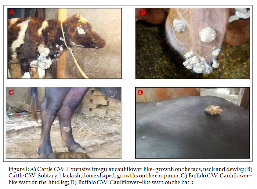
Figure 1: A) Cattle CW: Extensive irregular cauliflower like–growth on the face, neck and dewlap; B) Cattle CW: Solitary, blackish, dome shaped, growths on the ear pinna; C) Buffalo CW: Cauliflower–like wart on the hind leg; D) Buffalo CW: Cauliflower–like wart on the back

Histopathologically, most of cases (cattle–9 and buffaloes–5) diagnosed as fibropapilloma (exophytic) consisted of moderate to extensive degree of cornification (hyperkeratosis) with basket wave appearance, varying degree of parakeratosis, hyperplastic stratum spinosum with presence of many koilocytes and islands of dermal connective tissue surrounded by hyperplastic epidermal cell layers. Basal cell layer was hyperplastic with hyperchromatic nuclei, mild mitotic activity and occasionally, invasive growth pattern was seen. Below epidermis, the neoplastic stromal tissue consisted of large stellate shaped fibroblast cells and intense fibrocellular proliferative changes (Figure 2A–C). Comparable to cattle (with abundance of koilocytes), buffalo warts showed only few koilocytes in the upper layer of stratum spinosum. Some cases showed the similar histopathological features except the fact that they had long rete pegs extending towards the fibrous stoma and proliferated there extensively (fibropapilloma endophytic). Other histopathological types diagnosed were papilloma occult/ fibroblastic type (Figure 2D) and papilloma.

Polymerase Chain Reaction (PCR) Analysis
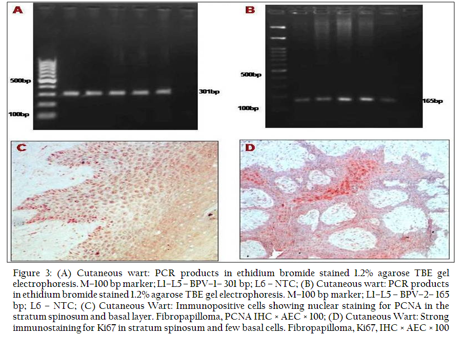
Out of 24 CW samples of cattle and buffaloes subjected to PCR, 14 samples of cattle were positive for BPV–2 while only 5 samples were found to have mixed infection as they were also positive for BPV–1. Out of 9 CW samples of buffaloes, 5 were found positive for BPV–1 and 3 for BPV–2. None of the sample was positive for BPV–5, –10 or any mixed infection. Detailed results are shown in Table–1. Gel electrophoresis showed the specific PCR product size of 301 and 165 bp for BPV–1 & –2, respectively (Figure 3A–B). PCR products of representative samples found positive for BPV–1 & –2 were purified and sequenced (HE603635, HE603636, HE600123 and HE600124) which revealed that the sequences generated in the present study has homology with earlier published sequences from India.
Results of SYBR Green Quantitative PCR assay
CW DNA samples positive for BPV–1 & –2 were utilized for determination of DNA copy number present in them by using SYBR Green Quantitative PCR. These samples were tested together with known standard samples (dilutions of known concentrations of plasmid DNA) along with “No Template Control” (NTC). DNA samples of the CWs of cattle revealed a copy number of BPV–1 between 3.17E+03 to 4.85E+05 while in buffalo CWs range vary from 8.48E+02 to 3.41E+05. All DNA samples of the cattle CWs were positive for the BPV–2 and the copy numbers detected in them vary from 4.69E+03 to 1.91E+16. Five CWs samples of the ear collected from the Gazaria Dairy Farm and Military Dairy Farm, Lucknow showed very high copy numbers i.e. 2.66E+09, 5.78E+15, 6.22E+15, 1.91E+16 and 1.75E+15. Cattle CWs from oC milnadu showed copy number of 5.55E+03 to 1.43E+04. Three DNA samples of the buffalo CWs showed copy number of 8.74E+04, 1.41E+04 and 1.41E+04.
Immunohistochemichemistry
CW samples were studied by IHC for PCNA and Ki67. A total of 9/17 (52.94%) CWs showed the immunopositivity for PCNA. Out of 10 fibropapilloma cases, only 4 (40%) showed immunoreactivity (PCNAmax–24.28±5.17; PCNAtot–15.02±8.85). Basal layer cells of the epidermis showed strong immunoreactivity while only few discrete cells in the parabasal and spinous layer showed weak immunostaining (Fig. 3C). Among 4 cases (cattle–3; buffalo–1) of fibropapilloma (endophytic), 2 cases of cattle showed the positivity for PCNA (PCNAmax–20.5±3.35; PCNAtot–14.45±6.42). One case each of cattle and buffalo of the occult papilloma/fibroblastic type were positive for PCNA and immunostaining was observed in the fibrous connective tissue (PCNAmax–22.33±2.64; PCNAtot–19.5±4.5). One papilloma case of buffalo was immunonegative for PCNA.
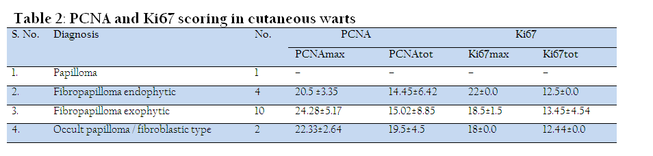
Ki67 immunopositivity was found in 4/17 (23.53%) samples, among them two were fibropapilloma (exophytic; Ki67max–22; Ki67tot–12.5), one each of fibropapilloma (endophytic; Ki67max–18.5±1.5; Ki67tot–13.45±4.54) and occult papilloma/fibroblastic type (Ki67max–18; Ki67tot–12.44). Immunostaining restricted mainly in the spinosum layer while discrete immunopositive cells were also observed in basaland parabasal layer. Few cells in fibrous stroma also showed positive reaction.
DISCUSSION
Clinically, CWs were observed in organised farms as well as rural areas in the cattle and buffaloes. The CWs are known to be transmitted from cattle to buffaloes and vice versa (Singh and Somvanshi, 2010). In Military Dairy Farm, Lucknow, UP warts was confined mostly to ear pinna which may be due the fact that these were transmitted from the infected animal to healthy one by tattooing machine. Association of tattooing with papilloma development on the ear of calves were reported earlier (Studdert et al., 1988). Histopathology revealed that fibropapilloma (exophytic) was the most common histopathological type observed in CWs of both cattle and buffaloes. Fibropapilloma (endophytic), papilloma occult/ fibroblastic type and papilloma were other types found in CWs. Similar histopathological types were also reported by earlier workers in the CWs (Pangty et al., 2010). Grossly, it was observed that few CW cases of cattle spread extensively to the adjacent areas forming large cauliflower–like masses but in buffaloes, almost all cases showed solitary growths. This finding was observed earlier also (Somvanshi, unpublished data).
PCR results revealed that DNA of CW of cattle was positive in 14 cases for BPV–2 while 5 were found to be positive for BPV–1. Five CW cases showed mixed infection as they were positive for both BPV–1 & –2. Out of 9 CW cases of buffaloes, 5 were found positive for BPV–1 and 3 for BPV–2. All samples were negative for BPV–5 and –10 which indicated that hairy skin is less prone to infection by these viruses although in earlier studies BPV–5 has been reported from the teat and facial skin (Lindholm et al., 1984; Bloch et al., 1994) and BPV–10 from the teat warts (Hatama et al., 2008; Rai et al., 2011). BPV–1, –2 and their mixed infections were also detected by previous workers in cattle (Leishangthem et al., 2008; Pangty et al., 2010; Pathania et al., 2011) and buffaloes (Silvestre et al., 2009; Singh and Somvanshi, 2010; Pangty et al., 2010) indicating that these two BPV types are prevalent in Indian cattle and buffalo population.
Real time PCR revealed a wide range of virus load in different samples for both BPV–1 & –2. Cattle CWs showed comparatively high viral DNA load of BPV–2 than buffaloes while for BPV–1 the CWs of both cattle and buffaloes had almost similar range. Earlier researchers also quantified the viral load of BPV–1 & –2 in cattle and buffalo CWs (Pangty et al., 2010; Nagarajan, 2011) but a comparative study is lacking. Determination of viral load and expression of the BPV E2, E5, E6 and E7 genes in four clinical types of equine sarcoid using quantitative real time PCR was performed earlier (Bogaert et al., 2007) and found that nodular sarcoid showed a significantly higher viral load than the other types.
In present study, PCNA positivity was observed mainly in basal cells and few discrete cells of parabasal and spinous layers of fibropapilloma and occult papilloma/fibroblastic. Similarly, PCNA was predominantly detected in the basal layer of the epidermis and in the superficial dermis in fibropapilloma of linea alba and teats in four heifers and the highest number of PCNA–positive nuclei was found in the basal layer of the epidermis (Jelinek and oC chezy, 2005). Lesions produced by human papilloma virus showed extensive PCNA expression in the epidermis, including the basal, parabasal and spinous layers (Penneys et al., 1992; Lu et al., 1999; Ozsoy et al., 2011). Ki67 immunopositivity was restricted mainly in the spinosum layers, although few positive cells were also detected in the basal and parabasal layer of the epidermis. Few comparative studies on the expression of PCNA and Ki67 in relation to HPV indicated that the expression of PCNA restricted mainly to the basal and parabasal layers while Ki67 was detected mainly in the spinous and upper layers of the epidermis along with HPV (Boon et al., 1993; Lu et al., 1996; Lu et al., 1999). Such findings were interpreted as Ki 67 expression was needed for the viral (HPV) replication and induction of both the markers were independent (Boon et al., 1993). Although present investigation also revealed similar expression behaviour of the PCNA and Ki67 in the bovine papillomatosis cases but to interpret the result in relation with BPV further studies needed.
ACKNOWLEDGEMENTS
We are thankful to Deputy Director General (Education), ICAR, New Delhi, for financial grant through ICAR–NAE Project and Director, IVRI, Izatnagar, UP, India for all necessary facilities for carrying out this research work.
REFERENCES
Bam J, Kumar P, Leishangthem GD, Saikia A and Somvanshi R (2012). Spontaneous cutaneous papillomatosis in yaks and detection and quantification of bovine papillomavirus -1 & -2. Transbound. Emerg. Dis. doi: 10.1111/j.1865-1682.2012.01361.x.
http://dx.doi.org/10.1111/j.1865-1682.2012.01361.x
Bloch N, Sutton RH and Spradbrow PB (1994). Bovine cutaneous papillomas associated with bovine papillomavirus type 5. Arch. Virol. 138: 373-377.
http://dx.doi.org/10.1007/BF01379140
PMid:7998842
Bogaert L, Van Poucke M, De Baere C, Dewulf J, Peelman L, Ducatelle R, Gasthuys F and Martens A (2007). Bovine papillomavirus load and mRNA expression, cell proliferation and p53 expression in four clinical types of equine sarcoid. J. Gen. Virol. 88: 2155 - 2161
http://dx.doi.org/10.1099/vir.0.82876-0
PMid:17622617
Boon ME, Howard CV and Velzen D (1993). PCNA independence of Ki67 expression in HPV infection. Cell Biol. Int. 17: 1001-1004.
http://dx.doi.org/10.1006/cbir.1993.1029
PMid:7906585
Culling CFA (1995). Hand book of Histological techniques. Second Edn. Butterworths and Co., London, UK
De Villiers EM, Faquet C, Brooker TR, Bernard HU and Hausen H (2004). Classification of papillomaviruses. Virol. 324: 17 - 27
http://dx.doi.org/10.1016/j.virol.2004.03.033
PMid:15183049
Hatama S, Ishihara R, Ueda Y, Kanno T and Uchida I (2011). Detection of a novel bovine papillomavirus type 11 (BPV-11) using Xipapillomavirus consensus polymerase chain reaction primers. Arch Virol. 156: 1281 - 1285
http://dx.doi.org/10.1007/s00705-011-0970-7
Hatama S, Nobumoto K and Kanno T (2008). Genomic and phylogenetic analysis of two novel bovine papilloma virus BPV-9 and BPV-10. J. Gen. Virol. 89: 158-163.
http://dx.doi.org/10.1099/vir.0.83334-0
PMid:18089739
Jelinek F and oC chezy R (2005). Cutaneous papillomatosis in cattle. J. Comp. Pathol. 132(1): 70 - 81.
http://dx.doi.org/10.1016/j.jcpa.2004.07.001
PMid:15629481
Leishangthem GD, Somvanshi R and Tiwari AK (2008). Detection of bovine papillomaviruses in cutaneous warts/papillomas in cattle. Indian J. Vet. Pathol. 32(1): 15 - 20
Lindholm I, Murphy J, O'Neil BW, Campo MS and Jarrett WFH (1984). Papilloma of the teat and udder of cattle and their causal virus. Vet. Rec. 115: 574
http://dx.doi.org/10.1136/vr.115.22.574
PMid:6098066
Lu S, Tiekso J, Hietanen S, Syrjaènen K, Havu VK and Syrjaenen S (1999). Expression of cell-cycle proteins p53, p21 (WAF-1), PCNA and Ki-67 in benign, premalignant and malignant skin lesions with implicated HPV involvement. Acta Derm. Venereol. 79: 268-273
http://dx.doi.org/10.1080/000155599750010634
PMid:10429981
Lu S, Syrjanen K, Havu VK and Syrjanen S (1996). Expression of PCNA is associated with the presence of HPV DNA in skin warts. Arch. Dermatol. Res. 289(1):35-39.
http://dx.doi.org/10.1007/s004030050149
PMid:9017133
Lunardi M, Alfieri AA, Otonel RA, de Alcantara BK, Rodrigues WB, de Miranda AB and Alfieri AF (2013). Genetic characterization of a novel bovine papillomavirus member of the Deltapapillomavirus genus. Vet. Microbiol. 162(1): 207-213
http://dx.doi.org/10.1016/j.vetmic.2012.08.030
PMid:22999523
Nagarajan N (2011). BPV-bracken fern interaction in buffaloes and laboratory model hamsters: Molecular and clinico-pathological studies. MVSc thesis (Veterinary Pathology), Deemed University, Indian Veterinary Research Institute, Izatnagar, Bareilly, UP, India
Nasir L and Campo MS (2008). Bovine papillomaviruses: Their role in the aetiology of cutaneous tumours of bovids and equids. Vet. Dermatol. 19: 243 - 254
http://dx.doi.org/10.1111/j.1365-3164.2008.00683.x
PMid:18927950
Ogawa T, Tomita Y, Okada M and Shirasawa H (2007). Complete genome and phylogenetic position of bovine papillomavirus type 7. J. Gen. Virol. 88(7): 1934 - 1938
http://dx.doi.org/10.1099/vir.0.82794-0
PMid:17554025
Ozsoy SY, Ozyildiz Z and Guzel M (2011). Clinical, pathological and immunohistochemical findings of bovine cutaneous papillomatosis. Ankara Univ. Vet. Fak. Derg. 58: 161-165
Pangty K, Singh S, Saikumar G, Goswami R and Somvanshi R (2010). Detection of BPV-1 and -2 and quantification of BPV-1 by real-time PCR in cutaneous warts in cattle and buffaloes. Transbound. Emerg. Dis. 57(3): 185 – 196
http://dx.doi.org/10.1111/j.1865-1682.2009.01096.x
PMid:20113447
Pathania S, Dhama K, Saikumar G, Shahi S and Somvanshi R (2011). Detection and quantification of bovine papilloma virus type 2 (BPV-2) by real-time PCR in urine and urinary bladder lesions in enzootic bovine haematuria (EBH)-affected cows. Transbound. Emerg. Dis. 59: 79 – 84
http://dx.doi.org/10.1111/j.1865-1682.2011.01248.x
PMid:21797988
Penneys NS, Bogaert M, Serfling U and Sisto M (1992). PCNA expression in cutaneous keratinous neoplasms and verruca vulgaris. Am. J. Pathol. 141(1): 139 - 142
PMid:1352943 PMCid:PMC1886566
Rai GK, Saxena M, Singh V, Somvanshi R and Sharma B (2011). Identification of bovine papilloma virus 10 in teat warts of cattle by DNase-SISPA. Vet. Microbiol. 147: 416-419
http://dx.doi.org/10.1016/j.vetmic.2010.07.015
PMid:20800979
Silvestre O, Borzacchiello G, Nava D, Iovane G, Russo V, Vecchio F, Ausilio D, Gault EA, Campo MS and Paciello O (2009). Bovine papillomavirus type 1 DNA and oncoprotein expression in water buffalo fibropapillomas. Vet. Pathol. 46: 636 - 641
http://dx.doi.org/10.1354/vp.08-VP-0222-P-FL
PMid:19276046
Singh V and Somvanshi R (2010). BPV-2 associated papillomatosis in Indian water buffaloes (Bubalus bubalis). Indian J. Anim. Sci. 80(10): 956 - 960
Singh V, Somvanshi R, and Tiwari AK (2009). Papillomatosis in Indian cattle: Occurrence and etiology. Indian J. Vet. Pathol. 33: 52 - 57
Stoimenov I and Helleday T (2009). PCNA on the crossroad of cancer. Biochem. Soc. Trans. 37: 605-613.
http://dx.doi.org/10.1042/BST0370605
PMid:19442257
Studdert MJ, Mccoy K, Allworth MB and Staples P (1988). Papilloma of the ears of calves following tattooing. Aus. Vet. J. 65(12): 399 http://dx.doi.org/10.1111/j.1751-0813.1988.tb14285.x PMid:2851971
Tomita Y, Literak L, Ogawa T, Jin Z and Shrivastava H (2007). Complete genomes and phylogenetic positions of bovine papillomavirus type 8 and a variant type from a European bison. Virus Genes, 35: 243 – 249
http://dx.doi.org/10.1007/s11262-006-0055-y
PMid:17265141
Woods AL, Hall PA, Shepherd NA, Hanby AM, Waseem NH, Lane DP and Levison DA (1991). The assessment of proliferating cell nuclear antigen (PCNA) immunostaining in primary gastrointestinal lymphomas and its relationship to histological grade, S + G2 + M phase fraction (flow cytometric analysis) and prognosis. Histopathol. 19: 21 – 27
http://dx.doi.org/10.1111/j.1365-2559.1991.tb00890.x
Zhu W, Dong J, Shimizu E, Hatama S, Kadota K, Goto Y and Haga T (2012). Characterization of novel bovine papillomavirus type 12 (BPV-12) causing epithelial papilloma. Arch. Virol. 157: 85 –91.
http://dx.doi.org/10.1007/s00705-011-1140-7
PMid:22033594


