Advances in Animal and Veterinary Sciences
Research Article
Pathological Affections of Lungs in Slaughtered Cattle and Buffaloes at Chittagong Metropolitan Area, Bangladesh
Md. Shafiqul Islam1, Shubhagata Das1, Md. Ariful Islam2, Md. Monjurul Islam Talukdar2, Md. Abul Hashem3, Sharmin Chowdhury1, Md. Masuduzzaman1
1Department of Pathology and Parasitology, Chittagong Veterinary and Animal Sciences University, Khulshi, Chittagong-4202, Bangladesh; 2MS, Microbiology, Bangladesh Agricultural University, Mymensingh, Bangladesh; 3Health department, Chittagong City Corporation, Chittagong, Bangladesh.
Abstract | The study was carried out to examine the pathological lesions in lungs of slaughtered cattle and buffalo carcasses in Chittagong Metropolitan area. A total of 882 (660 cattle and 222 buffalo) were examined in three different slaughterhouse where 128 (14.51%) cases were found with different pathological lesions in lungs. Among 128 (cattle 105 and buffalo 23) cases with lung lesions, the frequency of hydatid cyst was significantly (P< 0.05) higher in cattle (58.10%) than in buffaloes (34.78%), whereas the frequency of fibrous nodule (8.69%) and hemorrhages (17.39%) were significantly higher in buffalo lungs than cattle (1.9% and 0.06%). Different factors including the geographical and ecological aspects of the animal sources might have played significant role in the prevalence and comparative frequency of pathological lesions of lungs in cattle and buffaloes.
Keywords | Slaughterhouse, Lung, Pathology, Cattle, Buffalo
Editor | Kuldeep Dhama, Indian Veterinary Research Institute, Uttar Pradesh, India.
Received | June 28, 2014; Revised | December 02, 2014; Accepted | December 12, 2014; Published | December 15, 2014
*Correspondence | Md. Shafiqul Islam, Chittagong Veterinary and Animal Sciences University, Chittagong, Bangladesh; Email: si.mamun@ymail.com
Citation | Islam MS, Das S, Islam MA, Talukdar MMI, Hashem MA, Chowdhury S, Masuduzzaman M (2015). Pathological affections of lungs in slaughtered cattle and buffaloes at Chittagong Metropolitan Area, Bangladesh. Adv. Anim. Vet. Sci. 3(1): 27-33.
DOI | http://dx.doi.org/10.14737/journal.aavs/2015/3.1.27.33
ISSN (Online) | 2307-8316; ISSN (Print) | 2309-3331
Copyright © 2015 Islam et al. This is an open access article distributed under the Creative Commons Attribution License, which permits unrestricted use, distribution, and reproduction in any medium, provided the original work is properly cited.
Introduction
In Bangladesh most of the people are non-vegetarian and dependent largely on cattle and buffalo meat as protein source in diet. Food animal carcasses are the possible source of several trans-boundary infectious zoonotic diseases such as tuberculosis, leptospirosis, brucellosis, salmonellosis, anthrax, hydatidosis etc. (Scott et al., 1997). The emergence of anthrax has made the society more concerned about the health issues and quality of meat animal carcass (Akbar et al., 2007). Therefore, slaughter house inspection became a very important responsibility for the local veterinary authority and municipality to determine whether or not the animal was suffering from any disease or disease condition that makes the carcass inconsumable. In Bangladesh, traditional slaughtering places and slaughter slabs are present in municipalities and city corporations throughout the country, no slaughterhouses yet established with appropriate facilities. According to Torres (2007) an ideal slaughterhouse should have ante-mortem and postmortem inspection of carcasses, hygienic management and condemnation of diseased animal to ensure the best quality consumable animal products. Lung is a vital organ of the body and susceptible for several pathological affections indicative for disease conditions (Cullen and Maclachlan, 2001). Lung gets condemned for consumption if there is any pathological evidence of significant infectious diseases or disease conditions during carcass inspection. Parasitic diseases like hydatidosis, verminous pneumonia; bacterial diseases such as tuberculosis, abscess, viral diseases such as rhinitis, influenza and fungal diseases like mycoplasmosis, aspergillosis are the most common causes of condemnation of lungs. Besides, several other pathological affections like fibrosis, abscess, atelectasis, cysts, hemorrhages, congestion, edema, and pneumonia are common in lungs (Cullen and Maclachlan, 2001). Considering the above facts the present study was designed to investigate the gross and histopathological changes of localized or generalized diseases/disease conditions in the slaughtered animal carcasses.
MATERIALS AND METHODS
Study Area and Duration
This study was carried out by total 38 visits in three slaughterhouses (Firingibazar, Bohodderhat and Pahartali) of Chittagong Metropolitan area (CMA) from January to June 2012. All animals slaughtered in a particular visit day were taken into consideration for this study.
Sampling and Gross Examination of Slaughtered Carcasses
A total of 882 animals were carefully examined at the lairage (docking area) to record the demographic data. At slaughter slabs, when the carcasses are bled and completely opened, the visceral organs were thoroughly examined by close inspection and palpation. Lung suspected with pathological affections were isolated from the carcass and washed with water for detailed gross examination. Gross pathological lesions were detected and recorded with the aid of an experienced pathologist following FAO meat inspection manual (Herenda et al., 1994). From each and every suspected lung with pathological affections, samples were collected with the help of the butcher leader and city corporation meat inspector. The tissue samples were carried to Chittagong Veterinary and Animal Sciences University pathology laboratory in cool boxes as early as possible and fixed at 10% neutral buffer formalin for histopathological study.
Histopathological Study
For histopathological study formalin fixed tissue samples were washed and dehydrated in graded ethanol and embedded in paraffin wax. Fixed tissues were sectioned at 5μm thickness and stained with hematoxylin and eosin as per standard method (Luna, 1968).
Acid Fast (Ziehl–Neelsen) Staining for Detection of Mycobacterium Species
Lesions suspected for tuberculosis were stained with Ziehl–Neelsen acid fast staining to demonstrate Mycobacteria species as per the standard staining procedure.
Statistical Analysis
The obtained data form gross and microscopic examination was imported, stored and coded accordingly using Microsoft Excel-2000. Chi Square test was done for determination of association between the variables by using statistical software STATA/IC-11. Significance was determined in 95% confidence interval when P<0.05.
Results
In this study, the overall pathological conditions were 14.51% in different slaughterhouses whereas highest (25.00%) in Bohodderhat slaughter house (Table 1). The higher frequency of lung lesions was present in cattle (15.90%) than buffalo (10.36%) (Table 2). Out of 882 total carcasses examined, hydatid cysts was found in 69 (7.82%) cattle and buffalo which was the most prevalent lesions followed by emphysema 19 (2.15%), atelectasis 16 (1.81%), pulmonary hemorrhages 7 (0.79%), congestion 9 (1.02%), nodular lesions 4 (0.46%) and abscess 4 (0.46%) (Table 4). Among 128 (cattle 105 and buffalo 23) cases with lung lesions, the frequency of hydatid cyst was significantly higher in cattle (58.09%) lungs than buffalo (34.78%) lungs. (Table 3).
Gross and Histopathological Lesions in Affected Lungs
Hemorrhagic lesions were found as variable sized blood spots in the apical and middle lobe of lungs. Dark coloured extravasated blood spots were visible on the parietal surface with marked streaks of ribs impression. Hydatid cyst on lung parenchyma and pleura was most common and appeared as large watery or yellowish fluid filled space occupying lesion distributed over the organ. Multiple cysts on lung parenchyma made a polycystic appearance (Figure 1A). Cysts were often visible in clustered form in the pleura and thoracic cavity (Figure 1B). Firm to hard opaque cysts were found as a degenerated and calcified cyst (Figure 1C). Most of the cases of atelectasis were visible as a dark and depressed area. Distended pulmonary vessels with dark bluish coloration in middle and caudal lobe indicated hyperaemia and emphysema (Figure 1D). In some cases, partial hepatisation and consolidation was found (Figure 1E). Pulmonary abscesses were found in few buffalo lungs with solid elevated lesions in the parenchyma (Figure 1F).
Table 1: Overall Pathological lesions in the different slaughterhouse
|
Name Abattoirs |
No. of Carcass examined |
Cattle |
Buffaloes |
Total |
Overall % of lung lesions |
|
FiringiBazzar |
498 |
50 |
18 |
68 |
13.65% |
|
Bohodderhat |
152 |
35 |
3 |
38 |
25.00% |
|
Pahartali |
232 |
20 |
2 |
22 |
9.4% |
|
Total |
882 |
105 |
23 |
128 |
14.51% |
Table 2: Frequency of lung lesions in cattle and buffalo
|
Species |
Slaughtered animal |
Lung lesion |
Percentage |
|
Cattle |
660 |
105 |
15.90% |
|
Buffalo |
222 |
23 |
10.36% |
|
Total |
882 |
128 |
14.51% |
Table 3: Comparative frequency of lung affections in cattle and buffalo
|
Pathological Affections |
Cattle (N=105) |
Buffalo (N=23) |
P value |
||||
|
No |
(%) |
95% CI |
No |
(%) |
95% CI |
||
|
Hydatid Cyst |
61 |
58.10 |
48.52-67.67 |
8 |
34.79 |
14.69-54.88 |
0.0211 |
|
Atelectasis |
13 |
12.38 |
5.99-18.77 |
3 |
13.04 |
1.16-27.25 |
0.4653 |
|
Emphysema |
16 |
15.24 |
25.08-43.49 |
3 |
13.04 |
1.16-27.25 |
0.0225 |
|
Nodule |
2 |
1.90 |
0.75-4.56 |
2 |
8.69 |
3.19-20.58 |
0.0450 |
|
Congestion |
7 |
6.66 |
1.83-10-15 |
2 |
8.69 |
3.19-20.58 |
0.3652 |
|
Hemorrhage |
3 |
2.86 |
1.82-11.51 |
4 |
17.39 |
18.54-59.72 |
0.0001 |
|
Abscess |
3 |
2.86 |
1.82-11.51 |
1 |
4.35 |
4.26-12.95 |
0.0160 |
*=Means p<0.05
The lung samples with selected gross pathology were processed for histopathology and carefully examined for histopathological alterations. Hydatid cyst was characterized by the cyst wall was composed of proliferation of fibrous connective tissue with infiltration of mononuclear cells. Pulmonary emphysema was microscopically visible with severe enlargement of alveolar space with variable degree of pulmonary congestion (Figure 2A). Atelectatic lung showed collapse of the alveolar space with increased thickness of the septa. The blood vessel of the affected part was congested (Figure 2B). Purulent bronchopneumonia was characterized by massive infiltration of neutrophils that often occluded the lumen of the alveolar space and the terminal bronchi (Figure 2C). Accumulation of lymphocytes, macrophages and giant cells with copious eosinophilic exudates in the tissue section indicated the hepatisation and consolidation of lung parenchyma (Figure 2D). There was diffuse alveolar necrosis and edema in the inter-alveolar septa and sloughing of bronchial mucosa. Healed nodule was characterized by excessive proliferation of fibrous connective tissue indicating the feature of healing. Sub-acute pneumonia was characterized by the presence of immature collagen fibres associated with few reactive cells.
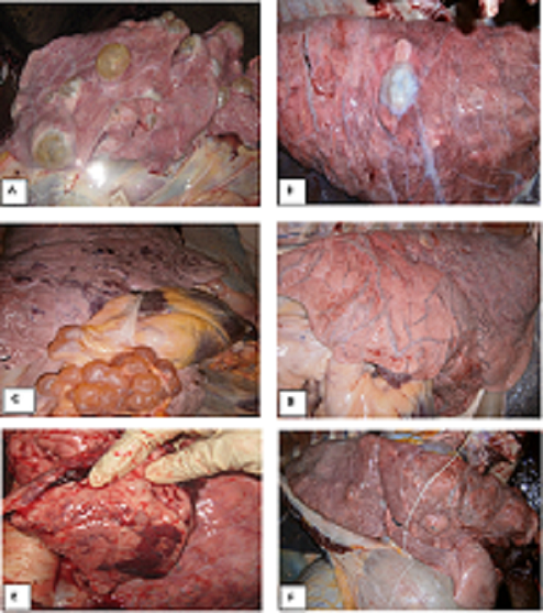
A. Polycystic lung containing large yellow fluid filled viable cyst and abscess; B. Old degenerated cyst and fibrosis in lung; C. Viable cyst in the pleura and lung in clustered appearance; D. Severe interstitial emphysema and marked distention of the veins (blue color) in middle and caudal lobe; E. Red Hepatization (consolidation) indicating pneumonia; F. Abscess in all over the lung parenchyma.
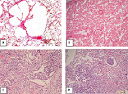
A. Pulmonary congestion and alveolar emphysema; B. Pulmonary atelectasis; C. Purulent bronchopneumonia in lung, accumulation of neutrophils in bronchiolar airway; D. Pulmonary hepatization (pneumonic lung) where alveolar space is filled with copious exudates.
Table 4: Percent prevalence of lung lesions in slaughtered Cattle and Buffalo in Chittagong Metropolitan Area
|
Pathological Affections |
Total lesion (Cattle and Buffalo) |
Prevalance % (Total animal = 882) |
|
Hydatid Cyst |
69 |
7.82 |
|
Atelectasis |
16 |
1.81 |
|
Emphysema |
19 |
2.15 |
|
Nodule |
4 |
0.46 |
|
Congestion |
9 |
1.02 |
|
Hemorrhage |
7 |
0.79 |
|
Abscess |
4 |
0.46 |
|
Total |
128 |
14.51 |
Discussion
Based on gross examination, among the 128 different cases, hydatid cysts were the most frequent lesions followed by emphysema, atelectasis, pulmonary hemorrhage, congestion, nodular lesions and abscess. This result come in close agreement with the findings of Belkhiri et al. (2009) who also reported that emphysema was the second most common bovine pulmonary lesions in slaughterhouses of Algeria. Rahman et al. (2006) reported that congestion, emphysema, hemorrhage, abscess and anthracosis as the other common affections in lung in Mymensingh. Similar lesions have been described earlier (Radostits et al., 2002).
Other lesions like black coloured lungs like anthracosis were observed by Radostits et al. (2002), but no such lesions were observed in the present investigation.
Statistically significant variation was observed between cattle and buffalo lungs in the frequency of pulmonary emphysema. Such distinction might be resulted from to the different physical factors of the buffalo and fewer samples in this study.
The higher frequency of hydatidosis, the most frequent pulmonary lesion in the present study is comparable with the findings of Belkhiri et al. (2009). High prevalence of hydatidosis in slaughtered carcasses was reported in Bangladesh by (Ahmedullah et al., 2007; Kabir et al., 2010; Basak et al., 2011). Akbar et al. (2007) reported 6.25% hydatid cyst from the slaughterhouses of Barisal sadar, Bangladesh. Pulmonary emphysema is often considered as the secondary lesion to traumatic pericarditis or pulmonary abscess (Blood and Henderson, 1976). Agonal emphysema is common in slaughtered animals resulted from agonal expiration (Cullen and Maclachlan, 2001).
Frequency of atelectasis was observed in 12.50% cases among all lung affections. According to Belkhiri et al. (2009) atelectasis is the collapse of certain portion of pulmonary tissue in absence of air content in alveoli. These lesions are classically localized in the apical and cardiac lobes, and more rarely in the diaphragmatic one. Pulmonary hemorrhages are related to severe septicemia or traumatic lesion to the lung. Agonal hemorrhages resulted from seizures and struggling during slaughter also results in pinpoint hemorrhage over lung surface particularly in the anterior lobes. The reason behind the higher rate of hemorrhagic lesion in buffalo is not clear at this moment and requires further investigation. The frequency of pulmonary congestion among the lung affections was 7.03% which is much less than the reports by Akbar et al. (2007) that showed 16.25% of pulmonary congestions in buffalo lungs by gross examination. Pulmonary congestion occurs due to the obstruction of the pulmonary vessels and passive which is sometimes followed by pulmonary edema (Blood and Henderson, 1976). Left-sided cardiac insufficiency, inflammation and degeneration of the myocardium or infarction of the heart muscle also cause such condition.
Abscess and nodular lesions were observed in lower frequency in this study. Nodular lesions in lung are observed in variety of conditions such as tuberculosis, granulomatous pneumonia, neoplastic growth etc. (Cullen and Maclachlan, 2001). Akbar et al. (2007) and Rahman et al. (2006) showed lower frequency of pulmonary nodules.
Acknowledgement
The authors are thankful to the authorities of University grand Commission (UGC) and to the director of Committee of higher education and research Chittagong Veterinary and Animal Sciences University for providing financial supports for this study.
References


