Advances in Animal and Veterinary Sciences
Research Article
Advances in Animal and Veterinary Sciences 2 (7): 414 – 417Preparation and Characterization of Chitosan Nanoparticles “Alternatively, Carrying Potential’’ for Cellular and Humoral Immune Responses
Vimal Kumar*, Satyabrata Dandapat, Ananda Kumar, Niranjan Kumar
-
Immunology Section, Indian Veterinary Research Institute (IVRI), Izatnagar, Bareilly, Uttar Pradesh. 243122
*Corresponding author:drvkyadava@gmail.com
ARTICLE CITATION:
Kumar V, Dandapat S, Kumar A, Kumar N (2014). Preparation and characterization of chitosan nanoparticles “alternatively, carrying potential’’ for cellular and humoral immune responses. Adv. Anim. Vet. Sci. 2 (7): 414 –417.
Received: 2014–06–15, Revised: 2014–08–21, Accepted: 2014–08–22
The electronic version of this article is the complete one and can be found online at
(
http://dx.doi.org/10.14737/journal.aavs/2014/2.7.414.417
)
which permits unrestricted use, distribution, and reproduction in any medium, provided the original work is properly cited
ABSTRACT
Chitosan is a natural polymer as well as biodegradable in nature and hence can be used safely for animal body system and can be a better delivery system for inactivated vaccines for which oil adjuvants are currently the sole option. The nanoparticles are prepared by ionotropic gelation technique which encapsulate the antigen and forms a complex which slowly releases the antigen resulting in prolonged immunity by the same mechanism as that of adjuvants. These nanaoparticles can be easily taken up by antigen presenting cells (APC) which process and present antigen to other immune cells. In the present study, we have taken bovine serum albumen (BSA) as model antigen. The encapsulation efficiency was found to be about 49%. The nanoparticles formed are approximately 205.8 nm in diameter as evident by the electron microscopy and zeta–sizer. The charge was 40 mV, which provides muco–adhesive property for mucosal delivery. The above characteristics makes easy uptake of nanoparticles–by the APC on which cellular as well as humoral response depend. These findings indicate the alternative potential of these nanoparticles over adjuvant for persistent and prolonged immune responses.
INTRODUCTION
Chitosan is a natural biodegradable polysaccharide, co–polymer of N–acetyl–d–glucosamine and d–glucosamine (Onishi and Machida, 1999) obtained by the alkaline deacetylation of chitin, which is a polysaccharide found in the exoskeleton of crustaceans of marine arthropods and insects (Romoren et al., 2002). Chitosan itself is also found in some microorganisms and in yeast and fungi (Watts et al., 1999). Chitosan is biodegradable in nature and its biocompatibility as well as physiochemical characteristics of hydrophobically modified glycol chitosan nanoparticles in murine osteosarcoma and osteoblast–like cells have been evaluated recently (Chin et al., 2014)
Chitosan is available in a broad range of molecular weights and its properties can be modified by changing the degree of deacetylation and the environment of the formulation (Davis and Illum, 2003). Due to its protonated amino functions in solution and its resultant polycationic nature, chitosan readily adheres to negatively charged surfaces, such as mucus and proteins. Numerous studies in mice and human (Davis and Illum, 2003; Moschos et al., 2004) have demonstrated that chitosan and their soluble derivatives are effective and safe adsorption enhancers to improve nasal and peroral delivery of hydrophilic antigens such as peptide and protein drugs and heparins. In these models, the absorption enhancing effect is caused by the opening of the intercellular tight junctions, thereby favouring the paracellular transport of macromolecular drugs, and by slowing down mucociliary clearance, thus maintaining the contact of antigen with the mucosa for a longer period of time, thereby facilitating higher drug and carrier bioavailability. In this present study, we prepared chitosan nanoparticles and characterized their encapsulation efficiency, size and charges which are important factors determining the uptake of antigen by APC and development of immune responses for sustained vaccine effects.
MATERIALS AND METHODS
Reputed National and International firms’ viz. Sigma Chemicals (USA), SRL (India) and Merck (India) were our sole source from where standard analytical grade of chemicals were procured for this research work. Chitosan and sodium tripolyphosphate (TTP) used in this study were purchased from Sigma, USA. Acetic acid was of HPLC grade procured from Merck. All other chemicals used in this study were of the molecular grade. We used bench top centrifuge– HermLe Z 360 K, Sorvall RC 5C plus high speed refrigerated centrifuge (Depont, USA), Micro centrifuge (Eppendorf, Germany), Magnetic stirrer– REMI, Micropipettes– Ependorff, Germany. Glass wares used were obtained from Borosil (India) and Schott Duran (Germany). Glass wares were thoroughly washed and sterilized wherever necessary following the recommended procedures.
Preparation of Blank Chitosan Nanoparticles
Chitosan nanoparticles were prepared according to the procedure developed by Zhang et al. (2004) based on the ionotropic gelation of chitosan (CS) with sodium tripolyphosphate (TPP) anions with slight modification. A total of 8.75 gm of CS was dissolved in 5mL of an aqueous solution of acetic acid (0.306 wt %) at concentrations of 0.175 wt %. Afterward, 2.0 mL of TPP solution (1mg/mL) was added drop wise to 5.0 mL of CS solution in a 20–mL glass vial under magnetic stirring at 600 rpm using an octagonal stirring bar. The mixture was then stirred for additional 10 min. The nanoparticles formed were centrifuged at 20000 rpm for 40 min. The pellet formed was resuspended in de–ionized water with constant shaking and then pipetting.
Antigen Encapsulation Efficiency
One mg of BSA was dissolved in 5mL of chitosan solution. Thereafter, the procedure for preparation of BSA loaded chitosan nanoparticles was same as described for the preparation blank nanoparticles. After preparation of BSA loaded nanoparticles, the solution was centrifuged at 24000g for 30 min at 10ºC. The concentration of antigen in the supernatant was measured using Bicinchoninic acid (BCA) method of protein estimation. Antigen encapsulation efficiency was determined by subtracting the antigen present in the supernatant from the total amount of antigen used for loading. Each batch was analyzed in triplicate.
EE= |
(A– B)/A |
× 100 |
Where A is the total amount of antigen added during preparation, B is the amount of antigen remaining in the supernatant.
Particle Size and Zeta Potential Analysis
The size distribution and zeta potential of nanoparticles were determined by quasi–elastic light scattering at 25 oC, at an angle of 900, using a Zeta Sizer 3000 HS (Malvern Instruments, UK). Highly diluted colloidal dispersions of nanoparticles in pure distilled water were used for size and zeta potential measurement as per the standard procedure followed at National Institute of Immunology (NII), New Delhi. All the measurements were performed in triplicate for 60 seconds at 1000 Hz, and an electric current of 3 mA with zero field correction.
Size and Morphology
Particle size and surface morphology of nanoparticles were determined by Scanning electron microscopy (SEM). Briefly, a small pinch of the air dried nanoparticles were coated on the metallic slub by ion sputtering or gold coating and then observed under different magnifications using Jeol JSM 840 x Scanning electron microscope.
Sterility Testing
The sterility of nanoparticles was checked by inoculating in nutrient broth, nutrient agar, blood agar and Robertson’s cooked meat media and observing for growth of any micro–organisms.
RESULTS
A 5 mL CS solution was prepared in acidic de–ionized water. The concentration of acetic acid was adjusted. A total of 1 mg of BSA antigen was dissolved in 5mL of CS solution. Afterward, 5mL of this solution was added to a 20 mL glass vial. BSA loaded CS–NP was formed spontaneously upon drop wise addition of 2 mL TPP (1mg/mL) in above solution under magnetic stirrer. Three batches were prepared and centrifuged. The pellet formed was resuspended in de–ionized water.
Antigen encapsulation efficiency was determined by subtracting the antigen present in the supernatant from the total amount of antigen used for loading. After preparation of BSA loaded nanoparticles, the solution was centrifuged at 24000g for 30 min at 10ºC. The concentration of BSA in the supernatant was measured using BCA method. Each batch was analyzed in triplicate.
EE= |
(A– B)/A |
× 100 |
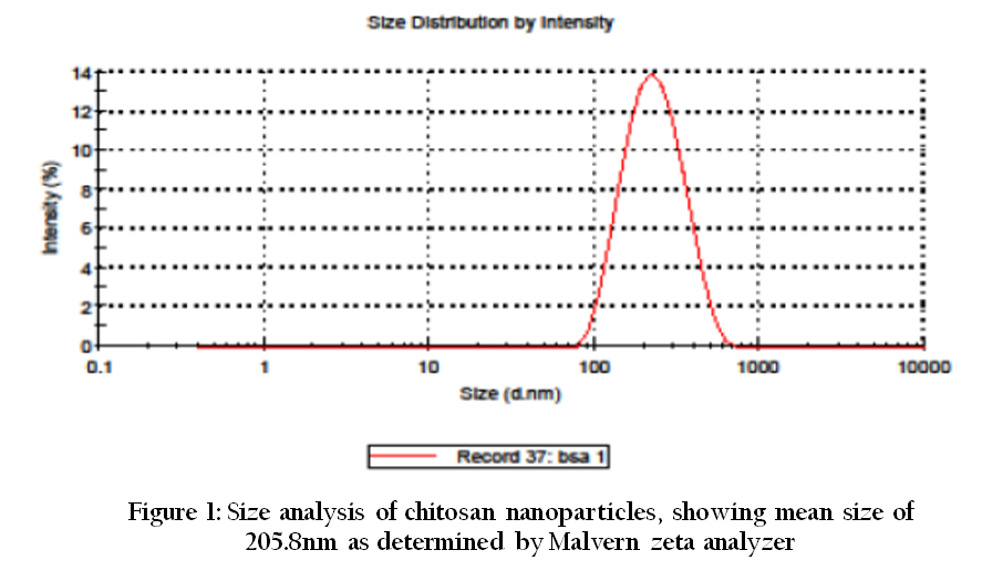
Figure 1: Size analysis of chitosan nanoparticles, showing mean size of 205.8nm as determined by Malvern zeta analyzer
Where A is the total amount of antigen added during preparation, B is the amount of antigen remaining in the supernatant. The encapsulation efficiency calculated was around 49%.
Nanoparticles made up of chitosan prepared by ionotropic gelation technique were comparatively smaller in size, ranging between 200 to 300 nm (mean size 205.8 nm) as determined by particle size analyzer (Figure 1). Nanoaprticles prepared by this method showed a mean positive charge of 40 mV (Figure 2). A homogenous population of nanoparticles having irregular shape was seen under scanning electron microscope. The sizes of the nanoparticles were ranged from 200–300 nm for BSA encapsulated nanoparticles (Figure 3).
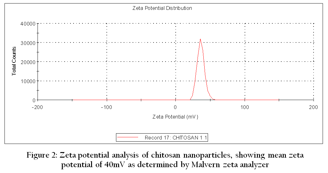
Figure 2: Zeta potential analysis of chitosan nanoparticles, showing mean zeta potential of 40mV as determined by Malvern zeta analyzer
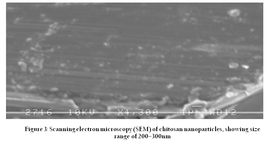
Figure 3: Scanning electron microscopy (SEM) of chitosan nanoparticles, showing size range of 200–300nm
The sterility of nanoparticles preparations was checked by inoculating in nutrient broth, nutrient agar, blood agar and Robertson’s cooked meat media and no growth of any micro–organisms was there.
DISCUSSION
Nanoparticles have received much attention for the delivery of molecular antigens, such as peptides, proteins and genes, due to their ability to protect the antigens from degradation by proteolytic enzymes (Sakuma et al., 2001). The potential use of polymeric nanoparticles as vaccine and drug carriers has led to the development of many different colloidal delivery vehicles. The main advantages of this kind of systems lie in their capacity to cross biological barriers, to protect molecules, such as peptides, proteins, oligonucleotides and genes from degradation in biological media and to deliver drugs or macromolecules to a target site following controlled release. In the past years, several synthetic as well as natural polymers have been examined for pharmaceutical applications. A basic requirement for these polymers to be used in humans or animals is that they have to degrade into molecules with no toxicity for biological environments. For this biocompatibility reason, a very limited numbers of polymers can be used to prepare biodegradable materials. Recently, the natural polymer, chitosan has attracted great attention in pharmaceutical and bio–medical fields (Paul and Sharma, 2000) because of its advantageous biological properties, such as biodegradability, biocompatibility, and non–toxicity. Chitosan is a cationic polysaccharide obtained by partial deacetylation of chitin, the major component of crustacean shells. In contrast to other polymers, chitosan is a hydrophilic polymer with positive charge that comes from weak basic groups, which give it special characteristics from the technological point of view.
In our study, we prepared BSA loaded chitosan nanoparticles. BSA was taken as the model antigen as its estimation is easy and also can compared easily with the standard protein estimation protocols. Encapsulation efficiency of the nanoparticles was calculated after subtracting the protein content of the supernatant from the total amount of antigen added during nanoparticle preparation and it was found to be 49 % (which was in accordance with findings of Zhang et al. (2004), who also got similar results depending upon the concentration of chitosan. Nanoparticles prepared by this method showed a mean size of 205.8 nm and mean positive charge of 40 mV when analyzed by Malvern zeta–sizer. These results were very much similar to that of Zhang et al. (2004). For all formulations the polydispersity index was less than 0.3, which indicates homogeneous nature of nanoparticles. Scanning electron microscopic (SEM) studies revealed that the size of nanoparticles, formed by ionotropic gelation technique, exhibited population of nanoparticles ranging from 200–300 nm and majority of them were of 200 nm in diameter which is consistent with the earlier findings of many workers (Calvo et al., 1997; Vila et al., 2004; Zhang et al., 2004). Nanoparticles were spherical in shape with solid and consistent structure. Earlier studies by Calvo et al. (1997) also demonstrated the same characteristics of nanoparticles. Nano–encapsulation of antigens by chitosan confers an adjuvant effect by improving uptake of antigens into the antigen presenting cells (APC) and by sustaining the release of antigenic material over a prolonged time. The adjuvant effect of chitosan of live vaccines have been evaluated in specific pathogen free (SPF) chickens immunized at day–old with an enterotropic NDV strain (Rauw et al., 2010) who demonstrated humoral, local antibody–mediated immune responses. Preparation and efficacy of a live Newcastle disease virus vaccine encapsulated in chitosan nanoparticles have been carried out recently (Zhao et al., 2012). The use of chitosan for delivery as well as an adjuvant for inacitivated antigen has to go a long way. In this study, we went for characterization of some of the parameters of chitosan nanoparticles that will lead to achieve the goal of chitosan nanoparticles based vaccine delivery system.
ACKNOWLEDGEMENT
We are thankful to joint directors and director of Indian Veterinary Research Institute (IVRI) for their kind support during my research work. We are also thankful to Dr Amulya Panda, Scientist in National Institute of Immunology (NII), New Delhi for providing Zeta–sizer facility.
REFERENCES
Calvo P, Remunan–Lopez C, Vila–Jato JL, Alonso MJ (1997). Novel Hydrophilic chitosan–polyethylene oxide nanoparticles as protein carriers. J. Appl. Polym. Sci. 63: 125 – 132.
http://dx.doi.org/10.1002/(SICI)1097-4628(19970103)63:1<125::AID-APP13>3.0.CO;2-4
Chin A, Suarato G, Meng Y (2014). Evaluation of physiochemical characteristics of hydrophobically modified glycol cells chitosan nanoparticles and their biocompatibility in murine osteosarcoma and osteoblast–like cells. J. Nanotech. Smart Mater. 1: 1–7
Davis SS, Illum L (2003). Absorption enhancers for nasal drug delivery. Clin. Pharmacokinet. 42: 1107 – 1128.
http://dx.doi.org/10.2165/00003088-200342130-00003
PMid:14531723
Moschos SA, Bramwell VW, Somavarapu S, Alpar HO (2004). Adjuvant synergy: the effects of nasal coadministration of adjuvants. Immunol. Cell. Biol. 82: 628 – 637.
http://dx.doi.org/10.1111/j.0818-9641.2004.01280.x
PMid:15550121
Onishi H, Machida Y (1999). Biodegradation and distribution of water–soluble chitosan in mice. Biomaterials.20: 175 – 182.
http://dx.doi.org/10.1016/S0142-9612(98)00159-8
Paul W, Sharma CP (2000). "Chiotosan, a drug carrier for the 21st century: a review," S.T.P. Pharma Sci. 10: 5 – 22.
Rauw F, Gardin Y, Palya V, Anbari S, Gonze M, Lemaire S, van den Berg T, Lambrecht B (2010).The positive adjuvant effect of on antigen specific cell mediated immunity after chickens vaccinated with live Newcastle disease vaccine . Vet. Immunol. Immunopathol. 134(3–4): 249 – 258.
http://dx.doi.org/10.1016/j.vetimm.2009.10.028
PMid:19939464
Romoren K, Thu BJ, Evensen O (2002). Immersion delivery of plasmid DNA. II. A study of the potentials of a chitosan based delivery system in rainbow trout (Oncorhynchus mykiss) fry. J. Control. Release. 85: 215 – 225.
http://dx.doi.org/10.1016/S0168-3659(02)00278-X
http://dx.doi.org/10.1016/S0168-3659(02)00279-1
Sakuma S, Hayashi M, Akashi M (2001). "Design of nanoparticles composed of graft copolymers for oral peptide delivery". Advanced Drug Delivery Reviews. 47(1): 21 – 37.
http://dx.doi.org/10.1016/S0169-409X(00)00119-8
Vila A, Sa’nchez A, Janes K, Behrens I, Kissel T, Jat JLV, Alonso MJ (2004). Low molecular weight chitosan nanaoparticles as new carriers for nasal vaccine delivery in mice. European Journal of Pharmaceutics and Biopharmaceutic. 57: 123 – 131.
http://dx.doi.org/10.1016/j.ejpb.2003.09.006
PMid:14729088
Watts TH, DeBenedette MA (1999). T cell costimulation other than CD28. Curr. Opin. Immunol. 11: 286 – 293.
http://dx.doi.org/10.1016/S0952-7915(99)80046-6
Zhang H, Allen C, Kumacheva E (2004). Monodisperse chitosan nanoparticles for mucosal drug delivery. Biomacromolecules. 5: 2461 – 2468.
http://dx.doi.org/10.1021/bm0496211
PMid:15530064
Zhao K, Chen G, Shi X, Gao T, Li W, Zhao Y, Zhang F, Wu J, Cui X, Wang YF (2012). Preparation and Efficacy of a Live Newcastle disease Virus Vaccine encapsulated in Chitosan Nanoparticles. PLOS ONE. 7: 1 – 10.


