Advances in Animal and Veterinary Sciences
Research Article
Advances in Animal and Veterinary Sciences 2 (7): 407 – 413Concurrent Administration of Curcumin Mitigates Arsenic – and Chlorpyrifos – induced Apoptosis in Rat Thymocytes
Atul Prakash1,2*, Saleem Khan2, Deepak Kumar3, Avinash G. Telang2, Jitendra Kumar Malik2
- Department of Pharmacology and Toxicology, College of Veterinary Science and Animal Husbandry, U. P. Pt. Deen Dayal Upadhyay Pashu Chikitsa Vigyan Vishwavidyalaya Evam Gau Anusandhan Sansthan (DUVASU), Mathura, India
- Division of Pharmacology and Toxicology, Indian Veterinary Research Institute, Izatnagar, Bareilly, India
- Division of Veterinary Biotechnology, Indian Veterinary Research Institute, Izatnagar, Bareilly
*Corresponding author:dratul2510@rediffmail.com
ARTICLE CITATION:
Prakash A, Khan S, Kumar D, Telang AG, Malik JK (2014). Concurrent administration of curcumin mitigates arsenic – and chlorpyrifos – induced apoptosis in rat thymocytes. Adv. Anim. Vet. Sci. 2 (7): 407 – 413.
Received: 2014–05–12, Revised: 2014–08–13, Accepted: 2014–08–15
The electronic version of this article is the complete one and can be found online at
(
http://dx.doi.org/10.14737/journal.aavs/2014/2.7.407.413
)
which permits unrestricted use, distribution, and reproduction in any medium, provided the original work is properly cited
ABSTRACT
The present study was undertaken to evaluate the arsenic and chlorpyrifos–induced apoptosis alone or in combination. In addition, the ameliorative efficacy of curcumin on as and/or chlorpyrifos–induced apoptosis in rat thymocytes in vivo. Adult male Wistar rats weighing 80–100g were divided into nine groups containing seven animals in each. The rats were fed with arsenic (20 ppm) in drinking water, chlorpyrifos (5 mg/kg body wt.), curcumin (100 mg/kg body wt.) and their combination for 28 days. After 28 days of exposure period, animals were sacrificed with cervical dislocation and thymus was collected to harvest the thymocytes and further reactive oxygen species (ROS) generation (by using non fluorescent DCFH–DA), % apoptotic DNA (by using propidium iodide) and mitochondrial transmembrane potential (by using DiOC6) were evaluated using Becton Dickenson Flowcytometer. The results showed that, the chlorpyrifos–induced per cent apoptotic DNA content (15.75±1.38), cells with loss of ψ Δm (16.70±1.33) and ROS generation (23.93±1.18) were significantly higher than the arsenic alone (10.95±1.05), (13.75±1.44) and (21.86±0.84) respectively, at given doses due to increased production of ROS. Arsenic and chlorpyrifos in combination potentiated the induction of apoptosis than their alone effect, however concurrent administration of curcumin considerably inhibited the induction of apoptosis by scavenging free radicals.
INTRODUCTION
It is becoming increasingly evident that environmental exposures in humans and animals are not just limited to single chemicals. Rather, they are exposed concurrently or sequentially to a large number of chemicals from a variety of sources. Arsenic is a well–recognized multisite human carcinogen presently affecting more than 24 countries around the world. In West Bengal, more than 6 million people in nine districts are endemically exposed to inorganic arsenic by drinking heavily contaminated groundwater from hand–pumped tube–wells (Chowdhury et al., 2001) while Ballia district of eastern Uttar Pradesh (GOI, 2011) and arsenic contamination in Ganga–Meghna– Brahmaputra (GMB) plain (Chakraborti et al., 2010) are heavily contaminated with arsenic. Arsenic concentrations in groundwater of these districts range from 50 to 1200 µg/l, far above the current maximum permissible limit of 10 µg/l laid down by World Health Organization (WHO, 1996; 2011). Soluble inorganic arsenic is acutely toxic. Intake of inorganic arsenic over a long period can lead to chronic arsenic poisoning (arsenicosis) and effects, take years to develop depending on the level of exposure, include skin lesions, peripheral neuropathy, gastrointestinal symptoms, diabetes, renal system effects, cardiovascular disease and cancer. The exact mechanism by which arsenic exerts its toxic effect is not clear. Indeed, some studies suggest that As III exert its toxicity by generating reactive oxygen species (ROS) and thereby oxidative stress (Chang et al., 2007) and ultimately apoptosis (Das et al., 2009; Prakash et al., 2011; Khan et al., 2013; Kumar et al., 2014).
Chlorpyrifos (CPF) is an effective organophosphate (OP) pesticide used heavily throughout the world for agriculture and domestic purposes. The health effects caused by this occupational exposure are enormous. Chlorpyriphos (O, O–diethyl–O– (3, 5, 6–trichloro–2–pyridinyl phosphorothioate) belongs to the phosphorothioate class of insecticides. It is classified as a moderately hazardous, Class II insecticide by the WHO (WHO, 1997). The main target of OP pesticides is acetylcholinesterase (AChE), which hydrolyses acetylcholine (ACh) in cholinergic synapses and at neuromuscular junctions. CPF elicits a number of other effects including hepatic dysfunction, immunological abnormalities, embryotoxicity, genotoxicity, teratogenicity, neurochemical, and neurobehavioral changes (Rahman et al., 2002; Harford et al., 2005; Verma et al., 2007). Acute and chronic exposure to chlorpyriphos has been shown to cause considerable liver damage. Chlorpyrifos induces apoptosis and DNA damage through generation of reactive oxygen species in drosophila (Gupta et al., 2010), human neuroblastoma cell line (Raszewski et al., 2014), PC–12 cells (Lee et al., 2012) and in primary culture of murine thymocytes (Prakash et al., 2009; 2010a; 2010b).
Plant products are known to exert their protective effects by scavenging free radicals and modulating antioxidant defense system. Curcumin (diferuloylmethane), an important constituent of turmeric (Curcuma longa L.) has been widely used for centuries as an indigenous medicine. Curcumin has been shown to possess a broad spectrum of pharmacological activities including antineoplastic, antimutagenic, anti–inflammatory and antioxidant (Naik et al., 2004). Curcumin is a potent scavenger of a variety of reactive oxygen species including superoxide anion radicals, hydroxyl radicals and inhibit lipid peroxidation and effectively block thiol depletion (Chattopadhyay et al., 2005). Furthermore, Curcumin was also found to be effective in preventing methylglyoxal (MG)–induced oxidative DNA damage, cell injury, apoptosis, and generation of ROS in mononuclear cells (Chan and Wu, 2006) .
The exposure to chemical mixture is environmental reality. In view of the potential increase in the drinking water contamination with arsenic and occupational exposure to pesticides, there is practicable possibility for the co–exposure of these two agents. Arsenic affects DNA repair mechanisms and also generates free radicals. On the other hand, CPF is known to induce DNA damage and free radicals generation. Although the toxicological information on arsenic (as) and chlorpyrifos (CPF) individually is available, but no information seems to be available about the possible toxicological interactions between them, till date. As both the compounds appear simultaneously to damage DNA/inhibit DNA repair, and generate free radicals, it is of notable importance to study their interaction. In the current study, we examined whether co–exposure to as and CPF could be more hazardous than the exposure to the individual agents to rats in vivo. The results of this study could clarify the role of the popular herbal drug curcumin in prevention of arsenic and chlorpyrifos induced toxicity.
MATERIALS AND METHODS
Animals
The study was conducted in adult male Wistar rats weighing 80–100 g procured from Laboratory Animal Resource Section, Indian Veterinary Research Institute, Izatnagar. Animals were maintained under standard managemental conditions as per the Institute animal ethics guidelines and caged in clean plastic cages containing wheat straw chips for bedding with 12 h of dark and light cycle, and given nutritionally adequate standard laboratory diet, and deionized water ad libitum. Before the start of the experiment, animals were kept in laboratory conditions for at least a period of seven days for acclimatization. All the experimental animals were kept under constant observation during the entire period of study.
Chemicals
Sodium arsenite and curcumin used in the present study were purchased from M/s Sigma Aldrich, USA. All other chemicals used were of analytical grade and purchased from Merck, India, Fluka and Sigma Aldrich, USA. Chlorpyrifos (95% purity, Technical grade) was generously supplied by Meghamani Organics Limite, Ahmedabad.
Experimental Design
Adult male Wistar rats were divided into nine groups namely: control (group I), vehicle control (group II), arsenic (group III), chlorpyrifos (group IV), arsenic plus chlorpyrifos (group V), curcumin (group VI), arsenic plus curcumin (group VII), chlorpyrifos plus curcumin (group VIII) and arsenic plus chlorpyrifos plus curcumin (group IX) comprising seven animals each. Chlorpyrifos (95% technical grade) @ 5 mG/kG body wt. and curcumin @ 100 mG/kG body wt. were given daily for 28 days by gastric intubation after proper dissolving in corn oil. The vehicle control group animals received an equal volume of corn oil in an identical manner. The arsenic containing water was prepared by adding 35 mG of sodium arsenite (Fluka) to one liter of deionized water to obtain 20 ppm of arsenic and given through drinking water. Control animals received only deionized water. Various toxicity end points were determined at the end of exposure period.
Sample Collection
At the end of the treatment period rats were sacrificed by cervical dislocation. Thymus were collected and finely chopped minced and thymocytes were harvested under cold condition for examining the induction of apoptosis.
Apoptosis Related Parameters (by Using Flowcytometry)
Reactive Oxygen Species (ROS) Generation (using DCFH–DA)
The procedure was followed as described by Wang et al., (1996). After the exposure period, thymocytes were collected in microcentrifuge tube; then cells were pelleted at 200 x g for 10 min at 20 °C. Cells were washed with PBS. DCFH–DA dye (final concentration 5 μM) was added to the sample and again kept for 15 min in the dark at room temperature. Flowcytometer analysis for DCF florescence was done as Counts vs FSC–H and 10000 thymocytes per event were taken. Per cent right shift in the florescence peak was recorded.
Apoptosis Detection (using Propidium Iodide)
After the exposure period thymocytes were isolated aseptically and washed with PBS and fixed by drop addition of ice cold 70% ethanol and stored at –20 o C overnight. The fixed cells were washed with PBS twice and suspended in 500µL of Propidium Iodide hypotonic buffer and incubated at 37oC for 15min in dark. The PI fluorescence was measured through FL–2 filter in Becton Dickenson Flowcytometer.
Mitochondrial Trans–membrane Potential (using DiOC 6 )
Mitochondrial transmembrane potential is analyzed according to the method of Castedo et al., (2002). Briefly, after the treatment period the cells are harvested and washed with PBS and dissolved in 0.5 ml of PBS and 25µL of DiOC6 is added. Tubes are incubated at 37o C for 10–15 min in dark. After the incubation, return the cells to ice and analyzed in Becton Dickinson flow cytometer through FL 1(emission: 530 nm).
DNA Laddering Assay in Thymocytes
After the exposure period, thymocytes were collected in microcentrifuge tube; cells were pelleted at 200 x g for 10 min at 20 oC. The pelleted thymocytes were lysed with 0.5 ml of hypotonic lysis buffer TTE (10mM Tris, 0.25% Triton X–100, 1 mM EDTA). Proteins were removed by phenol–chloroform isoamyl alcohol (25:24:1) followed by chloroformisoamyl alcohol (24:1) extraction. The DNA was precipitated with a 1/10 volume of 3 M sodium acetate solution and 2 volumes of cold absolute ethanol at –20°C overnight. DNA pellets were washed twice with 75% ethanol, dried under vacuum and resuspended in TE (10 mM Tris– HC1, pH 7.4, 1 mM EDTA). Sample containing approximately 1123 ng of DNA were subjected to electrophoresis in 15% agarose for 1–5 h at 60 V. The gels were stained with ethidium bromide and photographed under UV transillumination.
RESULTS AND DISCUSSION
Exposure to toxic metals and pesticides remains a wide–spread problem worldwide. Humans and animals are chronically exposed to arsenic that occurs in drinking water and is associated with immunotoxicity (Lemarie et al., 2006; Soto–Pena and Vega, 2008), reproductive toxicity (Chang et al., 2007, Jhala et al., 2008; Manna et al., 2008), tumors of skin, bladder, liver and lung and oxidative stress (Flora et al., 2005; Kinoshita et al., 2007). Chlorpyrifos (O,O–diethyl–O– (3,5,6–trichloro–2–pyridinyl phosphorothioate) is a broad–spectrum organophosphorus pesticide that is widely used throughout the world in agriculture and non–agricultural applications (Caroline, 1994; Richardson, 1993). The main toxicity of chlorpyrifos is neurotoxicity, which is caused by the inhibition of acetylcholinesterase (Whitney et al., 1995; Moser, 2000; Chanda and Pope, 1996; Chakraborti et al., 1993). On the other hand, it has been reported that chlorpyrifos caused immunologic abnormalities in animals (Blakley et al., 1999) and humans (Thrasher et al., 1993, 2002), Exposure to chlorpyrifos is associated with multiple chemical sensitivity (Berkson, 1994; Ziem and McTamney, 1997). The above findings strongly encouraged us to explore the possibility of arsenic– and chlorpyrifos–induced immunologic abnormalities. Thus, we investigated whether arsenic and chlorpyrifos induces apoptosis in rat thymocytes.
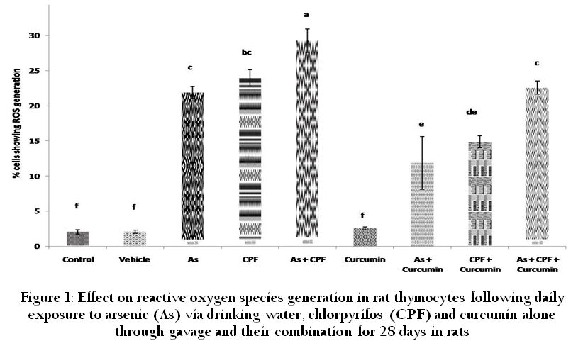
Figure 1: Effect on reactive oxygen species generation in rat thymocytes following daily exposure to arsenic (As) via drinking water, chlorpyrifos (CPF) and curcumin alone through gavage and their combination for 28 days in rats
Our study examined the effect of arsenic and chlorpyrifos on induction of apoptosis in rat thymocytes. After flowcytometry analysis we found that, As and CPF alone and in combination caused significant increase in the per cent cells involved in ROS generation, apoptotic DNA content and loss of ψ Δm as compared to control and vehicle control groups. Whereas, the group of rats received curcumin with As, CPF and As plus CPF showed ameliorative property and considerably reduced the per cent of cells involved in ROS generation (Figure 1), apoptosis (Figure 2), and loss of ψ Δm (Figure 3). Environmental stressors like heavy metals and pesticides are well known to induce oxidative stress and alterations in the cellular redox balance. An imbalance between the activities of an oxidant agent and the anti–oxidant system with in the cell leads to an oxidative damage i.e. generation of reactive oxygen species (ROS) viz., superoxide radical (O2–), hydrogen peroxide (H2O2) and hydroxyl radical (OH.) (Ercal et al., 2001). These xenobiotics acts as catalyst in the oxidative reaction of biological macromolecules (Stohs and Bagchi., 1993; Hultberg et al., 1999; 2001; Leonard et al., 2004; Flora et al., 2008). Arsenic (redox–inactive metal) has great affinity for –SH group of proteins (Quig, 1998), conjugate and forms covalent attachment with intracellular GSH and inhibit glutathione reductase activity as well as GSH synthesis, thus depleting intracellular thiols (Hansen et al., 2006) and has been reported to involve oxidative DNA damage, lipid peroxidation and change in calcium and sulfhydryl homeostasis (Valko et al., 2006; Hansen et al.,2006; Leonard et al., 2004). GSH depletion (data not shown) by As and CPF may be a trigger for the production of ROS that induce lipid, protein and DNA oxidation (El–desoky et al., 2013) leading to lipid peroxidation of mitochondrial membrane, resulting in loss of membrane integrity and finally cell necrosis or apoptosis (Jezek and Hlavata, 2005; Kaur et al., 2006). Apoptosis occurs when internal monitors recognize damage or malfunction and initiate signaling cascades that eventually activate caspases and endonucleases that kill the cell. Under the normal circumstances apoptosis is a naturally occurring process by which organisms intentionally eliminate damaged cells. The cell population control system disrupted by toxicant exposure (Liu et al., 2000). The excessive generation of intracellular ROS, which is often seen in metal toxicity, and its accumulation leads to activation of caspases (Chen et al., 1998) and ultimately lead to apoptosis. Arsenic has also been shown to induce apoptosis in in vivo studies (Aggarwal et al., 2006; 2008; Bashir et al., 2006, Khan et al., 2009, Prakash et al., 2011). In the present study, arsenic, CPF and arsenic plus CPF treatment produced significant increase in ROS generation, apoptotic DNA content and loss of mitochondrial transmembrane potential and characteristic DNA fragmentation. However, the induction of ROS generation, percent apoptotic cells loss of ψ Δm in CPF alone treatment is considerably higher than arsenic alone groups. This might be due to the ability of these insecticides to alter the enzyme activities associated with antioxidant defence mechanism (Ojha et al., 2011; Chiapella et al., 2013) along with increase formation of reactive oxygen species (Shadnia et al., 2007; Abdollahi et al., 2012). Curcumin also have chelating property and might have chelated with arsenic and helped to remove, this could be another reason of less toxic effect of arsenic than CPF alone. The induction of apoptosis in rat thymocytes by CPF is supported by the earlier studies carried out by Yu et al., (2008) in mouse retina in vivo via oxidative stress, in various studies in in vitro cultured cells (Nakadai et al., 2006; Saulsbury et al., 2007; Li et al., 2007) and in murine thymocytes (Prakash et al., 2009; 2010a; 2010b)
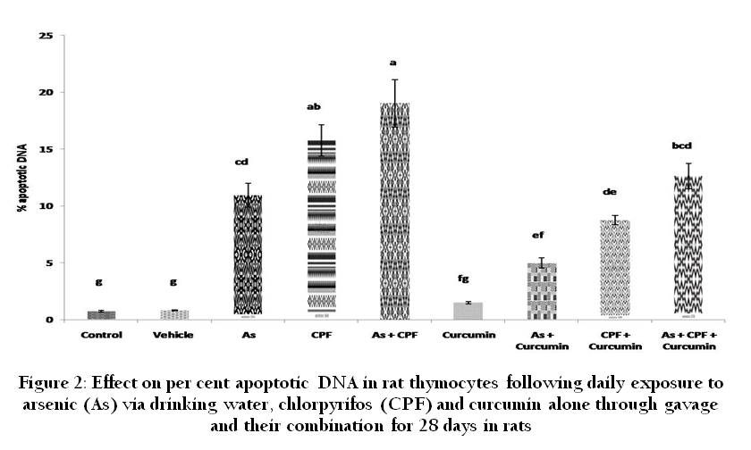
Figure 2: Effect on per cent apoptotic DNA in rat thymocytes following daily exposure to arsenic (As) via drinking water, chlorpyrifos (CPF) and curcumin alone through gavage and their combination for 28 days in rats
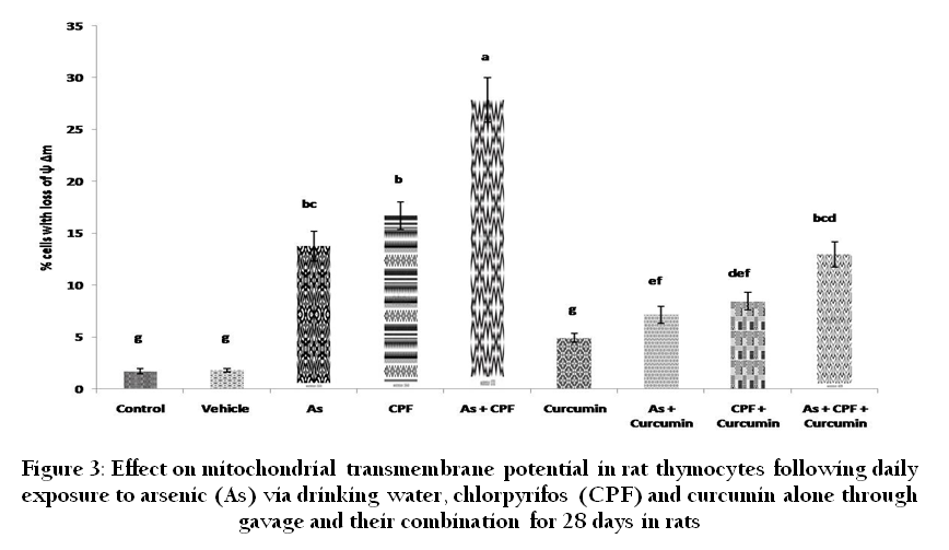
Figure 3: Effect on mitochondrial transmembrane potential in rat thymocytes following daily exposure to arsenic (As) via drinking water, chlorpyrifos (CPF) and curcumin alone through gavage and their combination for 28 days in rats
In the current study, DNA fragmentation exhibited a pattern of degradation compatible with inter nucleosomal fragmentation of the DNA in arsenic (group III), chlorpyrifos (group IV), arsenic plus chlorpyrifos (group V). DNA fragmentation corresponding to one to three nucleosomes was identified as strong bands by gel electrophoresis. In contrast, the DNA of control (group I), vehicle control (group II) and curcumin (group VI) treated thymocytes appeared as a single molecular weight species near the origin of migration indicating the intact DNA. Arsenic plus curcumin (group VII), Chlorpyrifos plus curcumin (group VIII) and Arsenic plus chlorpyrifos plus curcumin (group IX) showed lesser extent of DNA fragmentation indicating the ameliorative effect of curcumin in arsenic and Chlorpyrifos toxicity.
Curcumin, a yellow spice and pigment from Curcuma longa L. (Zingiberaceae), is by far known for its antioxidant, antiinflammatory and anticancer activities. Curcumin has proved not to have toxic, genotoxic or teratogenic properties, so this safe phytonutrient has been widely implied in preclinical and clinical studies. Curcumin and its derivatives have shown the ability of being free–radical scavenger, interacting with oxidative cascade, quenching oxygen and chelating and disarming oxidative properties of metal ions (Borsari et al., 2002). Curcumin has been demonstrated to inhibit the induction of apoptosis in previous studies. The inhibitory effects of curcumin on As–induced apoptosis and improvement in the antioxidant system and antioxidative enzymes (data not shown) is in the parallel with earlier reports in arsenic–induced oxidative stress (Shankaramurthy et al., 2007) who reported that curcumin inhibits excess ROS generation, apoptosis and ψ Δm in rat T lymphocytes and rat leydig cells of testes (Khan et al., 2013) and other antioxidant vitamins in mouse retina (Yu et al., 2008). The inhibition of ROS generation and ultimately apoptosis in the present study by antioxidants is in correlation with the earlier reports (Prakash et al., 2010b; 2011), however the antioxidants used were quercetin and catechin in an in vitro study in murine thymocytes. Curcumin, a phenolic compound, exhibits protective effects against oxidative damage and it is considered to be a potent cancer chemo–preventive agent (Duvoix et al., 2005). Curcumin exerts its protective effect by modulating ROS generation and augments antioxidant defense system. The ameliorative potential shown by curcumin might be due to its intrinsic antioxidant and chelation properties (Farombi and Ekor, 2006; Joe et al; 2004). Curcumin exhibits protective effects against oxidative damage by decreasing the level of free radicals, through its free radical–scavenging activity, particularly against oxygen radicals, which inhibits –SH group oxidation (Soudamini et al., 1992; Unnikrishnan and Rao, 1992; Manikandana et al., 2004). The antioxidant mechanism of curcumin was attributed to its conjugated structure which includes two methoxylated phenols and an enol form of β–diketone. The presence of electron donating groups like phenolic hydroxyl groups and a β–diketone structure is responsible for the typical radical trapping ability as a chain breaking antioxidant (Masuda et al., 2001) and inhibiting LPO (Osawa et al., 1995; Sreejayan and Rao, 1994; Wright, 2002). The preventive effect of curcumin is its ability to eliminate the hydroxyl radical (Reddy and Lokesh, 1994), superoxide radical (Sreejayan and Rao, 1994), single oxygen (Rao et al., 1995), nitrogen dioxide (Unnikrishnan and Rao, 1995) and NO (Sreejayan and Rao, 1994). In addition, curcumin primary metabolite, tetrahydro curcumin, a major antioxidant with β–diketone moiety, exhibited antioxidant activity by cleavage of the C–C bond at the active methylene carbon between the two carbonyls (Pan et al., 1999). These antioxidant properties seem to have a role in inhibiting singlet oxygen generation directly or indirectly.
In conclusion, this study indicates that chlorpyrifos alone at the given dose is more toxic than arsenic alone and their combination potentiate the induction of apoptosis and curcumin has ameliorative effect against arsenic and chlorpyrifos–induced apoptosis in rat thymocytes by its free radical scavenging activity. The mechanism of induction of apoptosis is might be due to ROS generation. The commonly available herb turmeric can be used to mitigate the toxic effects of arsenic particularly in heavily polluted areas where drinking water contamination is higher than the recommended level as well as can be used to reduce the toxicity produced by chlorpyrifos due to indiscriminate use of OPI compounds. Further study is required to elucidate the different pathways of induction of apoptosis by these xenobiotics.
ACKNOWLEDGEMENT
The Institutional Fellowship awarded to the first author by Indian Veterinary Research Institute, is gratefully acknowledged. The authors are thankful to the Director, Indian Veterinary Research Institute, for providing necessary facilities.
REFERENCES
Abdollahi M, Karami–Mohajeri S (2012). A comprehensive review on experimental and clinical findings in intermediate syndrome caused by organophosphate poisoning. Toxicol. Appl. Pharmacol. 258: 309 – 314.
http://dx.doi.org/10.1016/j.taap.2011.11.014
PMid:22177963
Aggarwal M, Malik JK, Kotresh AM, Gupta PK, Maurya P, Rao GS (2006). Induction of apoptosis in murine thymocytes by arsenic. Toxicol Lett. 20: 251
http://dx.doi.org/10.1016/j.toxlet.2006.07.183
Aggarwal M, Naraharisetti SB, Tiwari AK, Degen GH, Malik JK (2008). Assessment of apoptosis in peripheral blood lymphocytes and splenocytes of chickens simultaneously exposed to arsenite in drinking water and endosulfan in feed. Toxicol Lett. 5: 208
http://dx.doi.org/10.1016/j.toxlet.2008.06.114
Bashir S, Sharma Y, Irshad M, Nag TC, Tiwari M, Kabra M, Dogra TD (2006). Arsenic induced apoptosis in rat liver following repeated 60 days exposure. Toxicol. 217: 63 – 70.
http://dx.doi.org/10.1016/j.tox.2005.08.023
PMid:16288947
Berkson J (1994). Patient statement: a canary's tale. Toxicol. Ind. Health. 10: 323 – 326.
PMid:7539948
Borsari M, Ferrari E, Grandi R, Saladini Monica (2002). Curcuminoids as potential new iron–chelating agents: spectroscopic, polarographic and potentiometric study on their Fe (III) complexing ability. Inorg. Chimica Acta. 328: 61 – 68.
http://dx.doi.org/10.1016/S0020-1693(01)00687-9
Blakley BR, Yole MJ, Brousseau P, Boermans H, Fournier M, (1999). Effect of chlorpyrifos on immune function in rats. Vet. Hum. Toxicol. 41: 140 – 144.
PMid:10349701
Caroline C (1994). Insecticide fact sheet, Chlorpyrifos. Part 1. Toxicology. J. Pest. Reform. 14: 15 – 20.
Castedo M, Ferri K, Roumier T, Metivier D, Zamzaami M, Kromer G (2002). Qunatitation of mitochondrial alterations associated with apoptosis. J. Immunol. Meth. 266: 39 – 47.
http://dx.doi.org/10.1016/S0022-1759(02)00069-8
Chakraborti TK, Farrar JD, Pope CN (1993). Comparative neurochemical and neurobehavioral effects of repeated chlorpyrifos exposures in young and adult rats. Pharmacol. Biochem. Behav. 46: 219 – 224.
http://dx.doi.org/10.1016/0091-3057(93)90344-S
Chakraborti D, Rahman MM, Chowdhury UK, Paul K, Sengupta M, Lodh D, Basu GK, Chanda CR, Saha KC, Mukherjee SC (2010). Groundwater Arsenic contamination in Ganga–Meghna– Brahmaputra (GMB) plain and its health effects. Abstract Book of 5th International Conference on Arsenic Exposure and Health Effects.
Chan WH, Wu HJ (2006). Protective effects of curcumin on methylglyoxal–induced oxidative DNA damage and cell injury in human mononuclear cells. Acta. Pharmacol. Sin. 27:1192 – 1198.
http://dx.doi.org/10.1111/j.1745-7254.2006.00374.x
PMid:16923340
Chanda SM, Pope CN (1996). Neurochemical and neurobehavioral effects of repeated gestational exposure to chlorpyryfos in maternal and developing rats. Pharmacol. Biochem. Behav. 53: 771 – 776.
http://dx.doi.org/10.1016/0091-3057(95)02105-1
Chang SI, Jin B, Youn P, Park C, Park JD, Ryu DY (2007). Arsenic–induced toxicity and the protective role of ascorbic acid in mouse testis. Toxicol. Appl. Pharmacol. 218: 196 – 203.
http://dx.doi.org/10.1016/j.taap.2006.11.009
PMid:17188728
Chattopadhyay I, Bandyopadhyay U, Biswas K, Maity P, Banerjee RK (2005). Indomethacin inactivates gastric peroxidase to induce reactive–oxygen mediated gastric mucosal injury and curcumin protects it by preventing peroxidase inactivation and scavenging reactive oxygen. Free Radical Biol. Med. 40: 1397 – 1408.
http://dx.doi.org/10.1016/j.freeradbiomed.2005.12.016
PMid:16631530
Chen YC, Lin–Shiau SY, Lin JK (1998). Involvement of reactive oxygen species and caspase 3 activation in arsenite–induced apoptosis. J. Cell. Physiol. 177: 324 – 333.
http://dx.doi.org/10.1002/(SICI)1097-4652(199811)177:2<324::AID-JCP14>3.0.CO;2-9
Chiapella G, Flores–Martín J, Ridano ME, Reyna L, Magnarelli de Potas G, Panzetta–Dutari GM, Genti–Raimondi S (2013). The organophosphate chlorpyrifos disturbs redox balance and triggers antioxidant defense mechanisms in JEG–3 cells. Placenta. 34: 792 – 798
http://dx.doi.org/10.1016/j.placenta.2013.06.007
PMid:23850137
Chowdhury UK, Rahman MM, Mandal BK, Paul K, Lodh D, Basu GK, Chanda CR, Saha KC, Mukherjee SC, Roy S, Das R, Kaies I, Barua AK, Palit SK, Quamruzzaman Q, Chakraborty D (2001). Groundwater arsenic contamination and human suffering in West Bengal, India and Bangladesh. Environ. Sci. 8: 393 – 398.
Das J, Ghosh J, Manna P, Sinha M, Sil PC (2009). Taurine protects rat testes against NaAsO2–induced oxidative stress and apoptosis via mitochondrial dependent and independent pathways. Toxicol. Lett.18: 201 – 210.
http://dx.doi.org/10.1016/j.toxlet.2009.03.001
PMid:19429265
Duvoix A, Blasius R, Delhalle S, Schnekenburger M, Morceau F, Henry E (2005). Chemopreventive and therapeutic effects of curcumin. Cancer Lett. 223: 181 – 90.
http://dx.doi.org/10.1016/j.canlet.2004.09.041
PMid:15896452
El–Desoky GE, Bashandy SA, Alhazza IM, Al–Othman ZA, Aboul–Soud MAM (2013). Improvement of Mercuric Chloride–Induced Testis Injuries and Sperm Quality Deteriorations by Spirulina platensis in Rats. PLOS ONE. 8: 59177.
http://dx.doi.org/10.1371/journal.pone.0059177
PMid:23555627 PMCid:PMC3610915
Ercal N, Gurer–Orhan H, Aykin–Burns N (2001). Toxic metals and oxidative stress Part 1: mechanisms involved in metal induced oxidative damage. Curr. Topics Med. Chem. 1: 529 – 539.
http://dx.doi.org/10.2174/1568026013394831
PMid:11895129
Farombi EO, Ekor M (2006). Curcumin attenuates gentamicin–induced renal oxidative damage in rats. Food Chem. Toxic. 44: 1443 – 8.
http://dx.doi.org/10.1016/j.fct.2006.05.005
PMid:16814915
Flora SJ, Bhadauria S, Pant SC, Dhaked RK (2005). Arsenic induced blood and brain oxidative stress and its response to some thiol chelators in rats. Life Sci. 77: 2324 – 2337.
http://dx.doi.org/10.1016/j.lfs.2005.04.016
PMid:15964026
Flora SJS, Mittal M, Mehta A (2008). Heavy metal induced oxidative stress and its possible reversal by chelation therapy. Indian J. Med. Res. 128: 501 – 523.
PMid:19106443
GOI (2011). Water and Sanitation Report of Central Team on Arsenic mitigation in rural drinking water sources in Ballia district, Uttar Pradesh State. Ministry of Drinking Water and Sanitation. Government of India.
Gupta SC, Mishra M, Sharma A, Balaji TGRD, Kumar R, Mishra RK, Chowdhuri DK (2010). Chlorpyrifos induces apoptosis and DNA damage in Drosophila through generation of reactive oxygen species. Ecotoxicol. Environ. Safety. 73(6): 1415 – 1423.
http://dx.doi.org/10.1016/j.ecoenv.2010.05.013
PMid:20627310
Hansen BH, Romma S, Garmo A, Olsvika PA, Andersen RA (2006). Antioxidative stress proteins and their gene expression in brown trout (Salmo trutta) from three rivers with different heavy metal levels. Comp. Biochem. Physiol. C. 143: 263 – 274.
Harford AJ, O'Halloran K, Wright PF (2005). The effects of in vitro pesticide exposures on the phagocytic function of four native Australian freshwater fish. Aquat. Toxicol. 75: 330 – 42.
http://dx.doi.org/10.1016/j.aquatox.2005.09.005
PMid:16229903
Hultberg B, Anderson A, Isaksson A (1999). Thiol and redox reactive agents exerts different effects on glutathione metabolism in HeLa cell cultures. Clin. Chim. Acta. 283: 21 – 32.
http://dx.doi.org/10.1016/S0009-8981(99)00028-5
Hultberg B, Anderson A, Isaksson A (2001). Interaction of metals and thiols in cell damage and glutathione distribution: potentiation of mercury toxicity by dithiothreitol. Toxicol. 156: 93 – 100.
http://dx.doi.org/10.1016/S0300-483X(00)00331-0
Jezek P, Hlavata L (2005) Mitochondria in homeostasis of reactive oxygen species in cell, tissues, and organism. Int. J. Biochem. Cell. Biol. 37: 2478 – 2503.
http://dx.doi.org/10.1016/j.biocel.2005.05.013
PMid:16103002
Jhala DD, Chinoy NJ, Rao MV (2008). Mitigating effects of some antidotes on fluoride and arsenic induced free radical toxicity in mice ovary. Food Chem. Toxicol. 46: 1138 – 42.
http://dx.doi.org/10.1016/j.fct.2007.11.009
PMid:18187247
Joe B, Vijaykumar M, Lokesh BR (2004). Biological properties of curcumin cellular and molecular mechanisms of action. Crit. Rev. Food Sci. Nutr. 44: 97 – 111.
http://dx.doi.org/10.1128/MCB.24.22.9763-9770.2004
PMid:15509781 PMCid:PMC525473
Kaur P, Aschner M, Syversen T (2006). Glutathione modulation influences methyl mercury induced neurotoxicity in primary cell cultures of neurons and astrocytes. Neurotoxicol. 2: 492 – 500.
http://dx.doi.org/10.1016/j.neuro.2006.01.010
PMid:16513172
Khan S, Prakash A, Aggarwal M, Majhi CR, Telang AG, Malik JK (2009). Experimental exposure of arsenic induces apoptosis in murine thymocytes. Toxicol. Lett. 13: 225
http://dx.doi.org/10.1016/j.toxlet.2009.06.529
Khan S, Telang AG, Malik JK (2013). Arsenic–induced oxidative stress, apoptosis and alterations in testicular steroidogenesis and spermatogenesis in wistar rats: ameliorative effect of curcumin. WJPP. 2: 33 – 48.
Kinoshita A, Wanibuchi H, Wei M, Yunoki T, Fukushima S (2007). Elevation of 8–hydroxydeoxyguanosine and cell proliferation via generation of oxidative stress by organic arsenicals contributes to their carcinogenicity in the rat liver and bladder. Toxicol. Appl. Pharmacol. 221: 295 – 305.
http://dx.doi.org/10.1016/j.taap.2007.03.024
PMid:17481689
Kumar S, Yedjou CG, Tchounwou PB (2014). Arsenic trioxide induces oxidative stress, DNA damage, and mitochondrial pathway of apoptosis in human leukemia (HL–60) cells. J. Exp. Clin. Cancer Res. 33: 42 1 – 12.
Lee JE, Park JH, Shin IC, Koh HC (2012). Reactive oxygen species regulated mitochondria–mediated apoptosis in PC12 cells exposed to chlorpyrifos. Toxicol. Appl. Pharmacol. 263: 148 – 162.
http://dx.doi.org/10.1016/j.taap.2012.06.005
PMid:22714038
Lemarie A, Morzadec C, Bourdonnay E, Fardel O, Vernhet L (2006). Human macrophages constitute targets for immunotoxic inorganic arsenic. J. Immunol. 177: 3019 – 3027.
http://dx.doi.org/10.4049/jimmunol.177.5.3019
PMid:16920938
Li Q, Kobayashi M, Kawada T (2007). Organophosphorus pesticides induce apoptosis in human NK cells. Toxicol. 239: 89 – 95.
http://dx.doi.org/10.1016/j.tox.2007.06.100
PMid:17681413
Liu J, Liu YP, Goyer RA, Achanzar W, Waalkes MP (2000). Metallothionein–I/II null mice are more sensitive than wild–type mice to the hepatotoxic and nephrotoxic effects of chronic oral or injected inorganic arsenicals. Toxicol. Sci. 55: 460 – 467.
http://dx.doi.org/10.1093/toxsci/55.2.460
PMid:10828279
Manikandan P, Sumitra M, Aishwarya S, Manoharb BM, Lokanadamc B, Puvanakrishnan R (2004). Curcumin modulates free radical quenching in myocardial ischaemia in rats. J. Biochem. Cell Biol. 36: 1967 – 80.
http://dx.doi.org/10.1016/j.biocel.2004.01.030
PMid:15203111
Manna P, Sinha M, Sil PC (2008). Protection of arsenic–induced testicular oxidative stress by arjunolic acid. Redox Rep. 13: 67 – 77.
http://dx.doi.org/10.1179/135100008X259169
PMid:18339249
Masuda T, Maekawa T, Hidaka K (2001). Chemical studies on antioxidant mechanisms of curcumin: analysisof oxidative coupling products from curcumin and linoleate. J. Agric. Food. Chem. 49: 2539 – 2547.
http://dx.doi.org/10.1021/jf001442x
PMid:11368633
Moser VC (2000). Dose–response and time–course of neurobehavioral changes following oral chlorpyrifos in rats of different ages. Neurotoxicol. Teratol. 22: 713 – 723.
http://dx.doi.org/10.1016/S0892-0362(00)00087-8
Naik RS, Mujumdar AM, Ghaskadbi S (2004). Protection of liver cells from ethanol cytotoxicity by curcumin in liver slice culture in vitro. J. Ethnopharmacol. 95: 31 – 37.
http://dx.doi.org/10.1016/j.jep.2004.06.032
PMid:15374604
Nakadai A, Li Q, Kawada T (2006). Chlorpyrifos induces apoptosis in human monocyte cell line U937. Toxicol. 224: 202 – 209.
http://dx.doi.org/10.1016/j.tox.2006.04.055
PMid:16787693
Ojha A, Yaduvanshi SK, Pant SC, Lomash V, Srivastava N (2011). Evaluation of DNA damage and cytotoxicity induced by three commonly used organophosphate pesticides individually and in mixture, in rat tissues. Environ. Toxicol. http://dx.doi.org/10.1002/tox.20748.
http://dx.doi.org/10.1002/tox.20748
Osawa T, Sugiyama Y, Inayoshi M, Kawakishi S (1995). Antioxidant activity of tetrahydrocurcuminoids. Biosci. Biotechnol. Biochem. 59: 1609 – 12.
http://dx.doi.org/10.1271/bbb.59.1609
PMid:8520105
Pan M, Huang T, Lin J (1999). Biotransformation of curcumin through reduction and glucuronidation in mice. Drug. Metab. Dispos. 27: 486 – 94.
PMid:10101144
Prakash A, Khan S, Aggarwal M, Telang AG, Malik JK (2009). Chlorpyrifos induces apoptosis in murine thymocytes. Toxicol. Lett. 189: 83.
http://dx.doi.org/10.1016/j.toxlet.2009.06.251
Prakash A, Khan S, Aggarwal M, Telang A, Malik J (2010a). Quercetin and catechin attenuate chlorpyrifos–induced apoptosis in murine thymocytes. Toxicol. Lett. 196 Supplemet 1: 203.
Prakash A, Khan S, Telang AG, Malik JK (2010b). Modulation of chlorpyrifos–induced apoptosis in murine thymocytes by resveratrol. Med. Chem. Res. 19 (1/6): S55.
Prakash A, Khan S, Telang A, Malik J. (2011). Modulation of arsenic–induced apoptosis in murine thymocytes by quercetin and catechin. Toxicol. Lett. 28: 172.
http://dx.doi.org/10.1016/j.toxlet.2011.05.598
Quig D (1998). Cysteine metabolism and metal toxicity. Alter. Med. Rev. 3: 262 – 270.
PMid:9727078
Rahman MF, Mahboob M, Danadevi K, Saleha BB, Grover P (2002). Assessment of genotoxic effects of chloropyriphos and acephate by the comet assay in mice leucocytes. Mutat. Res. 516: 139 – 47.
http://dx.doi.org/10.1016/S1383-5718(02)00033-5
Raszewski G, Lemieszek MK, Łukawski K, Juszczak M, Rzeski Wojciech (2014). Chlorpyrifos and Cypermethrin Induce Apoptosis in Human Neuroblastoma Cell Line SH–SY5Y. Basic Clin. Pharmacol. Toxicol. DOI: 10.1111/bcpt.12285
http://dx.doi.org/10.1111/bcpt.12285
Reddy AC, Lokesh BR (1994). Studies on the inhibitory effects of curcumin and eugenol on the formation of reactive oxygen species and the oxidation of ferrous ion. Mol. Cell. Biochem. 137: 1 – 8.
http://dx.doi.org/10.1007/BF00926033
PMid:7845373
Leonard SS, Harris GK, Shi XL (2004). Metal–induced oxidative stress and signal transduction. Free Radic. Biol. Med. 37: 1921 – 1942.
http://dx.doi.org/10.1016/j.freeradbiomed.2004.09.010
PMid:15544913
Saulsbury MD, Heyliger SO, Wang K, Round D (2008). Characterization of chlorpyrifos–induced apoptosis in placental cells. Toxicol. 244: 98 – 110.
http://dx.doi.org/10.1016/j.tox.2007.10.020
PMid:18155347 PMCid:PMC2771452
Shadnia S, Dasgar M, Taghikhani S, Mohammadirad A, Khorasani R, Abdollahi M (2007). Protective effects of alpha–tocopherol and n–acetyl–cysteine on diazinon–induced oxidative stress and acetylcholinesterase inhibition in rats. Toxicol. Mech. Methods. 17: 109 – 115.
http://dx.doi.org/10.1080/15376510600860318
PMid:20020979
Shankaramurthy NC (2007). Studies on the effects of piperine and curcumin on arsenic–induced oxidative stress and apoptosis. M.V.Sc. thesis submitted to Deemed University, IVRI, Izatnagar, U.P., India.
Soto–Pe-a GA, Vega L (2008). Arsenic interferes with the signaling transduction pathway of T cell receptor activation by increasing basal and induced phosphorylation of Lck and Fyn in spleen cells. Toxicol. Appl. Pharmacol. 230: 216 – 26.
http://dx.doi.org/10.1016/j.taap.2008.02.029
PMid:18407307
Soudamini KK, Unnikrishnan MC, Soni KB, Kuttan R (1992). Inhibition of lipid peroxidation and cholesterol levels in mice by curcumin. Indian J. Physiol. Pharmacol. 36: 239 – 43.
PMid:1291474
Sreejayan N, Rao MN (1994). Curcuminoids as potent inhibitors of lipid peroxidation. J. Pharm. Pharmacol. 46: 1013 – 6.
http://dx.doi.org/10.1111/j.2042-7158.1994.tb03258.x
PMid:7714712
Stohs SJ, Bagchi D (1993). Oxidative mechanisms in the toxicity of the metal ions. Free Rad. Biol. Med. 18: 321 – 336.
http://dx.doi.org/10.1016/0891-5849(94)00159-H
Thrasher JD, Heuser G, Broughton A (2002). Immunological abnormalities in humans chronically exposed to chlorpyrifos. Arch. Environ. Health. 57: 181 – 187.
http://dx.doi.org/10.1080/00039890209602934
PMid:12507170
Thrasher JD, Madison R, Broughton A (1993). Immunologic abnormalities in humans exposed to chlorpyrifos: preliminary observations. Arch. Environ. Health. 48: 89 – 93.
http://dx.doi.org/10.1080/00039896.1993.9938400
PMid:7682805
Unnikrishnan MK, Rao MNA (1992). Curcumin prevents nitrite induced methhaemoglobin formation. FEBS. 301: 195 – 6.
http://dx.doi.org/10.1016/0014-5793(92)81246-I
Valko M, Rhodes CJ, Moncol J, Izakovic M, Mazur M (2006). Free radicals, metals and antioxidants in oxidative stress–induced cancer. Chem–Biol. Inter. 160: 1 – 40.
Verma RS, Mehta A, Srivastava N (2007). In vivo chlorpyrifos induced oxidative stress: Attenuation by antioxidant vitamins. Pest. Biochem. Physiol. 88: 191 – 196.
http://dx.doi.org/10.1016/j.pestbp.2006.11.002
Wang JE, Jerrells TR, Spitzer JJ (1996). Decreased production of reactive oxygen intermediates is an early event during in vitro apoptosis in rat thymocytes. Free Rad. Biol. Med. 20: 533 – 542.
http://dx.doi.org/10.1016/0891-5849(95)02085-3
Whitney KD, Seidler FJ, Slotkin TA (1995). Deveropmental neurotoxicity of chlorpyrifos: cellular mechanisms. Toxicol. Appl. Pharmacol. 134: 53 – 62.
http://dx.doi.org/10.1006/taap.1995.1168
PMid:7545834
WHO (1996). Guidelines for Drinking Water Quality, Health Criteria and Other Supporting Information. WHO, Geneva. 2(2): 940 – 994.
WHO (1997). The WHO Recommended Classification of Pesticides by Hazard. International Programme on Chemical Safety, WHO/IPCS/96.3.
WHO (2011). Guidelines for drinking–water quality, fourth edition. Edited by WHO. ISBN: 978 92 4 154815 1
Wright JS (2002). Predicting the antioxidant activity of curcumin and curcuminoids. J. Mol. Struct. 591: 207 – 17.
http://dx.doi.org/10.1016/S0166-1280(02)00242-7
Yu F, Wang Z, Ju B, Wang Y, Wang J, Bai D (2008). Apoptotic effect of organophosphorous insecticide chlorpyrifos on mouse retina in vivo via oxidative stress and protection of combination of vitamins C and E. Exp. Toxic. Pathol. 59: 415 – 423.
http://dx.doi.org/10.1016/j.etp.2007.11.007
PMid:18222074
Ziem G, McTamney J (1997). Profile of patients with chemical injury and sensitivity. Environ. Health Perspect., 105 (2): 417 – 436.
http://dx.doi.org/10.1289/ehp.97105s2417
http://dx.doi.org/10.2307/3433348
PMid:9167975 PMCid:PMC1469804


