Advances in Animal and Veterinary Sciences
Research Article
Advances in Animal and Veterinary Sciences 2 (7): 369 – 376Congenital Anomalies and their Surgical Correction in Ruminants
Anil Kumar Gangwar1,*, Khangembam Sangeeta Devi1, Ajit Kumar Singh1, Nitesh Katiyar1, Ghanshyam Pate2, Sushanta Srivastava2
- Department of Veterinary Surgery and Radiology, College of Veterinary Science and Animal Husbandry, Narendra Deva University of Agriculture and Technology, Kumarganj, Faizabad–224 229 (U.P.)
- Department of Veterinary Obstetrics and Gynaecology, College of Veterinary Science and Animal Husbandry, Narendra Deva University of Agriculture and Technology, Kumarganj, Faizabad–224 229 (U.P.) India
*Corresponding author:dranilvet2007@gmail.com
ARTICLE CITATION:
Gangwar AK, Devi KS, Singh AK, Katiyar N, Patel G, Srivastava S (2014). Congenital anomalies and their surgical correction in ruminants. Adv. Anim. Vet. Sci. 2 (7): 369 – 376.
Received: 2014–07–06, Revised: 2014–07–23, Accepted: 2014–07–24
The electronic version of this article is the complete one and can be found online at
(
http://dx.doi.org/10.14737/journal.aavs/2014/2.7.369.376
)
which permits unrestricted use, distribution, and reproduction in any medium, provided the original work is properly cited
ABSTRACT
This study included the congenital disorders in ruminants (cattle, buffalo, sheep and goat). During the period from Jan 2005 to Jan 2013, a total number of 112 cases of congenital malformations were presented. A total number of 94 cases of congenital malformations including atresia ani/atresia ani et recti/ atresia ani et recti et coli, atresia ani with rectovaginal fistula, cheiloschisis, arthrogryposis, contracted tendon, eventration of intestine, meningocele, unilateral and bilateral lateral patellar luxation, ocular dermoid and umbilical hernia were successfully corrected surgically. Other cases of congenital defect were not subjected to any surgical treatment.
INTRODUCTION
Abnormalities of structure and function, which are present at birth, are obviously congenital deformities (Badaway, 2011). Congenital malformations can result from defective genetics or environmental factors or a combination of both (Shukla et al., 2007). Pedigree analysis and breeding trials revealed that these anomalies are autosomal recessive diseases (Bryan et al., 1993). The environmental factors included consumption of toxic plants by the dam and maternal–fetal viral infections during early gestation (Bendemkiran et al., 2009). According to Sharma et al. (1986) chances of congenital abnormalities are more in animals having 4–8 weeks of pregnancy at the time of infection. Bovine viral diarrhoea–mucosal disease virus can induce congenital anomalies in the bovine fetus. This virus is capable of crossing both the placental and foetal blood brain barrier (Scott et al., 1993). There is no relationship between congenital anomalies and the time of lambing and phytoestrogens (Dennis, 1974). Maternal manganese deficiency is also responsible for these conditions (Staley et al., 1994). Atresia ani, arthrogryposis, contracted tendon, dermoid, polythelia, hypospadias and cryptorchidism are certain conditions common in ruminants. However, curved legs, microtia, deformed back, short mandible and/or cerebral deformities are more common in ovine in comparison to other ruminants (Elias and Benett, 1992). Atresia ani has been reported to be a heritable condition in pigs and calves (Kilic et al., 2004) and develops when a dorsal part of the cloacal plate fails to form (Magda and Youssef, 2007). It is characterized by persistence of the anal membrane covering the normal anal canal. Atresia ani with rectovginal fistula is characterized by communication between the dorsal wall of the vagina and ventral portion of the rectum, so that the vulva functions as common opening to the urogenital and gastrointestinal tract (Johnson et al., 1985). Brachygnathism or parrot mouth is a craniofacial defect caused by homozygous recessive gene with incomplete penetrance. Congenital patellar luxation is not a common condition in calf but it is common in dogs and cats. Silva et al., (2001) observed 8.86% patellar luxation in cattle raised under the extensive production system. Ocular dermoids are rare in cattle, with the prevalence between 0.002% and 0.4 percent (Kiliç et al., 2012).
This study records different congenital conditions and successful surgical treatment of some of these anomalies in farm animals.
MATERIALS AND METHODS
This study included the congenital disorders in ruminants (cattle, buffalo, sheep and goat) presented to the TVCC, College of Veterinary Sciences and Animal Husbandry, Kumarganj, Faizabad, Uttar Pradesh, India. The adult cases having congenital deformity without any complication were presented for treatment of other ailments like horn injury, upward fixation of patella etc. During the period from Jan 2005 to Jan 2013, a total number of 112 cases with the history of congenital malformations were presented.
Case History and Clinical Examination
Clinical examination was done for all the cases. Radiographic examination was performed in the cases of skeletal/tendon/ligament deformities and contrast radiography (barium sulfate enema) was performed in large intestinal deformities.
Atresia ani/atresia ani et recti/ atresia ani et recti et coli were presented with the history of absence of anal opening since birth. In cases of atresia ani with rectovaginal fistula, the feces were passed through the vulva.
A case of cheiloschisis was recorded in one female buffalo calf. Clinical examination revealed presence of nasal orifice over the gum/dental pad (Figure 1a, b).
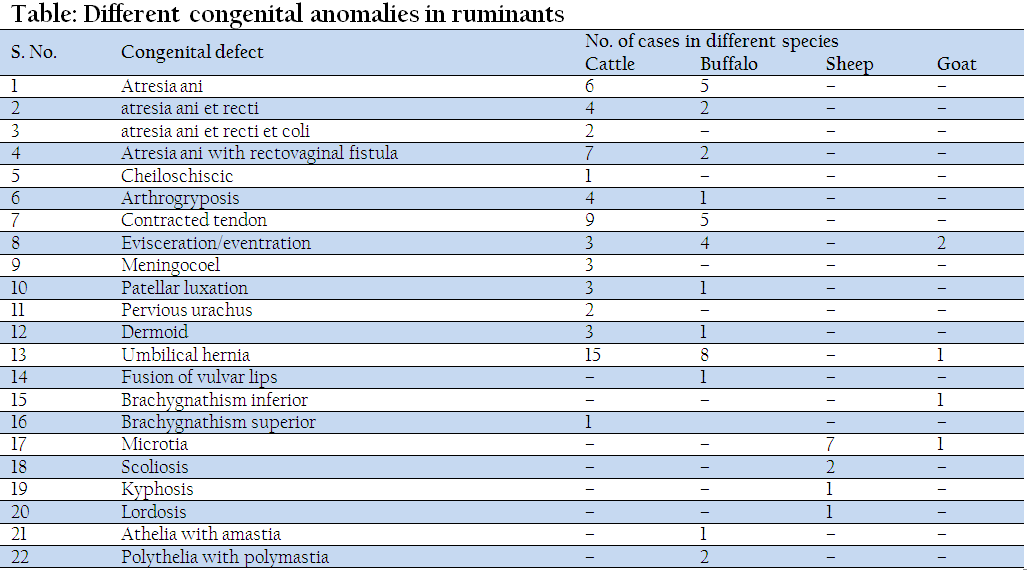
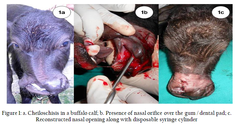
Figure 1: a. Cheiloschisis in a buffalo calf; b. Presence of nasal orifice over the gum / dental pad; c. Reconstructed nasal opening along with disposable syringe cylinder
Arthrogryposis and contracted tendon were reported in cattle and buffalo calves (Figure 2). The calves were presented with unilateral or bilateral congenital flexion of carpal and fetlock joint. In one case of congenital flexion, the limb below the carpal joint was abducted.
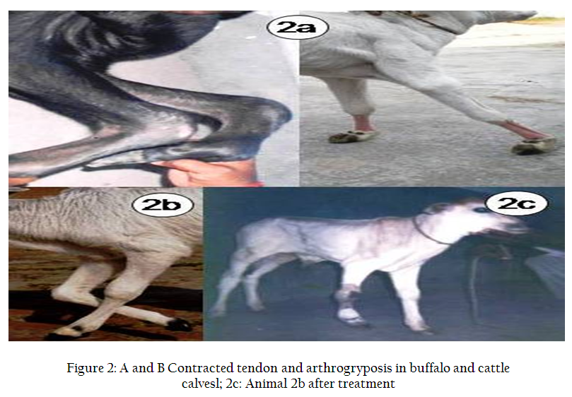
Figure 2: A and B Contracted tendon and arthrogryposis in buffalo and cattle calvesl; 2c: Animal 2b after treatment
Congenital eventration of intestine in calves and intestine and spleen in a kid were presented. In one case colon and rectum was eviscerated through the umbilicus and atresia ani was also present (Figure 3a). The exposed viscera were soiled. Contrast radiography revealed that the eviscerated part was rectum with blind end (Figure 3b).
Figure 3: a: Eviscerated colon and rectum through the umbilicus; b: Contrast radiography revealed blind end of rectum; c–e: Resection and temporary suturing of necrosed colon and rectum; f: Reconstruction of anal opening and pulling of sutured colon towards the anal opening; g: Suturing of colon to reconstructed anal opening
The cases of meningocele were brought with the history of large swelling over the head, dorsal cervical region (Figure 4) and lumbar region. The case with swelling over lumbar region was also having atresia ani and double vulva (Figure 5a). There was paresis of both hind limbs with swaying during movement. Radiological examination revealed hypoplastic lumbar vertebrae and protruded mass (Figure 5b). Contrast radiography could not be performed due to absence of contrast agent. The co–existence of three conditions (atresia ani, double vulva and spinal meningocele) in calves has not been reported so for in the literature as for as our knowledge is concerned.
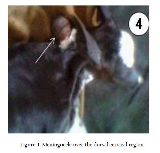
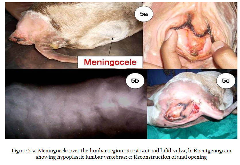
Figure 5: a: Meningocele over the lumbar region, atresia ani and bifid vulva; b: Roentgenogram showing hypoplastic lumbar vertebrae; c: Reconstruction of anal opening
Unilateral and bilateral lateral patellar luxation in cattle calves were presented with the history of no weight bearing by the affected limb/limbs since birth (Figure 6a).
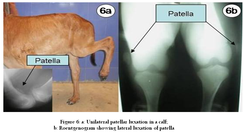
Figure 6: a: Unilateral patellar luxation in a calf; b: Roentgenogram showing lateral luxation of patella
The calves were parturated normally. In bilateral cases, the calves were in crouching stance with lowered pelvis, unable to extend its stifles and could not stand in upright position. On clinical examination it was observed that patellae were not in their normal position and could be palpated on the lateral aspect of the stifles. After extending the limb in full extension, the patellae could be placed in to their normal position. As soon as the limb flexed slightly, the patellae slip down into its abnormal position. The musculature of the pelvic limb was well developed which excludes the probability of bilateral femoral nerve paralysis. Lateral and cranio–caudal radiographic views of stifle joint showed complete lateral luxation of patella (Figure 6b). Trochlear grooves of both femurs were not as deep as normal calf of same age. The cases were diagnosed as a congenital lateral luxation of patella. Another case of unilateral luxation of patella was presented at the age of 2 months. The patella was fixed in position
The cases of pervious urachus were presented with the history of dribbling of urine through the umbilicus in female calves.
Ocular dermoid, a skin or skin like appendage, was reported in cattle calves. In clinical cases it was present on the palpebral conjunctiva, third eye lid and cornea (Figure 7a). Farm animals (24) were presented with the history of swelling over the umbilical region since birth (Figure 8a).All these cases were diagnosed as congenital umbilical hernias.
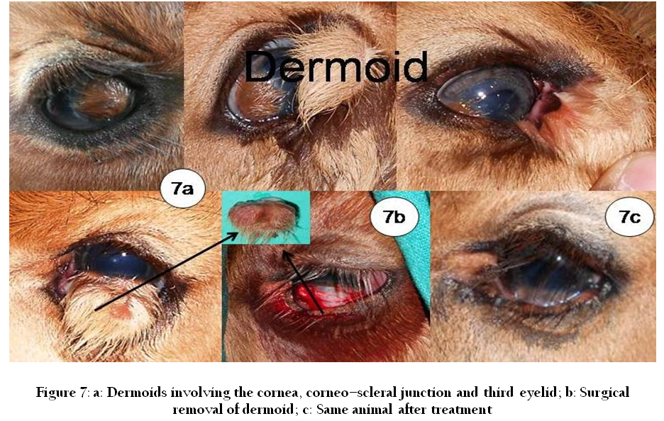
Figure 7: a: Dermoids involving the cornea, corneo–scleral junction and third eyelid; b: Surgical removal of dermoid; c: Same animal after treatment

Figure 8: : a: Umbilical hernia in a buffalo calf ; b: Reconstruction of hernia with acellular collagen matrices; c: Same animal after treatment
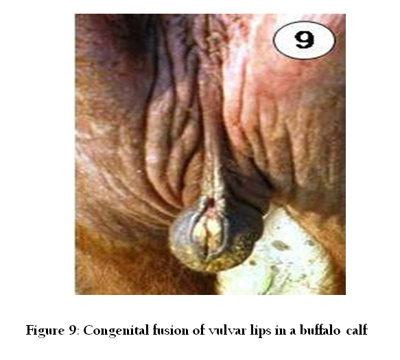
Congenital fusion of vulvar lips was reported in a buffalo calf (Figure 9). A small opening was present at the lowermost part of the vulva. The calf was urinating through that opening. Palpation revealed a cord like structure.
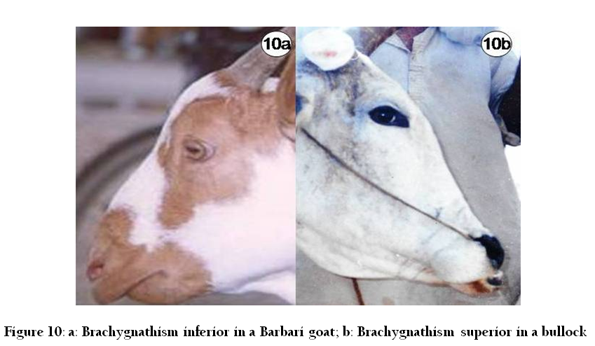
Brachygnathism inferior was reported in a Barbari goat (Figure 10a). The lower jaw was short and the animal was taking feed and water normally. Clinical sign included slight abrasion of hard palate.
A non–descript bullock, aged about eight years was presented to the polyclinic with the history of horn injury during fighting with another bullock. During clinical examination, it was observed that the upper jaw of the animal was short and all the incisors could be seen from a distance (Figure 10b). The animal was taking feed and water normally. The case was diagnosed as brachygnathism superior.
Microtia was reported in sheep and goat. History revealed that the parents and other progenies of these parents were also short eared.
A female Muzaffarnagri sheep was brought to the clinic from Teaching Livestock Farm with the history of dorsal curvature and lateral kinking of the thoracic vertebrae. History of parturition revealed that the animal was born after obstetrical mutation. The case was diagnosed as kypho–scoliosis in a sheep.
RESULTS
Surgical treatment was done for the 93 cases of congenital malformations. Other cases of congenital defect were not subjected to any surgical treatment.
Atresia ani and related surgical ailments were corrected under anterior epidural anesthesia. The cases of atresia ani and atresia ani et recti were treated by reconstruction of the anal opening using modified technique. A plus (+) shape incision was made over the skin where rectum was bulged after pressing the abdomen. Rectum was pulled out by alis tissue forceps. A nick incision was made on the rectum and meconium was evacuated. After irrigation with normal saline the skin flaps were sutured with the rectum in such a way that they invert into the rectum. All the cases recovered without any complication like contraction of reconstructed anal opening in the early post–operative days.
Atresia ani et recti et coli was treated by reconstruction of anal opening and distal blind end of the colon was identified through the ventral prepubic laparotomy and sutured to the skin of the anal opening as in cases of atresia ani.
Atresia ani with rectovaginal fistula was corrected by making a linear skin incision of 7–8 cm, extended horizontally, midway between the anus and vagina. The perineal tissue was separated by blunt dissection and rectal and vaginal walls were separated. The rectal and vaginal wall defects were sutured separately with chromic catgut no.1/0 by cushing suture pattern. Perineal tissue was sutured and the skin was closed in standard manner.
The case of cheiloschisis was recorded in one female buffalo calf. The nasal orifice was present over the gum/dental pad. The edges of the fissure in the muffle were trimmed and a nasal opening was reconstructed (Figure 1c). A disposable plastic syringe cylinder was cut and fixed within the reconstructed nostril to check the wound contraction and subsequent closure of nasal orifice. Remaining edges were sutured in position. The nasal opening over the gum was freshened and closed by using silk no. 2 using simple interrupted suture pattern. The case of cheiloschisis was corrected surgically without any complication.
Arthrogryposis and contracted tendon were initially treated by intravenous administration of oxytetracycline @ 20mg/kg b. wt. along with application of well padded bamboo splints. Oxytetracycline was repeated on alternate day and splint was also changed and tightened on the same day. More severe cases were corrected by tenotomy of the related tendons and subsequent application of plaster cast.
Congenital eventration of colon and rectum through the umbilicus was surgically corrected. Atresia ani was corrected by reconstruction of anal opening. The exposed part of colon and rectum was resected due to partial necrosis. The cut end of colon was temporarily sutured and a long thread was tied (Figure 3c). A long needle holder was passed through the reconstructed anal opening towards the laparotomy wound to hold and pull the sutured colon towards the anal opening. The cut end of the colon was sutured to the skin of the anal opening as in cases of atresia ani. Other cases of evisceration were surgically corrected using standard surgical technique. In one case of eventration, the intestinal loops were replaced in to the abdominal cavity after kelotomy. All the cases of evisceration were recovered successfully except one case in which the intestine was severely necrosed.
Frontal meningocele and cervical spinal meningocele in calves were surgically corrected. However, lumbar spinal meningocoel was not corrected due to hypoplasia of lumbar vertebrae.
After epidural anesthesia, lateral luxation of patella was surgically treated using medial imbrication technique (Fubini and Ducharme, 2004).
The cases of pervious urachus were surgically corrected using standard surgical technique.
The dermoids over the cornea, corneo–scleral junction and corneo–conjunctiva were peeled off and removed under retrobulbar, supraorbital and auriculopalpebral nerve block (Figure 7b). However, dermoid involving third eyelid was excised and removed.
Congenital umbilical hernias were corrected as per the size of the hernial ring. Small hernias were corrected by silk no.2 using vest over pant technique. However, large hernias were corrected using carbon mesh, carbon fibers and acellular collagen matrices (Figure 8b) using inlay technique. The cases of congenital umbilical hernia were successfully reconstructed with carbon mesh, carbon fibers, acellular dermal matrices. All the animals had an uneventful recovery without clinical signs of wound dehiscence, infection or recurrence of hernia.
Congenital fusion of vulvar lips was corrected surgically (vulvoplasty). After local anesthesia, the fused lips were separated by dorsal vertical enlargement of small opening using pointed B.P. blade. The separated lips were inverted and sutured in place using horizontal mattress suture pattern.
DISCUSSION
The congenital deformities may be lethal, semilethal or compatible with life causing aesthetic defects or having no effect on the animals (Badawy, 2011). We recorded a total no. of 112 ruminants suffering from congenital defects.
In present study all the cases of atresia ani alone or with rectovaginal fistula were diagnosed on the basis of clinical signs. Clinical signs and physical examination findings are sufficient to establish the diagnosis (Shakoor et al., 2012). However, radiographs are considered important to determine the position of the fistula (Rahal et al., 2007). Azizi et al., (2010) and Mahesh et al., (2014) described a good survival rate in response to atresia ani rectification by removing a circular skin piece and unifying the excised rectal loop with skin. In our study, the cases of atresia ani were corrected by using a modified technique without any complication like constriction of reconstructed anal opening after few days of surgery.
Cheiloschisis occurs due to failure of fusion of upper lip along the midline (Nolen–Walston et al., 2009) and are occasionally seen in cattle. Badawy (2011) reported a case of complete unilateral cleft upper lip with the absence the ventral boundary of the right nostril. In cattle and buffalo, upper front portion of the upper lip between the nostrils is known as muffle. In our opinion, the name of this condition might be muffloschisis because the anatomy of this region is different in this species (bovines).
Congenital anomalies of the distal part of the limb are common in animals (Smolec et al., 2010). Developmental defects like arthrogryposis, hydroencephaly, brachygnathism and scoliosis are common in ovines (Parsonson et al., 1981). Contracted flexor tendons and arthrogryposis are caused by autosomal recessive gene and it is the most prevalent abnormality in the new born calves (Shukla et al., 2007). The animals recovered completely after combined treatment with tenotomy, application of splint/cast and intravenous administration of oxytetracycline. Cihan et al., (2004) also treated the case of contracted tendon by intramuscular injection of oxytetracycline. Kidd and Barr (2002) suggested that the oxytetracycline has calcium ion chelating capacity which subsequently inhibits the muscle contraction.
Congenital intestinal prolapse through the persistant umbilical opening in the new born calf (Sharma, 2003) and kid (Jana, 2004) has been reported earlier. However, we reported a case of eventration of colon and blind end of rectum through the umbilicus.
Kohli and Naddaf (1998) described the cranial meningocele as the third most common congenital defect after umbilical hernia atresia ani in animals. Chandana et al., (1979) reported a case of congenital spinal meningocele and scoliosis in a day old kid. Hiraga and Abe (1987) reported segmental aplasia of the spinal cord in Holstein friesian calves.
Benesi et al., (2002) reported congenital bilateral patellar luxation in calves. In present study the lateral luxation of patella was due to hypoplastic trochlear ridge. Lateral luxation of patella might be attributed to hypoplasia and osteochondrosis of the lateral trochlea. Congenital or developmental defects could be responsible for this condition. This may be either due to malpositioning of the fetus in the uterus during development (Meagher, 1974) or change in effect of contraction of the quadriceps group of muscles. When the patella is displaced laterally, force of contraction of the quadriceps muscles no longer extends the stifle but rather flexes it. Winstanley and Gleeson (1974) suggested that the affected animals should not be used for breeding purpose.Treatment of such type of cases required lateral release, medial imbrication and recessive trochleoplasty (Ducharme, 2004). Winstanley and Gleeson (1974) surgically corrected this condition with the use of nylon prosthesis on the lateral trochlear ridge.
Dermoids usually arise on the limbus, conjunctiva and cornea (Ismail, 1994). Bilateral ocular dermoids are genetically transmitted defects in Hereford cattle (Alam and Rahman 2012). Roh et al., (2014) reported unusual bilateral ocular dermoids, characterized by a corneoconjunctival dermoid in the left eye and a nictitans dermoid in the right eye. Simon et al., (2010) and Kiliç et al., (2012) surgically corrected the dermoids by superficial lamellar keratectomy.
The acellular matrix possesses the appropriate mechanical properties and induces appropriate interaction with the host cells that resulted in the regeneration of functional tissue (Voytik–Harbin et al., 1998).
Narayanan et al., (2004) reported brachygnathism and agenesis of plate with musculo–skeletal dystrophy in sheep. Mihaly et al., (2002) reported disturbed feed intake and mastication with loss of condition of animal affected with brachygnathia inferior. However, in present case slight abrasions of hard palate were observed. Feed intake, mastication and condition of the animal was nearly normal. Dennis (1974) reported a case of microtia in sheep. However no such report is present in goats. Brachygnathism may be caused by teratogens and akabane virus infection (Parsonson et al., 1981). Mahajan et al., (2000) reported a case of prognathism inferior in jersey crossbred calf. Staley et al., (1994) reported congenital malformations in holstein calves with a dwarf like appearance, joint laxity, superior brachygnathism and domed fore heads. The cause of these skeletal defects was believed to be maternal manganese deficiency.
Congenital vertebral fusion and vertebral malformations were reported earlier in cattle, sheep and goat (Dennis, 1993). The present case of concurrent kyphosis and lordosis was initially treated by nervine tonics with slight improvement. After three months the animal was completely normal and no neurological disorder was noted up to 2 years. Rao et al., (2001) reported a calf having deviation of the vertebral column at lumbar region towards left side with ataxia. Thoraco–lumbar portion of the spine is usually affected in dogs (Hoskins, 1995). These vertebral malformations may occur with or without evidence of neurologic disorders. The neurologic symptom depends on degree of spinal compression. Kinky spine or scoliosis is caused by genetic or environmental factors such as intrauterine viral infection or some plant poisoning. Such type of malformations may occur alone or with other skeletal defects like ankylosed limbs and arthrogryposis. Linklater and Smith (1997) reported the lateral deviation of the vertebral column in lambs. Spinal column deformities may be associated with Akabane (Parsonson et al., 1981), blue tongue (Sharma et al, 1986) and in utero cache valley virus infection (Edward et al, 1989). Kumar et al, (2004) reported a case of scoliosis with ankylosed limbs in Nilgiri lambs.
Congenital malformations including atresia ani/atresia ani et recti/ atresia ani et recti et coli, atresia ani with rectovaginal fistula, cheiloschisis, arthrogryposis, contracted tendon, pervious urachus, eventration of intestine, meningocele, unilateral and bilateral lateral patellar luxation, ocular dermoid and umbilical hernia can be corrected surgically.
REFERENCES
Alam MM, Rahman MM (2012). A three years retrospective study on the nature and cause of ocular dermoids in cross–bred calves. Open Vet. J. 2: 10 – 14
Azizi S, Mohammadi R, Mohammadpour I (2010). Surgical repair and management of congenital intestinal atresia in 68 calves. Vet. Surg. 39: 115 – 120.
http://dx.doi.org/10.1111/j.1532-950X.2009.00611.x
PMid:20210955
Badawy AM (2011). Some congenital malformations in ruminants and equines with special reference to the surgical treatment of recto–vaginal and cysto–rectal fistulae. Benha Vet. Med. J. 1: 14 – 27.
Bendemkiran S, İcen H, Kurt D (2009). Congenital recto vaginal fistula with atresia ani in a heifer: a case report. Veteriner Fakultesi Dergisi. 20: 61 – 64.
Benesi FJ, Silva LCLC da, Coelho CS, Rios RL, Alvarenga J de (2002). Congenital bilateral patellar luxation in a jersey calf. Revista–de–Educacao–Continuada–do–CRMV–SP. 5: 288 – 292.
Bryan L, Schmutz S, Hodges SD, Snyder FF (1993). Bovine b–mannosidosis: Pathologic and genetic findings in salers calves. Vet. Pathol. 30: 130 – 139.
http://dx.doi.org/10.1177/030098589303000205
PMid:8470335
Chandana IS, Bhargava AK, Tyagi RPS (1979). Spinal meningocele and congenital scoliosis in a kid– a case report. Indian vet. J. 56: 240 – 241.
Cihan M, Atalan G, Ozba B, Ozaydin I (2004). Treatment of Congenital flexural tendon contracture oxytetracycline administration in calves. Indian vet. J. 81: 316.
Dennis SM (1974). A suyvey of congenital defects of sheep. Vet. Rec. 95: 488 – 490.
http://dx.doi.org/10.1136/vr.95.21.488
PMid:4155810
Dennis SM (1993). Congenital defects of sheep. Vet. Clin. North Am. Food Anim. Prac. 9: 203 – 217.
PMid:8457927
Ducharme NG (2004). Patellar luxation. In: Farm Animal Surgery, Fubini SL, Ducharme NG (eds). Saunders, Missouri. pp 497.
Edward JF, Livingstone CW, Chung SI, Collsion EC (1989). Ovine arthrogryposis and central nervous system malformations associated with in utero cache valley virus infection: Spontaneous disease. Vet. Pathol. 26: 33 – 39.
http://dx.doi.org/10.1177/030098588902600106
Elias E, Bennet R (1992). Congenital defects in Awassi fat tailed lamb. Small Rum. Res. 8: 141 – 150.
http://dx.doi.org/10.1016/0921-4488(92)90015-V
Hiraga T, Abe M (1987). Anatomical observation of six calves affected with segmental aplasia of the spinal cord. Anat. Rec. 219: 402 – 408.
http://dx.doi.org/10.1002/ar.1092190411
PMid:3448955
Hoskins JD (1995). Congenital defects in dog. In: Ettinger SJ, Keldman EC (edn): Textbook of Veterinary Internal Medicine. Diseases of dog and cats. 4th edn, W.B.Saunders Co. Philadelphia. pp 2115.
Ismail SF (1994). Ocular dermoids in some farm animals. Assuit Vet. Medical J. 30: 212 – 220.
Jana D (2004). Cingenital intestinal prolapse in a new born kid. Indian vet. J. 81: 78 – 79.
Johnson JL, Leipold TT, Hudson DB (1985). Prominant congenital defects in Nebraska beef cattle. Breed. Reprod. 4: 1 – 8.
Kidd JA, Barr ARS (2002). Flexural deformities in foals. Equine Vet. J. 14: 311.
http://dx.doi.org/10.1111/j.2042-3292.2002.tb00197.x
Kilic N, Sarierler M (2004). Congenital ıntestinal atresia in calves: 61 Cases (1999–2003). Revue Med. Vet. 155: 381 – 384.
Kiliç N, Toplu N, Epikmen ET (2012). Surgical treatment of corneal large dermoid in a simmental calf. Acta Sci. Veterinar. 40: 1041.
Kohli RN, Naddaf H (1998). Surgical treatment of cranial meningocele in Iranian calves. Vet. Rec. 142: 145.
http://dx.doi.org/10.1136/vr.142.6.145
PMid:9507651
Kumar RA, Srinivason P, Iyne M (2004). Kinky spine or scoliosis in Nilgiri lambs. Indian J. Anim. Reprod. 25: 72 – 73.
Linklater KA, Smith MC (1997). Diseases and disorder of the sheep and goat. Mosby–Wolfe. An imprint of Times Mirror International Publisher Limited. pp 10.
Magda MA, Youssef HA (2007). Surgical management of congenital malformations in ruminants. www.priory.com/vet/congenital_Malformations_ruminants.htm.
Mahajan A, Verma S, Katoch RC, Chahota R (2000). Prognathia–inferior in a jersey crossbred calf. Indian Veterinary Journal. 77: 347.
Mahesh R, Kamalakar G and Devi Prasad V (2014). Surgical management of atresia ani in a calf: a case report. Int. J. Agric. Sc and Vet. Med. 2(2):51 – 53.
Meagher DM (1974). Bilateral patellar luxation in calves. Canadian Vet. J. 15: 201 – 202.
PMid:4858732 PMCid:PMC1696565
Mihaly M, Ledicky V, Sevcik A, Capik I, Puzdar M (2002). Evaluation of dental problems in horses. Magyar Allator Vosok Lapja. 124: 82 – 85.
Narayanan K, Balachandran C, Ragapandi S, Murlimanohar B, Rajendran AS (2004). Brachygnathism and aplasia of palate with musculo–skeletal dystrophy in bharat merino lambs. Indian vet. J. 81: 948 – 949.
Nolen–Walston RD, Parente EJ, Madigan JE, David F, Knafo SE, Engiles JB (2009). Branchial remnant cysts of mature and juvenile horses. Equine Vet. J. 41: 918 – 923.
http://dx.doi.org/10.2746/042516409X452161
PMid:20383992
Parsonson IM, Della–Porta IA, Snowdon WA (1981). Akavane virus infection in pregnant ewe. II. Pathology of the foetus. Vet. Microbiol. 6: 209 – 224.
http://dx.doi.org/10.1016/0378-1135(81)90014-6
Rahal SC, Vicente CS, Mortari AC, Mamprim MJ, Caporalli EH (2007). Recto–vaginal fistula with anal atresia in 5 dogs. Canadian Vet. J. 48: 827 – 830.
PMid:17824325 PMCid:PMC1914316
Rao PPR, Rama Devi V, Kavita K, Klan PM (2001). A case of suspected copper deficiency in a buffalo neonate. Indian vet. J. 78: 1041 – 42.
Roh Y S, Gi D B, Lim C W and Kim B (2014). Asymmetrical ocular dermoid in native Korean cattle. J. Anim. Plant Sci. 24(3): 976 – 978.
Scott FW, Kahrs RF, Lahunta A de, Broen TT, Mc Entee K, Gillespie JH (1993). Virus induced congenital anomalies of the bovine fetus. I. Cerebellar degeneration (hypoplasia), ocular lesions and fetal mummification following experimental infection with bovine viral diarrhea–mucosal disease virus. Cornell Vet. 63: 536 – 560.
Shakoor A, Muhammad SA, Younus M, Kashif M (2012). Surgical repair of congenital recto–vaginal fistula with atresia ani in a cow calf. Pakistan Vet. J. 32: 298 – 300.
Sharma A (2003). Passage of abdominal viscera through persistant umbilical opening in a newly born female buffalo calf and its surgical correction. Intas Pol. 4: 335.
Sharma MM, Lonkar PS, Srivastava CP, Kalra DB (1986). Congenital anomalies in lambs as a sequelae of blue tongue infection of dams. Indian J. Anim. Sci. 56: 1055 – 59.
Shukla SP, Nema SP, Pandey AK, Garg UK (2007). Dystocia due to a bull dog calf in a she buffalo. Buff. Bull. 26: 104 – 105.
Silva LAF, Cunha PHJ, Fioravanti MCS, Borges NC, Eurides D, Moraes RR, Silva CA (2001). The prevalence of locomoter system diseases in cattle raised in extensive and semi–intensive production system from different regions of Goias State. Veter. Noticias. 7: 93 – 101.
Simon S, William MBJ, Velvan, Kannan TA, Kumar RS (2010). Ocular dermoid in calves and its surgical management–a review of five cases. Indian J. Field Vet. 6(1): 69 – 70.
Smolec O, Kos J, Vnuk D, Stejskal M, Bottegaro NB, Zobel R (2010). Multiple congenital malformation in a Simental female calf: a case report. Veterinarni Med. 55: 194 – 198.
Staley GP, Lugt JJ, Vander–Axsel G, Loock AH, Vander LJJ (1994). Congenital skeletal malformations in Holstein calves with putative manganese deficiency. J. South Afr. Vet. Assoc. 65: 73 – 78.
PMid:7776338
Voytik–Harbin SL, Brightman AO, Waisner BZ, Robinson JP, Lamar CH (1998). A tissue derived extracellular matrix that promote tissue growth and differentiation of cells in vitro. Tissue Eng. 4: 157 – 74.
http://dx.doi.org/10.1089/ten.1998.4.157
Winstanley EW, Gleeson LN (1974). Prosthetic trochlear ridge for treatment of pattellar luxation in a calf. J. Am. Vet. Med. Assoc. 164: 807 – 808.
PMid:4822494


