Advances in Animal and Veterinary Sciences
Research Article
Advances in Animal and Veterinary Sciences 2 (5S): 5 – 10Special Issue – 5 (2014) (Listeriosis and its public health concerns
Isolation and Identification of Pathogenic Listeria Monocytogenes from Diarrhoeal Cases in Human Infants and Young Animals
Mamta Negi11, Deepak Bhiwa Rawool1, Deepak Bhiwa Rawool1*, Jess Vergis1, Pankaj Dhaka1, Deepthi Vijay1, Vysakh Mohan1, Rahul Suryavanshi1, Satya Veer Singh Malik1, Ashok Kumar1, Sukhadeo Baliram Barbuddhe2, Pramod Wasudeo Ramteke3
- Division of Veterinary Public Health, Indian Veterinary Research Institute, Izatnagar, Uttar Pradesh, India, Pin- 243 122
- ICAR Research Complex for Goa, Ela, Old Goa, India
- Pin- 403 402, Sam Higginbottom Institute of Agriculture, Technology and Sciences Allahabad, Uttar Pradesh, India, Pin- 211007
*Corresponding author:deepak.rawool@yahoo.com
ARTICLE CITATION:
Negi M, Rawool DB, Vergis J, Dhaka P, Vijay D, Mohan V, Suryavanshi R, Malik SVS, Kumar A, Barbuddhe SB, Ramteke PW (2014). Isolation and Identification of pathogenic Listeria monocytogenes from diarrhoeal cases in human infants and young animals. Adv. Anim. Vet. Sci. 2 (5S): 5 – 10.
Received: 2014–06–18, Revised: 2014–07–08, Accepted: 2014–07–09
The electronic version of this article is the complete one and can be found online at
(
http://dx.doi.org/10.14737/journal.aavs/2014/2.5s.5.10
)
which permits unrestricted use, distribution, and reproduction in any medium, provided the original work is properly cited
ABSTRACT
Studies addressing isolation of Listeria monocytogenes from febrile gastroenteritis cases are very sparse. In the present study sporadic febrile diarrheal cases in human infants (less than 5 years of age) and young animals (0 - 6 months) were initially analyzed for isolation of L. monocytogenes. The isolates recovered were further characterized for their virulence potential using in vitro and in vivo pathogenicity tests and serogrouping by multiplex serotyping PCR. A total of 315 diarrhoeal samples (165 human infants and 150 young animals) were collected and analyzed for isolation of L. monocytogenes. On microbiological and biochemical analysis, four L. monocytogenes isolates were identified, two each from human infants and piglet respectively. The overall isolation rate of L. monocytogenes was very low in human infants (1.2%) and piglets (1.3%); however all the isolates were found to be highly pathogenic when assessed using in vitro and in vivo assays. Further, serotyping revealed that all the four L. monocytogenes isolates, belonged to serogroup 4b, 4d, 4e, which is a matter of great concern as serovar 4b in particular, is generally associated with listerial foodborne outbreaks worldwide. To conclude, isolation of pathogenic L. monocytogenes from sporadic febrile diarrheal cases (human infants and piglets) appears to be the first report in India. Thus, from future human and animal health perspective, due consideration should be imposed for either isolation or detection of L. monocytogenes from febrile gastroenteritis cases.
INTRODUCTION
In developed countries, food-borne pathogens are responsible for causing millions of gastrointestinal cases each year (Iyer et al., 2013), while in developing countries, these pathogens are responsible for causing approximately 1.8 million death annually (WHO 2013). The overall incidence of Listeria species is rather low if compared to other foodborne pathogens such as Salmonella species, Campylobacter species, Vibrio species and Shigella species (Barbuddhe and Chakraborty, 2009). However, the case fatality rate associated with listerial cases is as high as 30% (Liu, 2006). Listeriosis caused by Listeria monocytogenes, is a severe foodborne disease characterized by bacteremia and meningoencephalitis in neonates, individuals with impaired cell-mediated immunity, elderly persons, and immunosuppressed patients and pregnant woman (Barbuddhe, et al., 2012).
Listeriosis in general is considered to be invasive in nature exhibiting neural, visceral and reproductive disorders particularly in various species of animals and humans (Barbuddhe and Chakraborty, 2009). Although, antecedent diarrhoea has been reported in invasive form of listeriosis (Schwartz et al, 1989), however in recent past it has been convincingly proven that L. monocytogenes can cause a non-invasive acute, self-limiting, febrile gastroenteritis in healthy individuals including immunocompromised hosts (Ooi and Lorber, 2005). After ingestion of L. monocytogenes through contaminated food, the intestinal tract serve as a major portal of entry, wherein L. monocytogenes penetrate the mucosal tissue either directly, via invasion of enterocytes, or indirectly, via active penetration of the Peyer’s patches. Thus the onset of clinical symptoms generally occurs 24 h after ingestion of very high inoculum of L. monocytogenes and the symptoms usually lasts for 2 days (Barbuddhe and Chakraborty, 2009). The major clinical symptoms include diarrhoea, abdominal pain and cramps, headache, fever, muscle and joint pain. As the symptoms are often mild in nature, majority of listerial gastroenteritis either undergoes unnoticed or are often unreported. Even today, the incidence data of listerial gastroenteritis is unavailable with the food surveillance agencies such as FoodNet, because during routine investigation of stool cultures, L. monocytogenes is never considered as a vital pathogen responsible for febrile gastroenteritis. As a result there are only few reports of outbreaks of febrile listerial gastroenteritis (Dalton et al., 1994; Salamania et al, 1996; Riedo et al, 1994; Aureli et al, 2000; Ooi and Lorber, 2005). These outbreaks are generally involved due to ingestion of high doses of L. monocytogenes by otherwise healthy individuals or young infants (Barbuddhe and Chakraborty, 2009).
The present study appears to be the first report in India on isolation of pathogenic L. monocytogenes from human infants and piglets suffering from diarrhoea. The pathogenic potential of the recovered isolates was assessed using in vitro and in vivo tests. Also, serogroup identification of the isolates was carried using multiplex serotyping PCR.
MATERIALS AND METHODS
Bacteria
The standard culture of Listeria monocytogenes (MTCC 1143), was procured from Microbial Type Culture Collection Centre, Institute of Microbial Technology (IMTECH), Chandigarh, India. The standard L. monocytogenes (MTCC 1143) was assessed for its purity using recommended microbiological and biochemical tests. The standard strain was maintained in the laboratory by monthly sub culturing on Brain heart infusion slants.
Samples
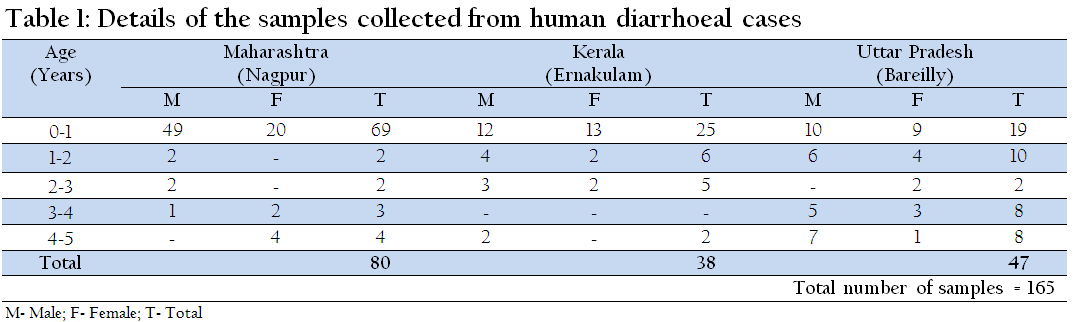
Diarrhoeal stool samples were collected from human infants and young animals of different species (canine, caprine, bovine, ovine, porcine and feline). The details of sample collected are presented in Table 1 and 2. A total of 315 stool samples were collected from 165 human infants and 150 young animals suffering from diarrheal problems. The stool samples were collected aseptically using Listeria Transport swabs (HiMedia Labs, Mumbai, India). All the collected samples were immediately transported to the laboratory under chilled conditions and processed further for the isolation and identification studies.

Isolation and Identification of L. monocytogenes
Isolation of Listeria spp. was attempted as per USDA–FSIS method (USDA, 2013). In brief the faecal swabs were aseptically inocualted in 10 ml of University of Vermont Medium (UVM)–1 supplemented with acriflavin and nalidixic acid, and incubated at 37oC for 18–24hours. Further, UVM-1 enriched inoculums (0.1ml) was inoculated in 10 ml of UVM2 and incubated again at 37oC for 24 hours. The enriched UVM-2 broth was then streaked on PALCAM agar (HiMedia Labs, Mumbai, India) plate for selective isolation of Listeria spp. colonies. The typical grayish-green glistening pin point colonies of about 0.5 mm diameter surrounded by a diffuse black zone of aesculin hydrolysis were presumptively identified as Listeria spp. The presumed colonies of Listeria spp. (at least 3/plate) were further confirmed by biochemical tests such as Gram’s staining, catalase reaction, tumbling motility at 20–25°C, methyl red-Voges Proskauer (MR-VP) reactions, nitrate reduction and fermentation of sugars (rhamnose, xylose, mannitol and α- methyl-D-mannopyranoside).
InVitro Pathogenicity Testing of L. monocytogenes Isolates
The biochemically confirmed L. monocytogenes isolates were subjected to in vitro pathogenicity tests which includes hemolytic titre assay, Phosphatidylinositol phospholipase C (PI-PLC) activity on Agar Listeria according to Ottaviani and Agosti medium (ALOA) and a multiplex PCR targeting crucial virulence associated genes (prfA, plcA, hly, actA).
Hemolytic titre assay
The haemolytic titre of the L. monocytogenes isolates were measured as per the protocol described by Young et al. (1986). The hemolytic units were expressed as the reciprocal of highest dilution of toxin that is required to lyse 50% of erythrocytes compared to lysis obtained by distilled water.
PI-PLC Activity on ALOA Medium
ALOA medium (Hi-media Labs, Mumbai, India) was prepared as per manufacturer’s instructions. On this medium all the Listeria spp. formed bluish green colonies due to the presence of a chromogenic compound X-glucosidase which detects β-glucosidase present in all Listeria spp. Pathogenic Listeria spp., can be distinguished from other Listeria spp. through the production of PI-PLC, which is detected by a substrate in the medium to form opaque halos around the colonies.
Multiplex PCR (mPCR) Targeting Virulence Associated Genes
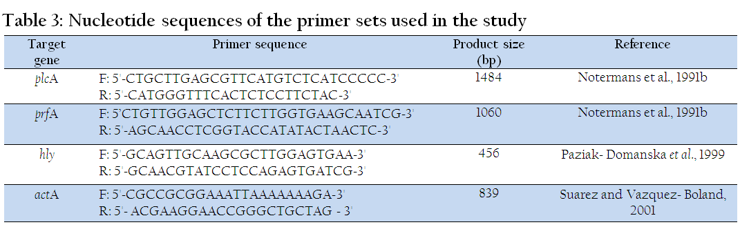
The primers used for the multiplex PCR targeting virulence associated genes were procured from Sigma-Aldrich, USA. The details of primers are presented in Table 3. The chemical used in PCR master mix were procured from 3B Black Bio, Spain. The mPCR targeting virulence associated genes (prfA, plcA, hly, actA) was performed as per the method described by Rawool et al., (2007) with suitable modifications. In brief, the multiplex PCR assay was standardized in two sets, each set was performed in a 25µl reaction volume; containing 2.5µl of 10X PCR buffer (100mM Tris-HCl buffer, pH 8.3 containing 500 mM KCl, 15 mM MgCl2 and 0.01% gelatin), 3µl of 10 mM dNTP mix (a final concentration of 1 mM), 3µl of 25 mM MgCl¬2 (final concentration 7.5 mM) and 30µM of three primer sets (set 1: plcA, actA and hly and Set 2: prfA, actA, hly) @ 10 µM of each primer set, 3 units of Taq DNA polymerase (3B Black Bio, Spain), a single colony of Listeria as DNA template and sterilized nuclease free water to make up the reaction volume. The reaction was performed in Mastercycler Pro Thermocycler (Eppendorf, Germany). The cycling conditions for PCR included an initial denaturation at 95°C for 5 minutes followed by 35 cycles each of 15 seconds denaturation at 95°C, annealing at 60°C for 30 seconds and extension at 72°C for 1 minute 30 second, followed by a final extension of 10 minutes at 72°C and hold at 4°C. The resultant PCR products (10 µl) were analysed by submarine gel electrophoresis, using 1.5% agarose gel stained with ethidium bromide (0.5μg/ml) and visualized and documented by UV Gel documentation system (UVP Gel Seq Software, England).
InVivo Chick Embryo Inoculation Test
The pathogenicity of the reference strains as well as the test isolates of Listeria monocytogenes was assessed by chick embryo inoculation test as per the method described by Notermans et al. (1991a). The isolates causing embryo mortality after 24 h of inoculation up till 5 days were considered to be pathogenic.
Multiplex Serotyping PCR
The multiplex serotyping PCR was performed for serogroup identification as per the protocol described by Doumith et al. (2004). The multiplex serotyping PCR divides L. monocytogenes strains into four distinct serogroup: 1/2a, 1/2c, 3a, 3c; 1/2c, 3c; 1/2b, 3b, 4b, 4d, 4e and 4b, 4d, 4e. The primers used for multiplex PCR serotyping were synthesized from Eurofins Genomics (Bengaluru, India).
RESULTS
On microbiological analysis of 315 diarrhoeal stool samples (165 human infants and 150 young animals), 80 human and 76 animal faecal swabs revealed typical colonies with aesculin hydrolysis on PALCAM agar plates. Further, on morphological and biochemical characterization, 4 L. monocytogenes isolates were identified. Of the four L. monocytogenes isolates, two were isolated from human faecal swabs and two from porcine faecal swabs. The overall isolation rate of L. monocytogenes in human infants and young animals was found to be 1.2% and 1.3% respectively. None of the samples from bovine, caprine, canine and felines were found positive for L. monocytogenes.
All the 4 L. monocytogenes isolates recovered in the present study were assessed for their pathogenic potential using in vitro as well as in vivo tests. The isolates were subjected to in vitro pathogenicity tests viz., hemolytic titre assay, PI-PLC activity on ALOA medium and a multiplex PCR targeting virulence associated genes (prfA, plcA, hly, actA) as well as to in vivo chick embryo inoculation test.
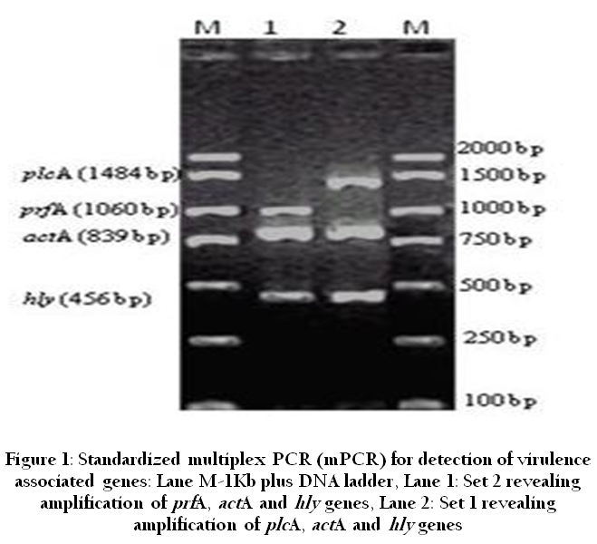
Figure 1: Standardized multiplex PCR (mPCR) for detection of virulence associated genes: Lane M-1Kb plus DNA ladder, Lane 1: Set 2 revealing amplification of prfA, actA and hly genes, Lane 2: Set 1 revealing amplification of plcA, actA and hly genes
On screening the 4 L. monocytogenes isolates (PKLM1, PKLM2, HMHM32 HGOM36) with hemolytic titer assay, of the two porcine isolates, one (PKLM1) was found to be highly hemolytic revealing a hemolytic unit of 32, whereas the other porcine isolate (PKLM2) was revealing a moderate (16) hemolytic unit. With regard to two human infant isolates, moderate (16) hemolytic unit was observed for one of the human isolate (HMHM32), while the other human isolate (HGOM36) revealed a hemolytic unit of 8. Similar results were also observed with regards to PI-PLC activity, as the porcine isolates (PKLM1 and PKLM2) and one human isolate (HMHM32) revealed prominent PI-PLC activity on ALOA medium within 24 hours of incubation, while one of the human isolate (HGOM36) showed moderate PI-PLC activity after 72 h of incubation (Table 4). On employing all the 4 L. monocytogenes isolates to the standardized multiplex PCR targeting virulence associated genes (Figure 1), all the 4 isolates revealed amplified products for all the virulence associated genes targeted in the present study (Table 4). L. monocytogenes MTCC 1143 was used as a positive control for all the tests. On comparing the results of in vitro pathogenicity tests with an in vivo chick embryo inoculation test, a good correlation was observed. The two porcine L. monocytogenes isolates (PKLM1, PKLM2) and one human isolate (HMHM32) killed the chick embryos within 24-36 h post inoculation, while the remaining one L. monocytogenes isolate from human (HGOM36) killed the chick embryo within 48-72h post inoculation (Table 4).
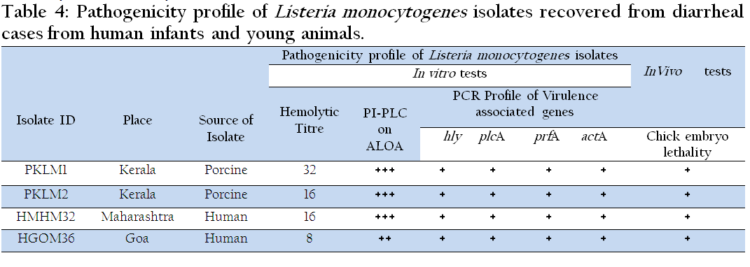
Table 4: Pathogenicity profile of Listeria monocytogenes isolates recovered from diarrheal cases from human infants and young animals.
The serogrouping of all the 4 L. monocytogenes isolates was carried out using multiplex serotyping PCR. On PCR analysis it was observed that all the isolates belonged to the serogroup 4b, 4d, and 4e. Among serogroup 4b, 4d and 4e, serovar 4b in particular have been widely reported to be associated with several listerial outbreaks.
DISCUSSION
L. monocytogenes has been associated with major food-borne outbreaks globally but yet there is insufficient data regarding isolation and identification of pathogenic L. monocytogenes from febrile listerial gastroenteritis. Recent investigations of food-borne outbreaks provided evidences that febrile gastroenteritis may indeed be the main clinical manifestation of infection caused by L. monocytogenes (Ooi and Lorber, 2005). Many patients experience antecedent diarrheoa in case of invasive listeriosis, however in recent past it was efficiently convinced that L. monocytogenes can cause an acute, self-limiting, febrile gastroenteritis in healthy individuals, which usually lasts for 2 days (Barbuddhe and Chakraborty, 2008). Over the last 20 years, so far only seven outbreaks of food-borne gastroenteritis due to L. monocytogenes infection have been reported (Schuppler and Loessner, 2010).
In India listeriosis is not a notifiable disease and also the epidemiological data available on listeriosis is not adequate for assessing the extent of infection in human beings and animals (Barbuddhe et al., 2012). The disease largely remains undiagnosed and under reported, largely due to the lack of a reliable, rapid and simple diagnostic test (Barbuddhe et al., 2012). Besides this, during routine stool sample examination L. monocytogenes is often ignored or not considered as a possible cause of febrile gastroenteritis. Therefore, the present study was undertaken to explore the isolation and identification of L. monocytogenes from spontaneous diarrheal cases in human infants and young animals.
In the present study, 4 L. monocytogenes isolates were isolated and identified. Of the 4 L. monocytogenes isolates, two were isolated from human infants and two from porcine respectively. Similar reports of isolation from febrile human listerial gastroenteritis cases have been reviewed (Ooi and Lorber, 2005; Schuppler and Loessner, 2010). Globally, there are no reports of isolation of L. monocytogenes from animal diarrheal cases. Thus, to the best of our knowledge, our finding appears to be the first report on isolation of L. monocytogenes from febrile diarrheal cases in piglets. Further, we were not successful in isolating L. monocytogenes from other species namely caprine, bovine, canine and feline, may be because the number of samples analyzed were very few or may be those species of animals were suffering from diarrheoa due to other etiological agents. Hence, before giving any recommendations, it would be wise to analyze more number of samples from above mentioned species having febrile gastroenteritis symptoms. The exact mechanism by which L. monocytogenes causes diarrheoa is yet unknown (Ooi and Lorber, 2005), however it has been reported that the organisms are well adapted to the conditions in the gastrointestinal tract and pursue different strategies to counteract changes in acidity, osmolarity, oxygen tension, or the challenging effects of antimicrobial peptides and bile. Besides this, L. monocytogenes are able to colonize and persist in the gallbladder for long-term resulting in chronic infections, which demonstrates their pathogenic ability to survive within the various microenvironments of the gastrointestinal tract (Schuppler and Loessner, 2010). Moreover, the adherence and invasive mechanism of L. monocytogenes responsible for causing febrile gastroenteritis has been extensively reviewed by Schuppler and Loessner (2010).
The occurrence of listeriae in nature is ubiquitous and therefore the possibility of cross contamination during collection of samples cannot be ruled out. Also L. monocytogenes exists and multiplies as a saprophytic organism in the soil, plants, sewage as well as river water (Anonymous, 1991; Farber and Peterkin, 1991). Besides, large number of Listeria isolates of various kinds (haemolytic and pathogenic, haemolytic and non-pathogenic as well as nonhemolytic and non-pathogenic), types (typical and atypical) and origin (clinical, food, environment, feed etc.) exits in clinical as well as surrounding environmental sources (Shoukat et al., 2013). Therefore, in order to link up the isolated L. monocytogenes as a possible cause of diarrhoea, they were assessed for their pathogenic potential. Usually the pathogenicity of L. monocytogenes depends on its ability to survive and multiply in the macrophages and other host cells (Vazquez-Boland et al., 2001). After internalization, virulent L. monocytogenes produces listeriolysin O (LLO) and PI-PLC which cause the lysis of phagosomal membrane and thus it comes in the cytosol from where it spreads from cell-to-cell with the aid of actin filaments (Vazquez-Boland, et al., 2001). Therefore, hemolysis is an important characteristic that seems to be directly related to the pathogenicity of listeriae since non-hemolytic Listeria spp. are considered as non-pathogenic (Courteiu, 1991). In view of above LLO being an important virulent marker, we assessed the hemolytic (LLO) titre of the recovered L. monocytogenes isolates. The hemolytic titre assay revealed that of the 4 L. monocytogenes isolates, three isolates (PKLM1, PKLM2 and HMHM32) were exhibiting high to moderate hemolytic titers except for one L. monocytogenes isolate (HGOM36) which suggests that these L. monocytogenes isolates may have the potential of causing febrile gastroenteritis. However, it is advisable to perform an alternative in vitro test such as detecting PI-PLC activity on ALOA medium. This method besides detecting PI-PLC activity also allows discrimination between the pathogenic species of Listeria from the non-pathogenic listeriae (Aurora et al., 2008). In the present investigation almost similar observation were seen for PI-PLC activity on ALOA medium as that observed for hemolytic titre for the recovered L. monocytogenes isolates. Similar results were also reported by several researchers (Shakuntala et al., 2006; Rawool et al., 2007; Kaur et al., 2009; Shoukat et al., 2012).
Studies addressing PCR based detection of a single virulence associated gene is neither sufficient to identify the isolate nor to reveal its true pathogenic potential (Nishibori et al., 1991; Shakuntala et al., 2006; Rawool et al., 2007a). Also detection of hly and plcA gene by PCR in L. monocytogenes isolates is not sufficient to elucidate the true pathogenic potential, because both the gene are regulated by a key regulator gene i.e., prfA (Shakuntala et al., 2006; Kaur et al., 2007; Rawool et al., 2007b; Aurora et al., 2008). In addition, other genes such as actA, internalins and several other virulent associated genes do play an essential role in pathogenesis of this bug. Practically, it is not possible to study all and hence in the present study four important virulence associated genes viz., prfA, plcA, actA, and hly were attempted in multiplex PCR format. In the present investigation all the four isolates of L. monocytogenes revealed amplification of all the four virulence associated genes, suggesting that the recovered isolates could be potentially pathogenic in nature. Similar observation has been reported, wherein L. monocytogenes isolates harboring prfA, plcA and hly are generally pathogenic in vivo model of infections (Shakuntala et al., 2006; Kaur et al., 2007; Rawool et al., 2007a; Aurora et al., 2008; Shoukat et al., 2012).
Virulence of Listeria spp. by in vivo method is often assessed by lethality in adult mice by intraperitoneal route (Chakraborty and Goebel, 1988) and/or by chick embryo inoculation by chorio-allantoic membrane (CAM) route (Notermans et al., 1991a). However, the mouse inoculation assay is objectionable from an ethical point of view; the virulence potential of recovered isolates was studied by chick embryo inoculation test. It was observed that all the four L. monocytogenes isolates were found to be pathogenic, as death was evidenced in all the inoculated chick embryos. The results are in completed agreement with earlier studies (Shakuntala et al., 2006; Kaur et al., 2007; Rawool et al., 2007a; Aurora et al., 2008; Shoukat et al., 2013).
Serotyping performed using molecular methods is an effective and an alternative choice to traditional seroagglutination methods, which are both time-consuming and expensive, with lower reproducibility of results (Zhang et al., 2004). In order to explore the serogroup of the recovered L. monocytogenes isolates, multiplex serotyping PCR was performed as described by Doumith et al., (2004). In the present study all the test isolates belonged to the serogroup 4b, 4d and 4e. In this serogroup, serovar 4b in particular has been widely reported in several outbreaks of foodborne listeriosis (Ward et al., 2010; Liu, 2006). Our results are also in agreement with the reports of febrile gastroenteritis outbreak in northern Italy which have revealed the presence of serotype 4b (Aureli et al., 1997).
To conclude, isolation of pathogenic L. monocytogenes from sporadic gastroenteritis cases suffering from diarrhoea appears to be the first report in India. Thus, from future human and animal health perspective, isolation or detection of L. monocytogenes must be considered while analyzing samples of febrile gastroenteritis. Besides this, the incidence rate and factors that govern the onset of this non-invasive form are yet unexplored, and therefore to have better understanding of febrile non-invasive gastroenteritis, proper risk assessment studies are required to address this key issue.
ACKNOWLEDGEMENT
The authors thank Director, Indian Veterinary Research Institute, Izatnagar for providing necessary facilities to undertake the research. The research grant No. BT/01/CEIB/11/VI/13 from the Department of Biotechnology, Government of India is duly acknowledged. We thank Mr. K. K. Bhatt for his excellent technical assistance.
REFERENCES
Anonymous (1991) Listeria monocytogenes Recommendation by the National Advisory Committee on microbiological criteria for foods. Intl. J. Food Microbiol. 14: 185.
PMid:1790101
Aureli P, Fiorucci GC, Caroli D, Marchiaro G, Novara O, Leone L, Salmaso S (2000). An outbreak of febrile gastroenteritis associated with corn contaminated by Listeria monocytogenes. N. Eng. J. Med. 342(17):1236 – 1241.
http://dx.doi.org/10.1056/NEJM200004273421702
PMid:10781619
Aurora R, Prakash A, Prakash S, Rawool DB, Barbuddhe SB (2008). Comparison of PI-PLC based assays and PCR along with in vivo pathogenicity tests for rapid detection of pathogenic Listeria monocytogenes. Food Cont. 19: 641 – 647.
http://dx.doi.org/10.1016/j.foodcont.2007.07.002
Barbuddhe SB, Chakraborty T (2009). Listeria as an enteroinvasive gastrointestinal pathogen. Curr. Top. Microbiol. Immunol. 337: 173 – 195.
http://dx.doi.org/10.1007/978-3-642-01846-6_6
PMid:19812983
Barbuddhe SB, Malik SVS, Kumar AJ, Kalorey DR, Chakraborty T (2012). Epidemiology and risk management of listeriosis in India. Intl. J. Food Microbiol. 154: 113 – 118.
http://dx.doi.org/10.1016/j.ijfoodmicro.2011.08.030
PMid:21955732
Barbuddhe SB, Hain T, Chakraborty T (2008). The Genus Listeria. In:Practical Handbook of Microbiology, CRC Press, Boca Raton. 533 –562.
http://dx.doi.org/10.1201/9781420009330.ch34
Chakraborty J, Goebel W (1988). Recent developments in the study of virulence in Listeria monocytogenes. Curr. Top. Microbiol. Immunol. 138: 41 - 58.
PMid:3143518
Courteiu AL (1991). Latest news on Listeriosis. Comp. Immunol. Microbiol. Infect. Dis. 14: 1 - 7.
http://dx.doi.org/10.1016/0147-9571(91)90035-C
Dalton CB, Austin CC, Sobel J, Hayes PS, Bibb WF, Graves LM, Swaminathan B, Procter ME, Griffin PM (1997) An outbreak of gastroenteritis and fever due to Listeria monocytogenes in milk. N. Eng. J. Med.336: 100 – 105.
http://dx.doi.org/10.1056/NEJM199701093360204
PMid:8988887
Doumith M, Buchrieser C, Glaser, P, Jacquet C, Martin P (2004). Differentiation of the major Listeria monocytogenes serovars bymultiplex PCR. J. Clin. Microbiol. 42 (8): 3819 – 3822.
http://dx.doi.org/10.1128/JCM.42.8.3819-3822.2004
PMid:15297538 PMCid:PMC497638
Farber JM, Peterkin PI (1991). Listeria monocytogenes, a food borne pathogen. Microbiol. Rev. 55: 476 - 511.
PMid:1943998 PMCid:PMC372831
Iyer A, Kumosani T, Yaghmoor S, Barbour E, Azhar E, Harakeh, S (2013). Escherichia coli and Salmonella spp. in meat in Jeddah, Saudi Arabia. J. Infect. Dev. Count. 7(11): 812 – 818.
Kaur S, Malik SVS, Bhilegaonkar KN, Vaidya VM, Barbuddhe SB (2009). Use of a phospholipase C assay, in vivo pathogenicity assays and PCR in assessing the virulence of Listeria spp. Vet. J. 184:366 - 370.
http://dx.doi.org/10.1016/j.tvjl.2009.03.032
PMid:19409824
Liu D (2006). Identification, subtyping and virulence determination of Listeria monocytogenes, an important foodborne pathogen. J. Med. Microbiol. 55(6): 645 – 659.
http://dx.doi.org/10.1099/jmm.0.46495-0
PMid:16687581
Nishibori T, Cooray K, Xiang H, Kawmuru I, Fujita M, Mitsuyama M (1995). Correlation between the presence of virulence associated genes as determined by PCR and actual virulence in mice in various strains of Listeria spp. Microbiol. Immunol. 39: 343 - 349.
http://dx.doi.org/10.1111/j.1348-0421.1995.tb02211.x
PMid:7565175
Notermans S, Dufrenne J, Chakraborty T, Steinmeyer S, Terplant G (1991a). The chick embryo test agrees with the mouse bio-assay for assessment of the pathogenicity of Listeria species. Lett. Appl. Microbiol. 13: 161-164.
http://dx.doi.org/10.1111/j.1472-765X.1991.tb00597.x
Notermans SHW, Dufrenne J, Leimeister-Wachter M, Domann E, Chakraborty T (1991b). Phosphatidylinositol-specific phospholipase C activity as a marker to distinguish between pathogenic and non-pathogenic Listeria species. Appl. Environ. Microbiol., 57: 2666 - 2670.
PMid:1662937 PMCid:PMC183637
Ooi ST, Lorber B (2005).Gastroenteritis due to Listeria monocytogenes. Clinical Infectious Diseases. 40 (9): 1327 – 1332.
http://dx.doi.org/10.1086/429324
PMid:15825036
Rawool DB, Malik SVS, Barbuddhe SB, Shakuntala I, Aurora R (2007a). A multiplex PCR for detection of virulence associated genes in Listeria monocytogenes. Intl. J. Food Safety. 9: 56 - 62.
Rawool DB, Malik SVS, Shakuntala I, Sahare AM, Barbuddhe SB (2007b) Detection of multiple virulence associated genes in pathogenic Listeria monocytogenes from bovines with mastitis. Intl. J. Food Microbiol. 113: 201 - 207.
http://dx.doi.org/10.1016/j.ijfoodmicro.2006.06.029
PMid:16979771
Riedo FX, Pinner RW, Tosca ML, Graves ML, Reeves MW, Weaver RE, Plikaytis BD, Broome CV (1994). A point-source foodborne listeriosis outbreak: documented incubation period and possible mild illness. J. Infect. Dis. 170: 693 – 696.
http://dx.doi.org/10.1093/infdis/170.3.693
PMid:8077731
Salamina G, Dalle DE, Niccolini A, Poda G, Cesaroni D, Bucci M, Fini R, Maldini M, Schuchat A, Swaminathan B, Bibb W, Rocourt J, Binkin N, Salmaso S (1996) A foodborne outbreak of gastroenteritis involving Listeria monocytogenes. Epidemiol. Infect. 117:429 – 436.
http://dx.doi.org/10.1017/S0950268800059082
PMid:8972666 PMCid:PMC2271639
Schwartz B, Hexter D, Broome CV, Hightower AW, Hirschhorn, RB, Porter JD, Hayes PS, Bibb WF, Lorber B, Faris DG (1989). Investigation of an outbreak of listeriosis: new hypotheses for the etiology of epidemic Listeria monocytogenes infections. J. Infect. Dis. 159:680 – 685.
http://dx.doi.org/10.1093/infdis/159.4.680
PMid:2494267
Shakuntala I, Malik SVS, Barbuddhe SB, Rawool DB (2006).Isolation of Listeria monocytogenes from buffaloes with reproductive disorders and its confirmation by polymerase chain reaction. Vet. Microbiol. 117: 229 - 234.
http://dx.doi.org/10.1016/j.vetmic.2006.06.018
PMid:16860946
Shoukat S, Malik SVS, Rawool DB, Kumar A, Kumar S, Shrivastava S, Barbuddhe SB, Das DP, Das S (2013).A Study on Detection of Pathogenic Listeria monocytogenes in Ovine's of Kashmir Region Having Abortion or History of Abortion. Proc. Natl. Acad. Sci., India, Sect. B Biol. Sci. DOI 10.1007/s40011 – 013 – 0228 - 0.
Schuppler M, Loessner MJ (2010).The Opportunistic Pathogen Listeria monocytogenes: Pathogenicity and Interaction with the Mucosal Immune System. Intl. J. Inflamm
http://dx.doi.org/10.4061/2010/704321
Todd J, Ward, Usgaard T, Evans P (2010). A Targeted Multilocus Genotyping Assay for Lineage, Serogroup, and Epidemic Clone Typing of Listeria monocytogenes. Appl. Environ. Microbiol. 76: 6680 – 6684.
http://dx.doi.org/10.1128/AEM.01008-10
PMid:20709839 PMCid:PMC2950480
USDA (2013). Laboratory Guidebook: Chapter MLG 8.09-Isolation and Identification of Listeria monocytogenes from Red Meat, Poultry and Egg Products, and Environmental Samples. USDA
Vazquez-Boland JA, Kuhn M, Berche P, Chakraborty T, Domı’nguez-Bernal G,Goebel W, Gonza’lez-Zorn B, Wehland J, Kreft J (2001). Listeria pathogenesis and molecular virulence determinants. Clin. Microbiol. Rev. 14: 584 – 640.
http://dx.doi.org/10.1128/CMR.14.3.584-640.2001
PMid:11432815 PMCid:PMC88991
WHO (2013). General information related to microbiological risks in food. Food saftey. December 14, 2013, from http://www.who.int/foodsafety/micro/general/en/.
Young JD, Leong LG, DiNome MA, Cohn ZA (1986) A semiautomated hemolysis microassay for membrane lytic proteins. Anal. Biochem. 154: 649 - 654.
http://dx.doi.org/10.1016/0003-2697(86)90042-4
Zhang W, Hughes A, Wilt G, Knabel SJ (2004). The BAX PCR assay for screening Listeria monocytogenes targets a partial putative gene lmo2234. J. Food Prot. 67:1507 – 1511.
PMid:15270511


