Advances in Animal and Veterinary Sciences
Research Article
Advances in Animal and Veterinary Sciences 1 (1): 35–40Comparative Potential of Traditional versus Modern Diagnostic Tests in Estimating Status of Caprine Johne’s Disease
Dilip Singh Barad1, Bharat Singh Chandel1, AbidaliI. Dadawala1, HarshadC. Chauhan1, Hemendra Singh Kher1, Sagar Shroff1, Abidali Gulamhaider Bhagat1, ShoorVir Singh2*, Pravin Kumar Singh2, Ajay Vir Singh2, Jagdip Singh Sohal2, Saurabh Gupta2, Kundan Kumar Chaubey2
- Department of Microbiology, College of Veterinary Science and A.H., SardarkrushinagarDantiwada Agricultural University, Sardarkrushinagar – 385 506, Dist. Banaskantha, Gujarat
- Microbiology Laboratory,Animal Health Division, Central Institute for Research on Goats, Makhdoom, PO –Farah, Dist.– Mathura, 281 122, Uttar Pradesh, India
*Corresponding author:shoorvir.singh@gmail.com; shoorvir_singh@rediffmail.com
ARTICLE CITATION:
Barad DB, Chandel BS, Dadawala AI, Chauhan HC, Kher HN, Shroff S, Bhagat AG, Singh SV, Singh PK, Singh AV, Sohal JS, Gupta S, Chaubey KK (2013). Comparative potential of traditional versus modern diagnostic tests in estimating incidence of caprine Johne’s disease. Adv. Anim. Vet. Sci. 1 (1): 35–40
Received: 2013–04–03, Revised: 2013–04–19, Accepted: 2013–04–20
The electronic version of this article is the complete one and can be found online at
(
http://www.nexusacademicpublishers.com/table_contents_detail/4/31/html
)
which permits unrestricted use, distribution, and reproduction in any medium, provided the original work is properly cited
ABSTRACT
Study aimed to compare easy to perform field based conventional tests {(fecal smear examination, Delayed Type Hypersensitivity (DTH), AGID} with respect to modern laboratory tests (IS900 PCR and Indigenous ELISA kit) in estimating the incidence of caprineJohne’s disease in two important breeds of goats (Mehsani and Surti) from Gujarat (Western India) in the year 2009. A total of 219 goats screened were categorized as Group–I (123 Mehsani goats), Group–II (76 Surti goats) and Group–III, (20 Non–descript goats). Percent positivity by fecal smear examination (fecal microscopy), Delayed Type Hypersensitivity (DTH), AGID, IS900 PCR and Indigenous ELISA kit was 9.2 (7/76), 21.9 (27/123), 10.9 (24/219), 12.5% (5/40) and 43.3 (95/219), respectively. Rectal pinch smear examination was carried out in 27 DTH positive goats and all smears were negative for the presence of acid fast bacilli. Screening tests (Indigenous ELISA and Delayed Type Hypersensitivity) showed very high incidence of MAP infection in the goat population.
INTRODUCTION
Mycobacterium avium subspecies paratuberculosis (MAP) is responsible for chronic granulomatous inflammation in animals known as Johne’s disease (JD) or paratuberculosis (Chiodiniet al., 1984). MAP infection leads to chronic enteritis of intestines, inflammation of lymph nodes especially mesenteric and lymph–angitis in domestic livestock and wild ruminants (Perez et al., 1996, Clarke, 1997, Manning and Collins, 2001, Singh et al., 2012). JD has been reported to be endemic in the animal population of world and India, where ever investigated. In India, limited studies (Pande, 1940; Singh et al., 1996, Tripathi et al., 2002, Singh et al., 2007a) reported variable incidence (2–18%) in domestic livestock. Animals may either receive infection from infected parents through semen, in–utero, milk and colostrums (Shankar et al., 2010) or pick up infection from contaminated environment soon after birth and then follows a protracted incubation period which may last few months to few years. Sharing of MAP between species (inter–species transmission) has been frequently reported (Singh et al., 2012). Clinical symptom being non–specific (weight loss and diarrhea) are not visible before disease gets fully established in the body (clinical) and progressive weakness ultimately leads to either un–timely death or culling. In small ruminants disease is clinically characterized by progressive weight loss and emaciation, soft pelleted feces in the terminal stages. Internally granulomatous enteritis leads to thickening of intestinal mucosa and regional lymphangitis and lymphadenitis (Chiodiniet al., 1984), however, corrugations are rare as animals goes out much before from the production system.
Johne’s is spectral disease therefore presents variable bacteriological, immunological and pathological picture which is responsible for variations in efficacy of the diagnostic methods employed at different points of time for detection of infection during the course of infection (Chiodini et al., 1984). Though, culture of bacilli samples (feces and tissues from ileo–caecal lymph nodes and small intestines) is specific but is time consuming due to long and variable incubation period which may take 12 weeks (Cocito et al., 1994) but sensitivity is poor (Sockett et al., 1992). For rapid diagnosis of JD, IS900 PCR may be used since it can detect MAP in feces, blood and tissues with sensitivity equivalent to culture (Tripathi et al., 2002, Singh et al., 2010). Animals infected with MAP elicit strong cellular immune (CMI) response in early stages of infection and in the later stages a strong humoral immune response (Clarke, 1997). Tests based on CMI response such as cutaneous testing with johnin PPD (DTH), gamma–interferon assay and lymphocyte stimulation test (LTT), though sensitive but have not been used frequently for the detection of early infection in sheep and goats (Molina et al., 1991; Storset et al., 2001; Kurade et al., 2004). Tests used for antibody detection include complement fixation test (CFT), agar gel immunodiffusion (AGID) and enzyme–linked immunosorbent assay (ELISA) tests. ELISA has been reported to be sensitive in clinical cases of JD but performs poorly in sub–clinical stages of infection (Stewart et al., 2006). The study aimed to evaluate the efficacy of easy to perform field based conventional tests {(fecal smear examination, Delayed Type Hypersensitivity (DTH), AGID)} with respect to modern laboratory tests (IS900 PCR and Indigenous ELISA kit) in estimating incidence of Mycobacterium avium subspecies paratuberculosis (MAP) in two important breeds of goats (Mehsani and Surti) from Gujarat (Western India) in the year 2009.
MATERIALS AND METHODS
Fecal Smear
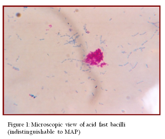
Approximately, 2 gram of fecal sample was finely ground in sterilized pestle and mortar with sterilized distilled water (10–12 ml). Finely ground samples were centrifuged at 2600 rpm for 45 min at room temperature (RT). Supernatant was discarded and from middle layer smears were prepared, stained by ZiehlNeelsen (ZN) staining and were examined under oil immersion (100X) for presence of acid–fast bacilli (AFB) indistinguishable to MAP. Positive fecal samples were stored at –20oCfor DNA extraction and culture.
Rectal Pinch Smear
Rectal pinch smear were examined in goats positive in Johnin test. Rectal pinch was collected with a sterilized Artificial Insemination sheath and smears were prepared, stained by ZiehlNeelsen (ZN) staining and were observed under oil immersion (100X) for presence of acid–fast bacilli (AFB) indistinguishable to MAP.
Delayed Type Hypersensitivity (DTH) / Johnin test
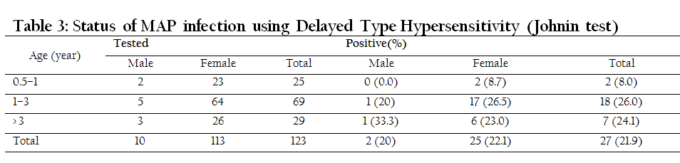
Single intra–dermal (DTH) test was performed on 123 goats of group–I by inoculation of 100 µg of Johnin PPD, obtained from Division of Biological Products, IVRI, Izatnagar (India) in the form of heat concentrated synthetic medium (HCSM) of Mycobacterium paratuberculosis. Test was carried out as per the instructions of manufacturer on the side of neck. Skin thickness was measured with Vernier Calipers at pre, 24, 48 and 72 hours post inoculation. Animals showing hot, edematous, painful skin thickness of more than 3 mm after 48 hours were considered positive.
Post Mortem and Histopathology
Goats died of natural infection were subjected to post mortem examination. Tissues (Ileocaecal junction of intestine, ileum and mesenteric lymph nodes) exhibiting gross lesions were collected and stored at –20oC without adding any preservative for DNA isolation and PCR, whereas, for histopathology tissues were stored in 10% formalin at room temperature. Formalin fixed tissues were cut into thin (2–3 mm) pieces and wash thoroughly with water for several hours before putting in ascending grades of alcohol for dehydration. Dehydrated tissues were cleared in xylene, embedded in paraffin blocks and 5 micron thick sections were prepared (Luna, 1968). Sections were stained with haematoxylin and eosin (H and E). However, smears made from tissues (Ileocaecal junction, mesenteric lymph nodes) were stained by ZiehlNeelsen (ZN) staining and were examined under oil immersion (100X) for presence of acid–fast bacilli (AFB) indistinguishable to MAP.
Agar Gel Immunodiffusion test (AGID)
Agar Gel Immunodiffusion test (AGID) was perfomed as per Ferreira et al. (2002). Briefly, Protoplasmic Antigen (Allied Monitor, USA) was used as antigen at the concentration of 10 mg/ml. 0.75% agarose was dissolved in 0.85% NaCl solution and buffered to pH 9.0 with 0.01 moltris (hydroxymethyl)–aminomethane and 0.02% sodium azide. Gel was poured in plates on a 4–mm thick layer and wells of 5 mm diameter were made in a hexagonal pattern of six peripheral wells for serum samples and a central well for antigen. Positive serum was placed into wells adjacent to test sera and were incubated at room temperature (RT). Plates were examined after 24 and 48 h of incubation and appearance of one or more clearly definable precipitation lines before or at 48 h constituted a positive test result. Absence of any precipitation lines was recorded as a negative test result. Serum from clinically infected animals was used as positive control.
DNA Isolation
After collection of tissues (mesenteric lymph nodes and intestines) were grounded and treated with 0.9% HPC (Hexadecylpyridinium chloride) overnight. The sediment (0.5–1.0 ml) was taken in 2.0 ml capacity eppendorf tubes and washed with PBS, 3–4 times by spinning and vortexing. Pellet was subjected to DNA isolation as per van Embedenet al. (1993) method with some modification. Briefly, washed pellet was suspended in 1.0 ml 1X TE buffer (pH 8.0) and centrifuged at 8000 rpm for 15 minutes. Pellet was re–suspended in 450 µl 1X TE buffer and subjected to freezing and thawing (heating to boiling and snap cooling at –20oC) repeat the process 3–4 times. Then, 40 µl of lysozyme (20 mg/ml) was added to tubes and incubated at 37oC for 2 hours. Proteinase K (6 µl) and 10% SDS (56 µl) were added and incubated at 65oC for 30 minutes. After that 64 µl of CTAB and 80 µl 5M NaCl were added and incubated at 65oC for 30 minutes. Equal volume of Chloroform and Isoamyl alcohol (24:1) was added, vortexed and centrifuged at 10,000 rpm for 10 minutes. DNA was precipitated by chilled absolute ethanol and washed with 70% chilled ethanol. DNA Pellet was re–suspended in 25 µl 1X TE buffer and stored at –20oC until further use.
Similarly, approximately, 2 gram of fecal sample was finely grounded in sterilized pestle and mortar with sterilized distilled water. Finely grounded samples were centrifuged at 2600 rpm for 45 min at room temperature (RT). Supernatant was discarded; middle layer was collected by sterilized swab and decontaminated in 0.9% HPC for 18–24 hours at room temperature. After discarding the decontaminated layer, 0.5–1.0 ml of sediment was washed with PBS and was subjected to DNA isolation as per method described by Van Embedenet al. (1993) with some modification.
Polymerase Chain Reaction (PCR)
DNA was isolated from decontaminated fecal and tissue samples were subjected to specific IS900 PCR using primers (Table 1)as described by Green et al. (1989) and Vary et al. (1990).
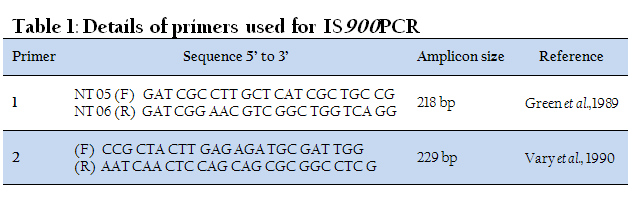
Briefly, PCR was set in volume of 25 µl, using 1.0–5.0 ng template DNA, 12.5 µl of 2X PCR Master Mix (Genei, Banglore) and 1.0 µl of each primer (10 pmole). Total of 37 cycles were performed in a thermocycler (MJ research) for complete amplification reaction. Thermal cycling conditions for primer 1 were: initial denaturation at 94oC for 3 min, followed by 35 cycles of denaturation at 94oC for 10 sec, annealing at 50oC for 10sec, extension at 72oC for 10 sec, and final extension at 72oC for 3 min. Thermal cycling conditions for primer 2 were: initial denaturation at 94oC for 4 min, followed by 35 cycles of denaturation at 94oC for 30 sec, annealing at 64oC for 30sec, extension at 72oC for 30 sec, and final extension at 72oC for 7 min. Presence and yield of specific PCR product was analyzed by 1.5% agarose gel electrophoresis.
Indigenous ELISA kit
Serum samples were screened by ‘indigenous ELISA kit’ as per the method of Singh et al (2007b). Briefly, 10µg per plate protoplasmic antigen of MAP ‘Indian Bison type’ was taken in 10 ml of antigen coating (carbonate–bicarbonate at pH–9.6) buffer and coated in 96 well flat bottom ELISA plates. Plates were incubated at 4oC overnight, blocked with 3% skimmed milk in PBS and incubated at 37oC for 1 hour. Plates were washed thrice with PBST (PBS with 0.05 % Tween 20) and stored at 4oC till further use. Then100µl of 1:50 diluted serum samples added in duplicate wells and incubated for 2 hrs at 37 oC. Plate was washed three times with PBST, 100 µl of optimally diluted (1:8000). Rabbit anti–goat horse radish peroxidase conjugate (Bangalore Genei) added to all wells and incubated for one hour at 37oC. Plate was washed thrice with PBST and 200 µl of freshly prepared OPD (5 mg / plate in substrate buffer at pH–5.0) was added to each well, incubated in dark for 30 min at 37oC. Absorbance was read at 450 nm.
Data Analysis
Data were managed in tables created using Microsoft Office Excel; Microsoft Corp., Redmond, Washington, USA. In ELISA, Positive and strong positive were considered as positive for MAP infection by converting OD values into sample–to–positive (S/P) ratio (using formulae; Sample OD – Negative OD / Positive OD – Negative OD in Excel cells) as per
Collins (2002).
RESULTS AND DISCUSSION
In the fecal smear examination of 56 goats of Group–I, 5 (8.9%) goats were positive, whereas, in Group–III, of 20 goats, 2 (10.0%) goats were positive. Of total 76 goats screened, 7 (9.2%) were positive (Table 2). Of 5 fecal positive goats of Group–I, 3 died during study. Clinical signs were observed in all the 7 fecal smear positive goats and were also positive in ELISA and AGID. Tripathiet et al. (2006) showed that of 36 known cases of caprineparatuberculosis diagnosed by clinical and fecal smear examination, 72.2% goats were shedding MAP bacilli (Figure 1). Examination of fecal smears by ZeihlNeelsen staining exhibited more number of positive cases as compared to culture and IS900 PCR (Munjalet al., 2007). Screening of 71 animals (55 goats and 16 sheep) belonging to Central Institute for Research on Goat (CIRG) located at Makhdoom (India) revealed that 40% goats and 31.2% sheep were shedding MAP in feces (Singh et al.,2007a, Singh et al., 2007b).
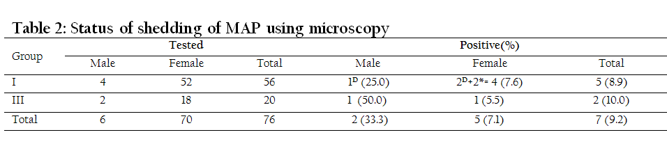
Rectal pinch smear examination was carried out in 27 DTH/Johnin positive goats. All rectal pinch smears were negative for the presence of MAP (Table 2). In accordance to this it was described in Ohio State University Fact Sheet for JD in small ruminants (sheep and goats) that smears from the biopsies of rectal mucosa are difficult to get and may not be very useful since in disease process it is less likely that it will involve rectum in cases of sheep and goats (http://ohioline.osu.edu/vme–fact/0003.html). Of the 123 goats of Group–I, 27 (21.9%) were positive in DTH test. Of 5 fecal positive goats which also showed clinical signs, 2 (3.5%) goats died during study were negative by Johnin (Table 3). Similar to these findings, Paliwal and Rajya (1982) and Rajukumar (1998) stated that sensitivity of Johnin test in goats ranged between 18–30% with least specificity in both preclinical and advanced stage of disease. Tripathiet al. (2006) reported that of 34 cases of caprineparatuberculosis, 73.5% goats were positive for Johnin test. In the present study, of 5 infected goats, 3 (60%) were positive in Johnin test.
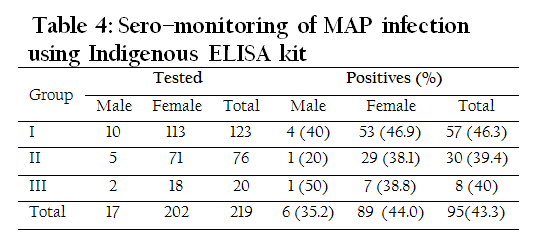
Of the 219 goats tested, 95 (43.3%) were positive by ELISA. Of 123 Mehsani goat breed (Group–I), positive reactors were higher 57(46.3%) and had history of recurrent diarrhea (Table 4). Sweeney et al.(1995) reported that sensitivity of ELISA is only 15.0% in animals excreting low quantities of MAP bacilli in their feces as compared to 87.0% in animals with clinical signs of Johne's disease. In this study similar results were obtained i.e. all the 7 goats showing clinical signs as well as fecal smear positivity were also positive by ELISA. Paolicchiet al.(2003) and Singh et al. (2009) observed that absorbed ELISA was useful to detect positive animals and those goats shedding MAP in feces. Similarly in the present study all animals shedding MAP in feces were positive in the ELISA test. As per OIE manual (2000), ELISA has been described as most sensitive and specific test for the detection of MAP infection however, in our study also Indigenous ELISA kit developed by CIRG, Makhdoom was found most sensitive and specific test for the detection of MAP infection.
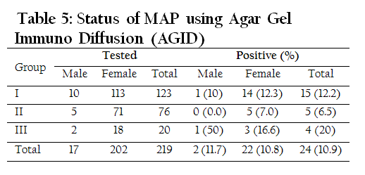
Of 219 goats tested, 24 (10.9%) were positive by AGID. Group–III consisting of 20 non–descript goats showed higher number (4 or 20%) of goats as positive reactors. All the 7 goats showed clinical signs and were positive in smear examination and AGID (Table 5). Similarly, Sherman et al. (1984) recorded that of 33 AGID positive cattle, 32 (96.9%) were confirmed for MAP infection by culture or necropsy. Similarly, Ferreira et al.(2002) found AGID specificity was 92.5% and sensitivity 57%. Raul et al. (1998) recorded 11.7% (64 of 546) sero–prevalence of MAP in sheep and goat flocks using AGID test.
PCR was carried out on fecal samples of suspected animals of Group–I and III with history of recurrent diarrhoea or pasty feces and tissue samples of three goats died in Group–I. Of 40 goats, 7 showed clinical signs and were also positive by faecal smear examination (Figure 2). Among these 7 goats faecal PCR detected 5 goats as positive while 2 were negative (Table 6). Munjal et al. (2007) reported that examination of fecal smears with ZiehlNeelsen staining detected more number of goats as positives as compared to culture and IS900 PCR. Some of the earlier studies have also reported higher prevalence of MAP infection in the farm herds as compared to farmer’s herds (Kumar et al., 2007). Tissue samples of all 3 goats died during study were positive by PCR. Similarly, Stabel (1997) reported that IS900 based PCR could be more sensitive in tissue samples in confirming diagnosis at necropsy.
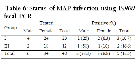
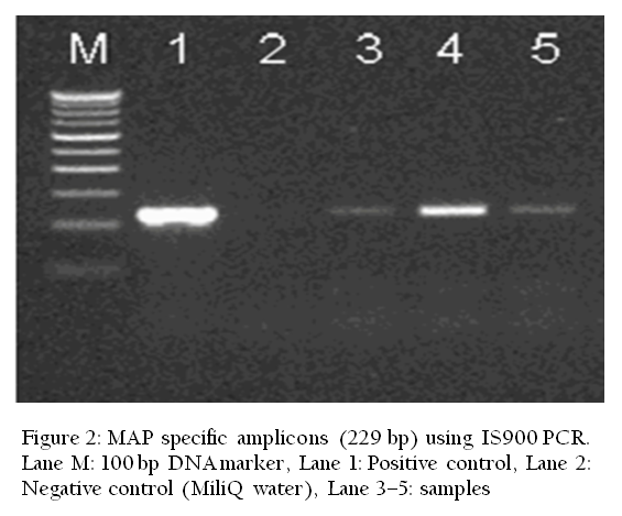
Figure 2: MAP specific amplicons (229 bp) using IS900 PCR. Lane M: 100 bp DNA marker, Lane 1: Positive control, Lane 2: Negative control (MiliQ water), Lane 3–5: samples
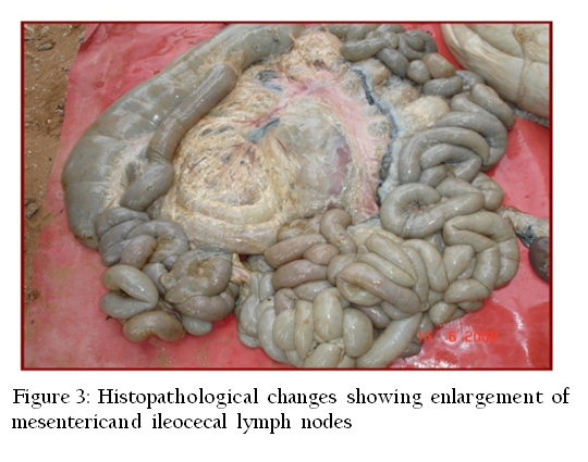
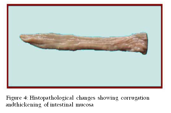
All three goats died during study in Group–I were subjected to post mortem examination, revealed enlargement of mesenteric and ileocecal lymph nodes and thickening of ileum and ileocecal junction wall (Figure 3 and 4). In one goat slight corrugation of ileal mucosa was observed. Carcass showed depletion of fat depots. Rajukumar (1998) observed similar lesions with enlarged, edematous and congested mesenteric and ileocecal lymph nodes. Corrugations in the ileal mucosa were observed in only one goat as has been described in the Ohio State University Fact Sheet for JD that small ruminants (goats and sheep) may reveal some lesions (gross and microscopic) that are not typical as those seen in cattle. Ridges and thickening of the small intestine and cecum are not always seen as is common in cattle.
Storsetet al. (2001) observed more number of goats with lesions in jejunum as compared to ileum while the lesions in ileum were detected in only one goat. In contrast to this lesions in jejunum were not observed in present study. Histopathological changes in goats died of JD revealed slightly congested intestinal mucosa with infiltration of mononuclear cells consisting of lymphocytes and macrophages in lamina propria. Villi were thickened and blunt at some places. Mesenteric lymph nodes exhibited infiltration of lymphocytes and macrophages. Munjalet al. (2005) observed similar lesions such as thickening of the intestinal villi with flat and wide tips, infiltration of lymphocytes, macrophages and epithelioid cells in lamina propria of intestine and inter–follicular area of mesenteric lymph nodes. Similar Histopathological changes observed by Paliwal and Rajya, (1982) and Rajkumar, (1998).
ACKNOWLEDGEMENT
Authors are thankful to Council of Science & Industrial Research, New Delhi for financial assistance and Head, Animal Health Division and Director, CIRG, Makhdoom for the technical assistance.
CONFLICT OF INTEREST
No conflict of interest to declare.
REFERENCES
Chiodini RJ, Van Kruiningen HJ and Merkal RS (1984). Ruminant paratuberculosis (Johne's disease): The current status and future prospects. Cornell Vet. 74(3): 218-262.
PMid:6375961
Clarke CJ (1997). The pathology and pathogenesis of paratuberculosis in ruminants and other species. J. Comp. Path. 116: 217-261.
http://dx.doi.org/10.1016/S0021-9975(97)80001-1
Cocito C, Gilot P, Coene M, de Kesel M, Poupart P and Vannuffel P (1994). Paratuberculosis. Clin. Microbiol. Rev. 7(3): 328-345.
PMid:7923053 PMCid:PMC358329
Collins MT (2002). Interpretation of a commercial bovine paratuberculosis enzyme-linked immunosorbent assay by using likelihood ratios. Clin. Diagn. Lab. Immunol. 9(6): 1367-1371.
PMid:12414776 PMCid:PMC130105
Ferreira R, Fonseca LS and Lilenbaum W (2002). Agar gel immunodiffusion test (AGID) evaluation for detection of bovine paratuberculosis in Rio de Janeiro, Brazil. Lett. Appl. Microbiol. 35(3): 173-175.
http://dx.doi.org/10.1046/j.1472-765X.2002.01149.x
PMid:12180935
Green EP, Tizzard MLV, Moss MT, Thompson J, Winterbourne DJ, Mc Fadden JJ and Hermon-Taylor J (1989). Sequence and characteristics of IS900, an insertion element identified in a human Crohn's disease isolate of Mycobacterium avium subsp. paratuberculosis. Nucleic acids Res. 17(22): 9063-9073.
http://dx.doi.org/10.1093/nar/17.22.9063
PMid:2555783 PMCid:PMC335114
Kumar P, Singh SV, Bhatiya AK, Sevilla I, Singh AV, Whittington RJ, Juste RA, Gupta VK, Singh PK, Sohal JS and Vihan VS (2007). Juvenile Capri-Paratuberculosis in India; Incidence and characterization of by six diagnostic tests. Small Rumin. Res. 73(1): 45-53
http://dx.doi.org/10.1016/j.smallrumres.2006.10.023
Kurade NP, Tripathi BN, Rajukumar K and Parihar NS (2004). Sequential development of histologiclesions and theirrelationship with bacterial isolation, fecal shedding, and immune responses during progressive stages of experimental infection of lambs with Mycobacteriumavium subsp. paratuberculosis. Vet Pathol. 41(4): 378–387.
http://dx.doi.org/10.1354/vp.41-4-378
PMid:15232138
Luna LG (1968). Manual of Histological Staining Methods of the Armed Forces Institute of Pathology. 3rd Edn. McGraw Hill Book Company, New York.
Manning EJB and Collins MT (2001). Mycobacterium avium subsp. paratuberculosis: pathogen, pathogenesis and diagnosis. Rev. Sci. Tech. off. Int. Epiz. 20(1): 133-150.
Molina A, Morere L and Lianes D (1991). Enzyme-linked immunosorbent assay for detection of antibodies against Mycobacterium paratuberculosis in goats. Am. J Vet. Res. 52(6): 863-868. PMid:1883088
Munjal SK Tripathi BN and Paliwal OP (2005). Progressive immunopathological changes during early stages of experimental infection of goats with Mycobacterium avium subspecies paratuberculosis. Vet. Pathol. 42(4): 427-436.
http://dx.doi.org/10.1354/vp.42-4-427
PMid:16006602
Munjal SK, Tripathi BN, Paliwal OP, Boehmer J and Homuth M (2007). Application of different methods for the diagnosis of experimental paratuberculosis in goats. Zoonosis Public Health. 54(3-4): 140-146.
http://dx.doi.org/10.1111/j.1863-2378.2007.01006.x
PMid:17456145
OIE Manual (2000). Johne's disease. pp. 30-35.
Paliwal OP and Rajya BS (1982). Evaluation of paratuberculosis in goats: Pathomorphological studies. Indian J. Vet. Path. 6: 29-34.
Pande P G (1940). Paratuberculosis (Johne 's disease) of cattle in Assam. Its invidence and epizootiology. Ind. J. Vet. Sci. 10: 40.
Paolicchi FA, Zumarraga MJ, Gioffre A, Zamorano P, Morsella C, Verna A, Cataldi A, Alito A and Romano M (2003). Application of different methods for the diagnosis of paratuberculosis in a dairy cattle herd in Argentina. J. Vet. Med. B. Infect. Dis. Vet. Public Health. 50(1): 20¬-26.
Perez V, Garcia-Marin JF and Badiola JJ (1996). Description and classification of different types of lesions associated with natural paratuberculosis infection in sheep. J. Comp. Path. 114: 107-122.
http://dx.doi.org/10.1016/S0021-9975(96)80001-6
Rajukumar K (1998). Pathology and diagnosis of caprine paratuberculosis. M.V.Sc. thesis. Deemed Unjversity. IVRI, Izatnagar.
Raul C, Mainar-Jaime and Vazquez-Boland JA (1998). Factors associated with seroprevalence to Mycobacterium paratuberculosis in small-ruminant farms in the Madrid region (Spain). Preventive Veterinary Medicine. 34 (4): 317-327.
http://dx.doi.org/10.1016/S0167-5877(97)00091-3
Shankar H, Singh SV, Singh PK, Singh AV, Sohal JS and Greenstein RJ (2010). Presence, characterization, and genotype profiles of Mycobacterium avium subspecies paratuberculosis from unpasteurized individual and pooled milk, commercial pasteurized milk, and milk products in India by culture, PCR, and PCR-REA methods. International J. Infect. Dis.14(2): e126-e126.
http://dx.doi.org/10.1016/j.ijid.2009.03.031
PMid:19576834
Sherman DM, Markham RJF and Bates F (1984). Agar gel immunodiffusion test for diagnosis of clinical paratuberculosis in cattle. J. Am. Vet. Med. Assoc. 185(2): 179¬-182.
Singh AV, Singh SV, Sohal JS and Singh PK (2009). Comparative potential of modified indigenous, indigenous and commercial ELISA kits for diagnosis of Mycobacterium avium subspecies paratuberculosis in goat and sheep. Indian J. Exp. Biol. 47(5): 379-382.
PMid:19579805
Singh N, Singh SV, Gupta VK, Sharma VD, Sharma RK and Katoch VM (1996). Isolation and identification of Mycobacterium avium subsp. paratuberculosis from naturally infected goatherds in India. Indian J. Vet. Path. 20: 104-108.
Singh PK, Singh SV, Kumar H, Sohal JS andSingh AV (2010). Diagnostic Application of IS900 PCR Using Blood as a Source Sample for the Detection of Mycobacterium avium subspecies paratuberculosis in Early and Subclinical Cases of Caprine Paratuberculosis. Veterinary Medicine International. doi:10.4061/2010/748621.
http://dx.doi.org/10.4061/2010/748621
Singh SV, Singh AV, Gupta S, Rajindran AS, Swain N, Singh PK, Singh H, Sohal JS and Kumar N (2012). Interspecies Sharing of 'Indian Bison Type' A Novel Predominant Genotype of Mycobacterium avium subspecies paratuberculosis between Naturally Infected and Endemic Flocks of Bharat Merino Sheep and a Colony of Rabbits (Oryctolagus Cuniculus) Raised on the Same Ecosystem in South India. Research and Reviews: A Journal of Life Sciences. 2(3): 1-8.
Singh SV, Singh AV, Singh PK, Sohal JS and Singh NP (2007b). Evaluation of an indigenous ELISA for diagnosis of Johne's disease and its comparison with commercial kits. Indian J. Microbiol. 46(3): 251-258.
http://dx.doi.org/10.1007/s12088-007-0046-2
PMid:23100673 PMCid:PMC3450340
Singh SV, Singh AV, Singh PK, Sohal JS, Vikram Y and Kharche SD (2007a). Sero-prevalence of Mycobacterium avium subspecies paratuberculosis infection in Barbari and Sirohi breeds of goats in their respective home tracts in semi-arid regions of North India. Haryana Vet. 46: 104-106.
Sockett DC, Carr DJ and Collins MT (1992). Evaluation of conventional and radiometric fecal culture and a commercial DNA probe for diagnosis of M. paratuberculosis infections in cattle. Can. J. Vet. Res. 56(2): 148-153.
PMid:1591658 PMCid:PMC1263523
Stabel JR (1997). Johne's disease - A hidden threat. J. Dairy Sci., 81: 283¬-288.
Stewart DJ, Vaughan JA, Stiles PL, Noske PJ, Tizard MLV, Prowse SJ, Michalski WP, Butler KL and Jones SL (2006). A long-term study in Angora goats experimentally infected with Mycobacterium avium subsp. paratuberculosis: Clinical disease, fecal culture and immunological studies. Vet. Microbiol. 113 (1-2): 13-24.
http://dx.doi.org/10.1016/j.vetmic.2005.09.015
PMid:16310981
Storset AK, Hasvold HJ, Valheim M, Brun-Hansen H, Bernstsen G, Whist SK, Djonne B, Press CM, Holstad G and Larsen HJ (2001). Subclinical paratuberculosis in goats following experimental infection. An immunological and microbiological study. Vet. lmmunol. Immunopathol. 80(3-4): 271-287.
http://dx.doi.org/10.1016/S0165-2427(01)00294-X
Sweeney RW, Whitlock RH, Buckley CL and Spencer PA (1995). Evaluation of a commercial enzyme-linked imunosorbant assay for the diagnosis of paratuberculosis in dairy cattle. J. Vet. Diagn. Invest. 7: 488-493.
http://dx.doi.org/10.1177/104063879500700411
PMid:8580170
Tripathi BN, Munjal SK and Paliwal OP (2002). An overview of paratuberculosis (Johne's disease) in animals. Ind. J. Vet. Pathol. 26(1-2): 1-10.
Tripathi BN, Sivakumar P, Paliwal OP and Singh N (2006). Comparison of IS900 tissue PCR, bacterial culture, Johnin and serological tests for diagnosis of naturally occurring paratuberculosis in goats. Vet. Microbiol. 116(1-3): 129-137.
http://dx.doi.org/10.1016/j.vetmic.2006.03.017
PMid:16698200
Van Embden JDA, Cave D, Crawford JT, Dale JW, Eisenach KD and Gicquel B (1993). Strain identification of Mycobacterium tuberculosis by DNA fingerprinting: recommendations for a standardized methodology. J Clin. Microbiol.31(2): 406-9.
PMid:8381814 PMCid:PMC262774
Vary PH, Andersen PR and Green E, Hermon Talyor J and McFadden JJ (1990). Use of highly specific DNA probes and the polymerase chain reaction to detect Mycobacterium paratuberculosis in Johne's disease. J. Clin. Microbiol. 28(5): 933-937.
PMid:2351737 PMCid:PMC267840


