Advances in Animal and Veterinary Sciences
Research Article
Advances in Animal and Veterinary Sciences 2 (2S): 35 – 38Special Issue–2 (2014) (Advances in Diagnosis and Control of Infectious Diseases of Animals)
PCR Based Screening of Bulls for BoHV–1 Infection in Haryana
Aman Kumar1*, Kanisht Batra1, Arnab Ghosh1, Narender Singh Maan2, Sunayna1, Trilok Nanda1, Sushila Maan1*
- Department of Animal Biotechnology, College of Veterinary Sciences, LLR University of Veterinary and Animal Sciences, Hisar, 125 004, Haryana, India
- Department of Animal Nutrition, Resource faculty Dept. of ABT, College of Veterinary Sciences, LLR University of Veterinary and Animal Sciences, Hisar, 125 004, Haryana, India
*Corresponding author 1: sushilamaan105@gmail.com; *Corresponding author 2: amankumar34237@gmail.com
ARTICLE CITATION:
Kumar A, Batra K, Ghosh A, Maan NS, Sunayna, Nanda T, Maan S (2014). PCR based screening of bulls for BoHV–1 infection in Haryana. Adv. Anim. Vet. Sci. 2 (2S): 35 – 38.
Received: 2014–03–08, Revised: 2014–05–12, Accepted: 2014–05–12
The electronic version of this article is the complete one and can be found online at
(
http://dx.doi.org/10.14737/journal.aavs/2014/2.2s.35.38
)
which permits unrestricted use, distribution, and reproduction in any medium, provided the original work is properly cited
ABSTRACT
The aim of the present study was to identify the prevalence of Bovine Herpesvirus–1 (BoHV–1) in bovine bulls using PCR technology as molecular diagnostic tool. A total of 366 biological samples including 162 semen and 204 blood from buffalo bulls and cow bulls collected during July 2010 to December 2013 from different semen banks of Haryana were processed for identification of BoHV–1 using conventional and TaqMan probe based real–time PCR. No semen sample was found positive by gB and gC gene based conventional or real–time PCR for the presence of BoHV–1 DNA, however, only one (0.4%) out of 204 blood samples was found positive for the same. From the study it was concluded that the prevalence of BoHV–1 in Haryana is very less as compared to its surrounding states.
INTRODUCTION
Bovine herpesvirus 1 (BoHV–1) is the causative agent of a diverse clinical syndromes, including infectious bovine rhinotracheitis (IBR), infectious pustularvulvovaginitis (IPV) and infectious balanoposthitisabortion, infertility, conjunctivitis and encephalitis in bovine species (Gibbs and Rweyemamu, 1977). It belongs to family Herpesviridae, subfamily Alphaherpesvirinae and genus Varicellovirus. It was first reported in India by Mehrotra et al., (1976). It is transmitted directly by aerosol or by close contact among infected animals and indirectly via contaminated semen from virus–shedding bulls (Afshar and Eaglesome, 1990). Clinical stages in the pathogenesis of herpesvirus infections are acute disease, latency, and reactivation. After an acute infection, the virus may spread in the infected host by viraemia, affecting different tissues and organs and causing a variety of disease symptoms. It is an important viral pathogen of cattle and buffalo, responsible for great economic loss to livestock sector in terms of milk production and reproduction (Gibbs and Rweyemamu, 1977).
The most important feature of disease is latency of virus in nervous system. It remains dormant in sacral ganglia (Ackermann and Wyler, 1984) and can be reactivated when animal is exposed to several predisposing factors. The latent viral infection is responsible for many genetic disorders such as endometritis, infertility and abortion (Elazhary et al., 1984) and act as source of transmission through natural as well as artificial insemination.
The commonly used method for the detection of BHV–1 in bovine semen is virus isolation in cell culture from various tissues, but this is expensive and time consuming. The cell culture techniques also lack sensitivity because semen is routinely diluted to reduce cytotoxicity (Weiblen et al., 1992) and over–dilution of semen does not permit the detection of viable virus on cell culture. Moreover, depending on the number of blind passages in tissue culture, the procedure may take 1–3 weeks to be completed.
Nowadays, molecular techniques such as PCR and real–time PCR (qPCR) for the detection of viruses in clinical specimens are in routine use for diagnostic services. Various PCR assays for the recognition of BHV–1 have already been described and used for the detection of viral genomes in different biological and clinical specimens (semen or peripheral blood), which are more sensitive and rapid than virus isolation (Van Engelenburg et al., 1995, Wiedmann et al., 1993, Vilcek et al., 1994, Wagter et al., 1996). The Government of India has decided to screen breeding bulls for the BHV–1 infection before using semen for artificial insemination purposes. The seroprevalence of BHV‑1 infections has been reported repeatedly from different parts of the country (Singh et al., 2010). However, studies on PCR based screening of blood and semen of breeding bulls for BHV‑1 status are scanty. Thus the purpose of this study is to find out the prevalence of BHV–1 among bovine bull population in Haryana.
MATERIALS AND METHODS
Samples
A total of 366 biological samples including 162 semen and 204 blood from buffalo and cow bulls of different semen banks of Haryana that were submitted in the Department of Animal Biotechnology, LUVAS, Hisar, during July 2010 to December 2013 were used for testing of BoHV–1. Available details of samples are provided in Table 1.

Processing of Samples
Blood samples were collected in EDTA and were kept at 4C for DNA extraction. Semen straws were thawed at 37oC, diluted to 1:10 in sterile normal saline solution followed by pelleting by centrifugation at 10,000 rpm for 10 minutes.
DNA Isolation
Supernatant of semen and whole blood were subjected for the extraction of nucleic acid (DNA) using purelink genomic DNA extraction kit as per kit protocol (Catalog no. 51304, Invitrogen). The purified DNA samples were kept at –20oC for downstream applications.
Polymerase Chain Reactions (PCR)
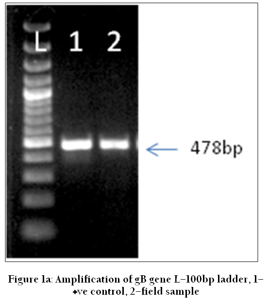
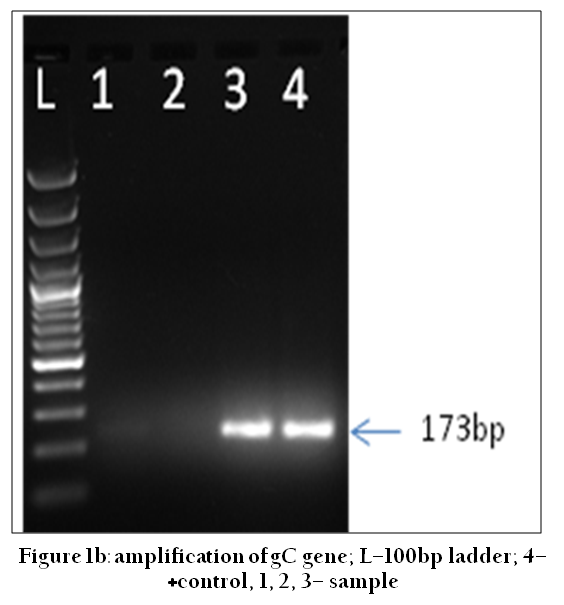
Glycoprotein gB gene specific PCR was carried out using published gB1/gB2 primer pair (Fuchs et al., 1999). This primer pair was designed from the published DNA sequence of glycoproteins gB gene of BHV–1.1 Cooper strain (accession no. M21474). The expected amplicon size is 478 base pairs (bp). The second PCR uses gC1/gC2 primer pairs designed from glycoprotein C (gC) gene sequences of the BHV–1 (EngelenburgEngelenburg et al., 1993) where the expected amplicon size is of 173bp.
Cycling Conditions for PCR
PCR was performed in a 25 µL volume containing 12.5 µL of Top Taq master mix (Qiagen), 1 µL of each primer (10 mM), 4 µL of extracted DNA and 6.5 µL of DNAse free water. The amplification was performed in a thermal cycler. The cycling conditions consisted of an initial denaturation step at 95oC for 5 minutes, followed by 35 cycles of 94oC for 1 minute, 62oC for 45 sec, 72oC for 45 sec and a final extension at 72oC for 10 minutes for gB gene specific PCR and gC specific PCR except the annealing conditions were 60oC for 45 sec in the latter case. Appropriate positive and negative controls were kept in the PCR. The negative control consisted of nuclease free water instead of DNA template while positive control consist of DNA extracted from the Mardin Darby Bovine Kidney (MDBK) cell line adapted reference strain of BHV–1. After amplification, 5 µL of the reaction mixture was electrophoratically analysed in a 2.0 % agarose gel, having ethidium bromide (0.5mg/mL). The amplified product was visualized as a band of expected size under UV light and documented by a gel documentation system (BioRad).
Real– Time PCR
Real–time PCR was performed using a pair of gB sequence–specific primers for amplification of target viral DNA and a 5’–nuclease oligo probe (TaqMan) for the detection of amplified products (Abril et al. 2004). The oligo probe was a single, sequence–specific oligonucleotide, labelled with two different flurophores, the reporter, 5–carboxyfluorescein (FAM) at the 5’end, and the acceptor/quencher 6–carboxytetramethylrhodamine (TAMRA) at the 3’end. This real–time PCR assay is expected to detect the viral DNA from all BHV–1 strains, including subtype 1 and 2, from bovine semen as well as from other clinical samples. Details of the primers and probes are given in Table 2.

Reaction Mixtures and Cycling Conditions for Real Time PCR
The PCR reaction mixtures were prepared in a clean laboratory room and in a dedicated qPCR work station to prevent contamination. All the reagents, except the test samples, were mixed before distribution to each individual reaction tube. Non templates control (NTC) was included in each test. The PCR amplifications were carried out in a volume of 25 μL containing : 2× PCR super mix (ABI) 12.5 μL, Forward primer (gB–F, 10μM) 1 μL, Reverse primer (gB–R, 10μM) 1 μL, Probe (5μM) and 7.5μL of nuclease free water. 2μL of the DNA isolated from cell culture supernatants /clinical blood/semen samples was added to the PCR reagent mixture to make final volume of 25 μL.
RESULTS AND DISCUSSION
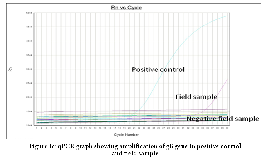
BHV–1 is one of the major infectious agents which can be transmitted between cattle and buffalo by natural service or through artificial insemination (AI). BoHV–1 establishes latent, life–lasting, and periodically reactivating infections in the host. It is the most harmful herpes virus property, which makes the use of semen from BoHV–1 seropositive bulls a major concern. In this study, partial sequence of gB and gC gene were successfully amplified in positive control having product of 478 bp and 173 bp respectively (Figure 1a and 1b). A total of 366 biological samples including 162 semen and 204 blood from buffalo bulls and cow bulls collected during July 2010 to December 2013 from different semen banks of Haryana were processed for identification of BoHV–1 using conventional and real–time PCR. No semen sample was found positive by gB and gC gene based on conventional or real–time PCR for the presence of BoHV–1 DNA (Figure 1a, 1b and 1c). However, out of 204 blood samples only one sample from Yamunanagar district was found positive for BoHV–1 (Table 1).
It revealed the quite low prevalence (0.4%) of disease among bull population of organized farm in the state. It was earlier reported that sero–prevalence of BoHV–1 in the neighboring state Punjab is 0 to 80% (Mweene, 2003). In four southern state of India, 523 semen samples were screened by virus isolation in two different cell lines (Bovine turbinate [BT] and MDBK). This study revealed that virus could only be isolated in four semen samples reflecting the low prevalence of BoHV–1 (Chandranaik, 2010). Although the method used by Chandranaik, (2010) was different to that of this study but the findings of both studies showing low prevalence of BoHV–1corroborated well. A recent study by Dehkordi (2013) in Iran using real–time PCR assay detected the presence of BoHV–1 DNA in 14.68%, 15.55%, and 12.65% of aborted bovine, buffalo and camel fetuses, respectively.
CONCLUSION
In the present study breeding bulls were tested for the presence of BoHV–1 infection by PCR, and qPCR. We conclude that none of the semen samples screened using PCR, yielded positive PCR/qPCR results indicating that the most of the farms of the state screened was free from BoHV–1 infection. However, one positive blood sample from Yamunanagar district is a cause for concern and preventive measures need to be taken to control its spread. The finding presumed worth as far as farm hygiene and management is concerned because BoHV–1 infection in farm is deadly and if established cannot be eliminated easily since virus persists in affected animals as latent infection. This study also showed the value of these PCR assays as a fast, easy and valuable diagnostic tool to diagnose and control the spread of BoHV–1 infection.
AUTHOR’S CONTRIBUTIONS
SM, NSM, AK, TN: conception and design of experiments, drafting the manuscript, provided reagents and facilities. Sunayna, AG, KB, AK: acquisition and analysis of data. All authors have read and approved the manuscript.
ACKNOWLEDGEMENTS
We are highly thankful to field veterinarians and other colleagues who have provided samples and helped directly and indirectly for these studies. We are also thankful to non–teaching staff of the department for their all help. This work was partly funded by Rastriya Krishi Vikas Yojna scheme no. 4011/C (g) ABT–4–0A.
COMPETING INTERESTS
The authors declared that they have no competing interests.
REFERENCES
Abril C, Engels M, Liman A, Hilbe M, Albini S, Franchini M, Suter M, Ackermann M (2004). Both viral and host factors contribute to neurovirulence of bovine herpesviruses 1 and 5 in interferon receptordeficient mice. J. Virol. 78, 3644–3653.
http://dx.doi.org/10.1128/JVI.78.7.3644-3653.2004
PMCid:PMC371052
Ackermann M, Wyler R (1984). The DNA of an IPV strain of bovid herpesvirus 1 in sacral ganglia during latency after intravaginal infection. Vet. Microbiol. 9: 53–63.
http://dx.doi.org/10.1016/0378-1135(84)90078-6
Afshar A, Eaglesome MD (1990). Viruses associated with bovine semen. Vet. Bull. 60:93–109.
Chandranaik BM, Shivaraj C, Kumar S, Renukaprasad C (2010). Isolation of BHV–1 from bovine semen and application of real–time PCR for diagnosis of IBR/IPV from clinical samples. Vet. Arhiv. 80, 467–475.
Dehkordi F, Safarpoor N, Haghighi H, Momtaz M, Rafsanjani S, Momeni M (2013). Conventional Vs Real–Time PCR for detection of bovine herpes virus type 1 in aborted bovine, buffalo and camel fetuses. Bulg. J. Vet. Medicine. 16 (2): 102−111
Engelenburg FAC, van Maes RK, Oirschot JT van, Rijsewijk FAM (1993) Development of a rapid and sensitive polymerase chain reaction assay for detection of bovine herpesvirus type 1 in bovine semen. J. Clin. Microbiol. 31 (12): 3129–3135.
PMid:8308103 PMCid:PMC266363
Gibbs EP, Rweyemamu MM (1977). Bovine herpesviruses, part 1. Vet. Bull. 47: 317–343.
Fuchs M, Hubert P, Detterer J, Rziha HJ (1999). Detection of bovine herpesvirus type 1 in blood from naturally infected cattle by using a sensitive PCR that discriminates between wild–type virus and virus lacking glycoprotein E. J. Clin. Microbiol. 37: 2498–2507.
PMid:10405392 PMCid:PMC85268
Mehrotra ML, Rajya BS, Kumar S (1976). Infectious bovine rhinotracheitis (IBR) keratoconjunctivitis in calves. Indian J. Vet.Pathol. 1: 70–73.
Mweene AS, Fukushi H, Pandey GS, Syakalima M, Simuunza M, Malamo M, Nambota A, Samui KL, Tsubota T, Nakazato Y, Onuma M, Yasuda J (2003) The prevalence of bovine herpesvirus–1 in traditional cattle in Southern Province, Zambia. Rev. Sci. Tech. Off. Int. Epiz., 22 (3): 873–877.
Van Engelenburg FA, Maes RK, van Oirschot JT and Rijsewijk FA (1993) Development of a rapid and sensitive polymerase chain reaction assay for detection of bovine herpesvirus type 1 in bovine semen. J. Clin. Microbiol. 31: 3129–3135.
PMid:8308103 PMCid:PMC266363
Vilcek S. (1993) Detection of bovine herpesvirus–1 BHV–1 genome by PCR. J. Virolog. Meth. 41: 245–247
http://dx.doi.org/10.1016/0166-0934(93)90132-B
Weiblen R, Kreutz LC, Canaborro TF, Schuch LF, Rebelatto MC (1992) Isolation of bovine herpesvirus 1 from preputial swabs and semen of bulls with balanoposthitis. J. Vet. Diagn. Invest. 4: 341–343.
http://dx.doi.org/10.1177/104063879200400321
PMid:1325195
Wiedmann M, Brandon R, Wagner P, Dubovi EJ, Batt CA (1993) Detection of bovine herpesvirus–1 in bovine semen by a nested PCR assay. J. Virol. Methods. 44: 129–139.
http://dx.doi.org/10.1016/0166-0934(93)90015-J


