Advances in Animal and Veterinary Sciences
Research Article
Advances in Animal and Veterinary Sciences2 (2S): 31 – 34Special Issue 2 (2014) (Advances in Diagnosis and Control of Infectious Diseases of Animals)
Molecular detection and phylogenetic analysis of sheeppox virus in Al – Hassa of Eastern Province of Saudi Arabia
Abdulkarem Al-Shabebi1, Ibrahim El-Sabagh2,3*, Eltayb Abu-Elzein1, Ahmed Zaghawa1, Abdelmohsen Al-Naeem1, Fahdel Housawi1
- Department of Clinical Studies, College of Veterinary Medicine and Animal Resources, King Faisal University, Al-Hufof, 31982, Saudi Arabia
- Central Biotechnology Laboratory, College of Veterinary Medicine and Animal Resources, King Faisal University, Al-Hufof, 31982, Saudi Arabia
- Department of Virology, Faculty of Veterinary Medicine, Cairo University, Giza, 12211, Egypt
*Corresponding author:ibrahimelsabagh@yahoo.com
ARTICLE CITATION:
Al- Shabebi AA, El-Sabagh IM, Abu-Elzein EM, Zaghawa AA, Al-Naeem AA, Housawi FM (2014). Molecular detection and phylogenetic analysis of Sheep pox virus in Al – Hassa of Eastern Province of Saudi Arabia. Adv. Anim. Vet. Sci. 2 (2S): 31 – 34.
Received: 2014–04–09, Revised: 2014–04–19, Accepted: 2014–04–20
The electronic version of this article is the complete one and can be found online at
(
http://dx.doi.org/10.14737/journal.aavs/2014/2.2s.31.34
)
which permits unrestricted use, distribution, and reproduction in any medium, provided the original work is properly cited
ABSTRACT
The current study was conducted to molecularly identify Sheep pox virus (SPPV) from recent outbreak affected sheep farms in Al-Hassa of Eastern Province of Saudi Arabia during the period between the winter and the spring, 2013. Thirty skin lesions of crusted scab samples were collected from six clinically infected SPPV sheep showing average morbidity and mortality rates of 80% and 15%, respectively. The collected samples were screened for presence of SPPV DNAs using KS-1.5/KS-1.6 and InS-1.1/InS-1.1/-based multiplex PCR. The P32 gene of selected two positive samples was sequenced and aligned with different SPPV, GTPV and LSDV available in GenBank. The phylogenetic analysis revealed that the Sheep pox virus strain; SPPV/Al-Hassa/2013/Saudi Arabia (accession number, KF204447) clustered on SPPV clade with SPPVs from India and China. This finding elucidated that the causative agent of this recent sheep pox outbreak is SPPV.
INTRODUCTION
Sheep pox virus (SPPV) is an enveloped and double-stranded DNA virus belongs to Genus Capripoxvirus of the Family Poxviridae. It is classified in this genus and family with goatpox virus (GTPV) and lumpy skin disease virus (LSDV) (Buller et al., 2005). SPPV is a notifiable disease, listed in group A diseases of the OIE and causes a severe and highly contagious disease (World Animal Health, 1996). The geographical distribution of SPPV includes Central and northern Africa, the Middle East, Turkey, Central Asia, India and China (OIE, 2010). The disease is characterized by pyrexia, rhinitis, conjunctivitis, excessive salivation, generalized multifocal necrotic lesions in the skin and internal organs including the lungs, liver and gastrointestinal tract and lymphadenopathy (Babiuk et al., 2008). The Sheep pox disease is associated with high morbidity and mortality rates especially in young animals. The mortality rate in young animals can exceed 50% but in naïve animals can reach 100% (Bhanuprakash et al., 2006).
Capripoxviruses (CaPVs) are mainly host-specific, means that SPPV only infected sheep while GTPV only infected goats, but some recent researchers recorded that some strains of SPPV and GTPV could infect both sheep and goats (Bhanuprakash et al., 2006; Bhanuprakash et al., 2010). Conventional serological assays could not distinguish SPPV, GTPV and LSDV due to the close antigenic and virulence relationship (Balinsky et al., 2008). Characterization of these viruses needs molecular techniques targeting CaPVs specific genes like P32, GPCR and RPO30 genes (Zhou et al., 2010; Yan et al., 2012).
Unfortunately, the knowledge on the current situation of Capripoxvirus infection of sheep and goats in Saudi Arabia is very scanty. The only published paper was carried out by Abu-Elzein et al., 2003, which recoded the first isolation of a virulent field Capripoxvirus from diseased goats. The current study described an outbreak of sheep pox disease in Al-Hassa of the Eastern Province of Saudi Arabia during 2013. The causative agent was molecularly identified as SPPV by KS-1.5/KS-1.6 and InS-1.1/InS-1.1/-based multiplex PCR and P32 gene-based sequencing and phylogenetic tree analysis.
MATERIALS AND METHODS
Clinical Specimens
Thirty skin samples of crusted scab lesions were collected from six different sheep unvaccinated farms in Al-Hassa Governorate in the Eastern Province of Saudi Arabia between January and March, 2013. Samples were transferred in sterile cups to the Central Biotechnology Laboratory at the College of Veterinary Medicine and Animal Resources, King Faisal University, Saudi Arabia and stored at -80ºC until used.
DNA Extraction
Total DNA was extracted from up to 25 mg tissue samples as well as commercial live attenuated Sheep pox virus as positive control using DNeasy Blood and Tissue Kit (QIAGEN, USA). After complete lysis of the specimens by ATL buffer and proteinase K, absolute ethanol was added, then the mixture was transferred to a spin column according to manufacturer`s protocol. Purified DNAs were recovered in 150 µl AE buffer and stored at -20ºC for further testing.
Oligonucleotides Primers
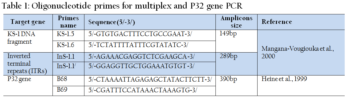
The primers used in PCR assays were KS-1.5/KS-1.6 and InS-1.1/InS-1.1/, which used in multiplex PCR for SPPV screening and B68/B69 primers that used for partial sequencing of P32 gene. These primers were analyzed by OligoAnalyzer 3.1 Integrated DNA Technologies, USA and synthesized by Metabion International AG, Germany. The complete data of primers are shown in Table 1.
Multiplex PCR for Detection of Sheep Pox Virus
The extracted DNAs were screened for presence of SPPV by multiplex PCR using HotStartTaq® Plus Master Mix Kit (QIAGEN, USA). 2 µl sample of each purified genomic DNAs were amplified in 20 µl of the final volume of a 2X HotStartTaq Plus Master Mix containing 1.5 mM MgCl2, 200 µM of each dNTP and 1 unit HotStartTaq Plus DNA polymerase and 10 µM of each KS-1.5/KS-1.6 and InS-1.1/InS-1.1/ primers. Thermocycling conditions were enzyme activation and initial denaturation at 95 ºC for 5 minutes followed by 35 cycles of 94 ºC for 30 second, 43 ºC for 30 second and 72 ºC for 30 second and a final extension step at 72 ºC for 10 minutes. The amplified PCR products were electrophoresed in 1.5% agarose gel stained with ethidium bromide and documented using ultraviolet gel documentation system (BIORAD).
Sequencing and Construction of Phylogenetic Tree
A 390 bp of P32 gene (envelope protein) of some detected SPPV were amplified using B68/ B69 primers in HotStartTaq® Plus Master Mix Kit (QIAGEN, USA). The PCR master mix and thermocycling conditions were similar to that in multiplex PCR except the annealing temperature was 48 ºC. The 390 bp PCR specific band was excised from the agarose gel, purified using Montàge DNA gel extraction kit (Millipore, USA) and sequenced in an automated ABI 3730 DNA sequencer (Applied Biosystems, USA). The obtained sequence was analyzed using online BLAST server and compared with capripoxviruses sequences available in GenBank (Table 2). A phylogenetic tree was constructed using MEGA version 5.20 software.

Genbank Accession Number
The obtained P32 gene sequence of one of the detected SPPV was submitted to the GenBank database with the accession number (KF204447), Sheep pox virus strain SPPV/Al-Hassa/2013/Saudi Arabia.
RESULTS AND DISCUSSION
Sheeppox Virus Outbreak 2013
Between January and March, 2013 a Pox-like disease was observed in different sheep farms in the Eastern Province of Saudi Arabia. The infected sheep suffered from pyrexia accompanied with cutaneous papules and vesicles especially in wool-less areas of skin (Figure. 1). The average morbidity rate was 80%, whereas the mortality rate was 15%. Post-mortem examination of dead sheep showed no nodules in the lungs.
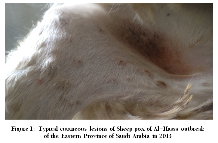
Figure 1: Typical cutaneous lesions of Sheep pox of Al-Hassa outbreak of the Eastern Province of Saudi Arabia in 2013

Figure 2: Agarose gel electrophoresis of multiplex PCR amplified products of sheep pox disease field samples. Lanes M, molecular weight markers; Lanes 1-11 field samples; Lane 12, Sheep pox virus vaccine strain served as positive control and Lane 13, water served as negative control
Multiplex PCR Testing of Suspicious Samples
Thirty papules/scabs suspected Sheep pox virus infected samples were positive for viral DNAs in multiplex PCR. Approximately 149 and 289 bp individual sharp bands were observed in gel electrophoresis for positive samples in comparison with vaccinal strain as positive control and non-template negative control (Figure 2).

Figure 3: Agarose gel electrophoresis of the PCR products of the P32 gene. Lane M, molecular weight marker; Lanes, 1 and 2 field samples; Lane 3, Sheep pox virus vaccine served as positive control and Lane 4, water served as negative control
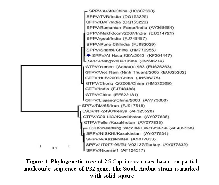
Figure 4: Phylogenetic tree of 26 Capripoxviruses based on partial nucleotide sequence of P32 gene. The Saudi Arabia strain is marked with solid square
Sequencing and Phylogenetic Tree Analysis
A 390 bp from P32 gene were successfully amplified from two selected positive Sheep pox virus field samples (Figure.3). For further confirmation of the identity of the detected viruses, the amplified products were sequenced and the entire sequence of P32 gene was analyzed. The results of sequence analysis revealed that the two amplicons were identical. One of these sequences designated Sheep poxvirus strain SPPV/Al-Hassa/2013/Saudi Arabia was submitted to GenBank under accession number of KF204447. BLAST program analysis showing 100% and 98%maximum homology of this nucleotide sequence of the detected SPPV with Sheep pox and goatpox P32 genes available in GenBank, respectively. Based on alignment and phylogenetic tree analysis of P32 gene, the SPPV field strain (SPPV/Al-Hassa/2013/Saudi Arabia) was clustered with different SPPV strains from China and India (Figure. 4).
CONCLUSION
This study records the first molecular detection, identification and epidemiological investigation of SPPV in Saudi Arabia. Based on sequence and phylogenetic analysis of P32 gene, this paper elucidated genetic relationship between identified Saudi Arabia SPPV with other viruses detected in China and India. These results provide new information on the epidemiology of sheep pox in Saudi Arabia and that further investigations on the epidemiology of SPPV are required.
ACKNOWLEDGMENTS
This study was funded by the College of Veterinary Medicine and Animal Resources, King Faisal University, Al-Hufof, Kingdom of Saudi Arabia.
CONFLICT OF INTEREST
The authors declare that they have no conflict of interest.
REFERENCES
Abu-Elzein E, Housawi F, Ramadan O, Gameel A, Al-Afaleq A, Al-Gundi, O (2003). Observations on natural and experimental infection of sheep and goats with a virulent field Capripoxvirus with high affinity to goats. Veterinarski Arhiv, 73(3), 119 - 131.
Babiuk S, Bowden TR, Boyle DB, Wallace DB, Kitching, RP (2008). Capripoxviruses: an emerging worldwide threat to sheep, goats and cattle. Transboundary and Emerging Diseases, 55(7):263 - 272.
http://dx.doi.org/10.1111/j.1865-1682.2008.01043.x
PMid:18774991
Balinsky CA, Delhon G, Smoliga G, Prarat M, French RA, Geary SJ, Rock DL, Rodriguez LL (2008). Rapid preclinical detection of Sheep pox virus by a real-time PCR assay. J Clin Microbiol, 5, 438 – 442.
http://dx.doi.org/10.1128/JCM.01953-07
PMid:18032617 PMCid:PMC2238129
Bhanuprakash V, Indrani BK, Hosamani M, Singh RK (2006). The current status of sheep pox disease. Comp Immunol Microbiol Infect Dis, 29(1):27 - 60.
http://dx.doi.org/10.1016/j.cimid.2005.12.001
PMid:16458357
Bhanuprakash V, Venkatesan G, Balamurugan V, Hosamani M, Yogisharadhya R, Chauhan RS, Pande A, Mondal, B Singh RK (2010). Pox outbreaks in sheep and goats at Makhdoom (Uttar Pradesh). India: evidence of Sheep pox virus infection in goats. Transboundary and Emerging Diseases, 57, 375 – 382.
http://dx.doi.org/10.1111/j.1865-1682.2010.01158.x
PMid:20673232
Buller RM, Arif BM, Black DN, Dumbell KR, Esposito JJ, Lefkowitz EJ, McFadden G, Moss B, Mercer AA, Moyer RW, Skinner MA, Tripathy, DN (2005). Family Poxviridae. in: C. M. Fauquet, M. A. Mayo, J. Maniloff, U. Desselberger and Ball, LA (Eds) Virus Taxonomy: Eighth Report of the International Committee on Taxonomy of Viruses, San Diego: Elsevier Academic Press. pp. 117 – 133.
Heine HG, Stevens MP, Foord AJ, Boyle DB (1999). A Capripoxvirus detection PCR and antibody ELISA based on the major antigen P32, the homolog of the vaccinia virus H3L gene. J. Immunol. Methods, 227, 187 – 196.
http://dx.doi.org/10.1016/S0022-1759(99)00072-1
Mangana-Vougiouka O, Markoulatos P, Koptopoulos G, Nomikou K, Bakandritsos N, Papadopoulos O (2000). Sheep poxvirus identification from clinical specimens by PCR, cell culture, immunofluorescence and agar gel immunoprecipitation assay. Mol Cell Probe, 14(5), 305 – 310.
http://dx.doi.org/10.1006/mcpr.2000.0319
PMid:11040094
OIE (2010). Sheep Pox and Goat Pox. Manual of Diagnostic Tests and Vaccines for Terrestrial Animals. Chapter 2.7.14.1 - 12, Online:http://www.oie.int./fileadmin/Home/eng/Health_standards/tahm/2.07.14_S_Pox_G_ROX.Pdf.
World Animal Health (1996-1997). In: Reports on the animal health status and disease control methods and list A disease outbreaks. Statistics OIE, Paris, France, P.4.
Yan X-M, Chu Y-F, Wu G-H, Zhao Z-X, Li J, Zhu H-X, Zhang Q (2012). An outbreak of sheep pox associated with goat poxvirus in Gansu Province of China. Vet Microbiol, 156, 425 - 428.
http://dx.doi.org/10.1016/j.vetmic.2011.11.015
PMid:22169434
Zhou T, Jia H, Chen G, He X, Fang Y, Wang X, Guan Q, Zeng S, Cui Q, Jing Z (2012). Phylogenetic analysis of Chinese Sheep pox and goatpox virus isolates. Virol J, 9 - 25.


