Advances in Animal and Veterinary Sciences
Review Article
Advances in Animal and Veterinary Sciences 2 (4S): 24 – 32Special Issue-4 (2014) (Reviews on Frontiers in Animal and Veterinary Sciences)
Emerging Food–Borne Parasitic Zoonoses: a bird’s eye view
Alok Kumar Singh1, Amit Kumar Verma2, Amit Kumar Jaiswal1, Vikrant Sudan1, Kuldeep Dhama3
- Department of Veterinary Parasitology, Uttar Pradesh Pandit Deen Dayal Upadhayaya Pashu Chikitsa Vigyan Vishwavidyalay Evum Go-Anusandhan Sansthan (DUVASU), Mathura, India
- Department of Veterinary Epidemiology and Preventive Medicine, Uttar Pradesh Pandit Deen Dayal Upadhayaya Pashu Chikitsa Vigyan Vishwavidyalay Evum Go-Anusandhan Sansthan (DUVASU), Mathura, India
- Division of Pathology, Indian Veterinary Research Institute (IVRI), Izatnagar, Bareilly (U.P.) – 243122, India
*Corresponding author:dr_amitverma@sify.com
ARTICLE CITATION:
Singh AK, Verma AK, Jaiswal AK, Sudan V, Dhama K (2014). Emerging food-borne parasitic zoonoses: a bird’s eye view. Adv. Anim. Vet. Sci. 2 (4S): 24 – 32.
Received: 2014–04–16, Revised: 2014–04–23, Accepted: 2014–04–23
The electronic version of this article is the complete one and can be found online at
(
http://dx.doi.org/10.14737/journal.aavs/2014/2.4s.24.32
)
which permits unrestricted use, distribution, and reproduction in any medium, provided the original work is properly cited
ABSTRACT
Zoonotic transmission of parasitism is an underreported and under recognized, worldwide distributed entity. Humans acquire these infections either through food, water, soil or close contact with animals. Mostly parasitic zoonoses are those of neglected diseases but with more demand of the food supply, increased travelling and increased ratio of highly susceptible persons coupled with changes in culinary practices with simultaneous improvement in diagnostic tools as well as communication facilities. These conditions are emerging at an alarming rate. Global sourcing of food, coupled with changing consumer vogues, including the consumption of raw vegetables and undercooking to retain the natural taste and preserve heat-labile nutrients, can increase the risk of foodborne transmission. The increasing demand for raw or under-cooked food is also considered as one of the major reasons causing food borne infections, especially waterborne parasitic diseases, in the last decade. The present review will discuss the factor responsible for transmission and occurrence of zoonotic diseases along with different helminthes and protozoan parasites that are considered to be as important food borne zoonoses. A greater awareness of parasite contamination of our environment and its impact on health has precipitated the development of better detection methods. Overall, there is an urgent need for better monitoring and control of food-borne parasites using new technologies. Robust, efficient detection, viability and typing methods are required to assess risks and to further epidemiological understanding. This paper reviews the most important emerging food-borne parasites, with emphasis on transmission routes.
INTRODUCTION
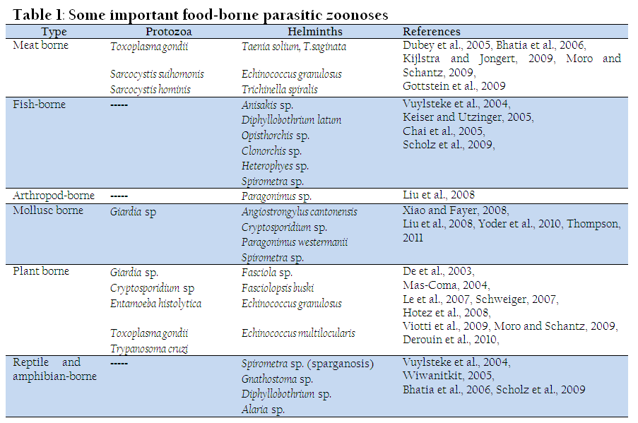
Food-borne parasitic zoonoses include both helminthic and protozoan infections. Amongst one thousand five hundred known infectious agents for human being, 66 are protozoa and 287 are helminths (Chomel, 2008; Taylor et al., 2001) and amongst them majority (60.3%) of the emerging infectious diseases are zoonotic (Jones et al., 2008). Zoonoses represent a large burden of disease and there are changing patterns of disease burdens with disease emergence (Dhama et al., 2013a, 2013b). Unlike bacteria and viruses, parasites generally have unique features which often make them remarkably suited for survival in the environment and dissemination by water. Many of these parasites are difficult to detect and control. Some have the potential to cause severe public health problems, while others are capable of inflicting considerable losses in livestock. The resulting production and economic losses can be extensive. Human population growth and socioeconomic changes have resulted in the migration of populations into new ecological niches and changes in animal husbandry practices have tremendously impacted on disease emergence and disease burden (Macpherson, 2005). In addition, improved diagnostics are demonstrating that many zoonoses have a higher burden then previously recognized ones. Some new syndromes are also being attributed to parasitic zoonoses and hence add to the disease burden. Environmental changes and ecological disturbances, due to both natural phenomena and human intervention, have exerted and can be expected to continue to exert a marked influence on the emergence and proliferation of zoonotic parasitic diseases. Global warming may change the transmission dynamics of parasitic zoonoses in endemic areas and enable some parasites to transmit in regions where they were previously absent. A range of parasites are well adapted and have coevolved with their hosts so to persist in relationships which may be sub-clinical or even mutualistic in their nature: this would guarantee the survival both of the host and of the parasite populations. This may be true if a population is constantly exposed to a parasite, where young individuals acquire tolerance gradually by, for example, consumption of the “local” contaminated food. Such balance between a foodborne parasitic disease and a host population was usually confined to specific environments and host populations or geographic areas. Nowadays, due to the increased globalization and movement of people and food commodities, this geographical segregation is not necessarily evident any more. Food and waterborne infections have received considerable attention in the last decade and some of them are considered as emerging diseases (WHO, 2002; Singh et al., 2014). Although humans may serve as host of around 300 species of parasitic worms and over 70 species of protozoa (Cox, 2002), only around 100 species are known to be food borne (Orlandi et al., 2002). Human and society behavior plays a fundamental role in the epidemiology, emergence and spread of parasitic zoonoses. Host-parasite relationships are intricately linked; each component is important and may determine the dynamics and outcome of disease transmission and control (Azim et al., 2008). During the last decades there have been changes in food preferences and eating habits; there is a growing market for more ready-to-eat fresh and healthy food, as well as novel, ethnic food products, which have created new situations where pathogens may be introduced into food and then to populations. The present review will discuss the factor responsible for transmission and occurrence of zoonotic diseases along with different helminthes and protozoan parasites that are considered to be as important food borne zoonoses.
FACTORS RESPONSIBLE FOR TRANSMISSION AND OCCURRENCE OF PARASITIC ZOONOSES
Lack of personal hygiene viz., improper or non-washing of hands before meal and after defecation, long nails fingers.
Consumption of raw or under cooked food.
Defecation in open area near water bodies
Lack of education, poor living standards and poverty
Unavailability of clean potable water
Environmental factors facilitating the survival of parasites and their development stages
PARASITES TRANSMITTED BY CONTAMINATED FOOD AND WATER
Water is a one of the major sources for parasites and their different environmental stages, which contaminate the food stuffs viz., fruits and vegetables (Mahima et al., 2013; Rahal et al., 2014). It is difficult to associate an outbreak with a particular food item, and therefore, if the food-borne route is suspected, it means how, identify the food implicated became contaminated (Slifko et al., 2000). Due to these difficulties, the acquisition of parasitic infections via the food-borne route is almost certainly poorly detected by a factor 10 or more (Casemore, 1990). Of the emerging waterborne parasitic infections that may be acquired by food are Cyclospora cayetanensis, Cryptosporidium spp., Fasciola spp. and Fasciolopsis spp.
CYCLOSPORA SP.
Cyclospora cayetanensis is a coccidian parasite that can infect small intestine and clinically leads to watery diarrhoea ranging 4-8 stools per day, nausea and vomition in man. In Unites States and Canada, approximately 1500 cases were reported by consuming raspberries from Guatemala (Herwaldt and Ackers, 1997; CDC, 1997). Cyclospora organisms have also been isolated from patients suffering with AIDS and chronic diarrhoea (Long et al., 1990; Ortega et al., 1993; Pape et al., 1994; Cox, 2002; Orlandi et al., 2002; Vuong et al., 2007).
CRYPTOSPORIDIUM SP.
Cryptosporidium is also major cause of diarrhoeic disorders in man all over the world. Water-borne, food-borne and infected animals may act as the major routes of transmission of this zoonotic disease (Slifko et al., 2000; Smith et al., 2007) especially in patients that were immune-suppressed, suffereing with HIV, chronic diseases and sometimes infection may become fatal too. In immune-competent patients, cryptosporidiosis is usually a self-limiting disease. Till there are 16 species of Cryptosporidium species and 33 genotypes, but only few of them are zoonotic (Xiao and Fayer, 2008). Cryptosporidium parvum is the major zoonotic species, whose reservoir is calves. Water-borne outbreaks and infections occur through handling of infected animals, contact with children and through contaminated recreational waters. Oocysts may also contaminate soft fruits, salad, vegetables, shell fish viz., oysters and mussels (Anh et al., 2007; Schets et al., 2007). In an epidemiological survey in UK, about 38.5% of human cryptosporidiosis cases were due to C. parvum out of these 25% is through direct contact with cattle, while in France 54% of cryptosporidiosis were reported due to C. parvum (Derouin et al., 2010). These zoonotic Cryptosporidium cases were more in high income countries as compared to low income countries. Children and HIV patients have a higher prevalence of Crytosporidium especially C. meleagridis, C. canis, C. felis, and C. muris. But most human infections reported with C. canis from low income countries. Moreover, other interesting feature is that most human infections are due to the anthroponotic of C. parvum. Thus, where C. parvum does occur in such countries it is more likely to be of anthroponotic rather than zoonotic in origin (Xiao and Feng, 2008). The burden of disease in high income countries due to zoonotic cryptosporidiosis is low. Although there are a small number of fatalities (Scallan et al., 2011), the disease generally has an acute, non fatal and outcome with few long term sequaelae. In contrast there are reports from lower income countries of significant fatality rates in young children and long term growth retardation has been observed (Chalmers and Davies, 2010).
GIARDIA SP.
Giardia is worldwide in distribution and considered as most common intestinal protozoan parasites of man and animals (Thompson, 2011). However, its zoonotic transmission is uncertain and known to affect at least 40 species of vertebrates, including humans. Due to host specificity of G. duodenalis, its role in zoonoses is controversial. Only two assemblages (A and B) appears to be zoonotic (Monis et al., 2009). The clinical signs of giardiosis in man vary greatly (Thompson, 2011) and some of them are acute short-lasting diarrhoea or chronic syndromes associated with nutritional disorders, malabsorption of fat and weight loss. A chronic syndrome is more common and supposed to cause more harm. In low and middle income countries about 200 million persons have symptomatic giardiosis and new cases are added at the rate of 500,000 cases per year (Savioli et al., 2006). Various studies supported that direct transmission is more important than water-borne, food-borne or zoonotic transmission (Hunter and Thompson, 2005). Livestock such as cattle are frequently infected with giardiosis, but molecular evidence suggests its zoonotic transmission useless. However, molecular studies support the zoonotic transmission of giardia from dog (Leonhard et al., 2007).
ENTAMOEBA SP.
Entamoeba histolytica is of highest clinical importance and considered as the only amoeba parasitic in the intestine of human beings (Schuster and Visvesvara, 2004). Dogs are considered to be an important source of infections to human beings (Barr, 1998). E. histolytica is also found in non-human primates (Schuster and Visvesvara, 2004). Other potentially zoonotic but nonpathogenic and commensals in nature of Entamoeba are E. colii, E. polecki and E. hartmanni (Meloni et al., 1993; Stensvold et al., 2011). Of these, E. colii is one of the most commonly reported human protozoa (Chunge et al., 1991; Youn, 2009; Boeke et al., 2010). It has also been recovered dogs, nonhuman primates (Howells et al., 2011), and marsupials (Campos-Filho et al., 2008; Youn, 2009). Previous report suggested that E. polecki to be zoonotic but recent molecular diagnosis suggested that this species is restricted to humans only whereas E. hartmanni is potentially zoonotic and reported in non-human primates (Stensvold et al., 2011).
FASCIOLA SPP.
Fasciolosis is caused by two species of liver fluke i.e. Fasciola hepatica and Fasciola gigantica. Among these, F. hepatica is cosmopolitan in distribution and has the capacity to infect variety of host species. Its intermediate host (snail) is adapted to a wide range of ecological niches (Bhatia et al., 2006). F. gigantica has a more restricted distribution because of reduced ability of the aquatic snail to invade new niches and is generally found in tropical regions of Africa, South and East Asia and the Middle East continents. Man is always acquiring the infection with local affected animals, although the distribution of infection in man and level of prevalence may not always correlate with that observed in animals (Mas-Coma, 2004; Mas-Coma et al., 1999a). Infection is usually acquired by the ingestion of various freshwater aquatic plants, such as watercress, on which the metacercariae have settled. Farm management practices and the culturing of edible aquatic plants in greenhouses has limited the extent of human infection in industrialized regions, but in some developing countries wild aquatic plants or plants grown in fields where infected animals can easily roam form a regular part of the daily diet. Metacercariae may also associate with floating in water and, therefore, infection can be acquired through drinking water. The importance of human fasciolosis has recently been recognized. Earlier in 1992 total cases of human fasciolosis was estimated to be less than 3000, but recently this data have reached to 2.4 million (Rim et al., 1994; Curtale et al., 2005) and 17 million (Hopkins, 1992). Clinically disease occurs due to migration of the young or immature flukes through the liver parenchyma leading to abdominal discomfort, diarrhoea, weight loss and malaise. Sometimes, erratic migrations of Fasciola may also occur (Le et al., 2007). Fasciolosis is emerging as a major plantborne zoonotic disease in high altitude Andean rural regions of Bolivia and Peru (Mas-Coma et al., 1999b), northern provinces of Iran bordering Caspian sea (Rokni et al., 2002; Moghaddam et al., 2004), Nile Delta region of Egypt (Esteban et al., 2003) and Central provinces of Vietnam (De et al., 2003).
FASCIOLOPSIS SPP
Fasciolopsis buski is commonly known as ‘giant intestinal fluke’ and present in small intestine of man and pigs, transmitted by consumption or handling of metacercariae infected aquatic plants viz., water chestnut, water hyacinth and water morning glory etc or by drinking or using untreated water (Bhatia, 2003). Man may be infected when peeling off the hull or skin of infected plants using their teeth. Pigs are the only prevalent reservoir, although they harbor few flukes. The infection occurs focally and linked to freshwater habitats with stagnant or slow moving waters, and is associated with common social and agricultural practices and promiscuous defecation. The disease may lead to erosions of intestine, abdominal pain, yellow offensive stools, ulceration, haemorrhage, abscesses and catarrhal inflammation (Mas-Coma et al., 2005).
PARASITES TRANSMITTED BY FAECAL CONTAMINATION OF FOOD AND DRINKS
Infective stages of some parasites come out through faeces and contaminate foodstuffs such as vegetables, fruits of fruit juices and edible raw materials. Human infection with Toxoplasma gondii, a cosmopolitan zoonotic protozoan parasite may be congenital or acquired (Flegr et al., 2002, 2009). Infection can be acquired either through ingestion of raw or under-cooked meat containing tissue cyst or ingestion of sporulated oocysts along with food or water. Several studies have found that Echinococcosis increases public health concern (Moro and Schantz, 2009). Dogs are increasingly recognized as important animal reservoirs for Trypanosoma cruzi (Hotez et al., 2008) and transmitted through oral infection via fruit juices, contaminated with faeces from infected bugs (Rodriguez-Morales, 2008).
TRYPANOSOMA CRUZI
It causes chagas disease in man, affecting young ones and neonates. Disease is prevalent in South America, United State of America and Canada and transmitted by blood-sucking Reduvid bugs living in cracks, crevices and roofs of poor quality dwellings in rural and unhygienic areas. Disease may also be transmitted by blood transfusion, organ transplantation and congenital infection. Various animals may be infected and serve as reservoirs like dogs, cats, pigs, foxes, ferrets, squirrels, opossum and monkey (Hotez et al., 2008). Recent outbreaks in Brazil and Venezuela suggested its transmission through consumption of fruit juices contaminated with faeces of infected bugs. These outbreaks are of significant importance in periurban areas of non-endemic regions (Rodriguez-Morales, 2008). The chronic Chagas disease is the usual form seen in adults and clinical manifestations depend on the location of the organisms but cardiac form is more common.
ECHINOCOCCUS
Echinococcosis in humans occurs as a result of infection by accidental ingestion of contaminated food and water with eggs of the taeniid cestodes of the genus Echinococcus. These eggs may contaminate raw vegetables and fruits resulting in a food-borne infection. Four species viz., Echinococcus granulosus (causes cystic echinococcosis), Echinococcus multilocularis (causes alveolar echinococcosis), Echinococcus oligarthus and Echinococcus vogeli (causes polycystic echinococcosis) have been considered as important from public health point of view (Moro and Schantz, 2009). Humans are accidental intermediate and dead end host. Liver is the most prevalent site for the hydatid cyst development, followed by lungs or any other part of the body of ungulates such as spleen, kidneys, heart, bone and central nervous system. Cysts may reach upto 10cm in diameter per year, and may take many years before showing any clinical signs. If a cysts ruptures, the sudden release of its contents causes serious allergic or immunological reactions from mild to fatal anaphylaxis (Das et al., 2003, Moro and Schantz, 2009). Control of stray dog populations, treatment of dogs with anti-cestodal drugs, prohibition of home slaughter of sheep and correct slaughter practices and disposal of carcass, supported by health education programmes, are some of effective ways to break its life cycle.
MEAT-BORNE PARASITE INFECTIONS
Meatborne parasitic zoonoses are important cause of illness and economic loss, globally (Roberts et al., 1994, Murrell, 1995, Gamble et al., 1998). Among the meat-borne parasitic infections Toxoplasma gondii, Taenia spp., Sarcocystis spp., and Trichinella spp are of significant socio-economic importance. Man get infection by eating raw or undercooked meat infected with cyst of these parasites. Meat inspection for cysticercosis has very low sensitivity, so that resulting in a high number of infected carcasses entering the food chain. However, in developing countries a large proportion of the carcass escapes meat inspection because it is not a routine practice or the animals are not slaughtered in abattoirs. For sarcocystosis and toxoplasmosis no any specific meat inspection is done. Cooking is effective criteria for killing the parasites if the appropriate temperature is reached in the core of the meat product. Freezing, drying, smoking and curing are other effective process to reduce the risk of infection by consuming contaminated meat, except for some species of Trichinella, which show resistance upto some extent. Recently, all Trichinella infections occurring in animals and humans were associated to Trichinella spiralis. Presently, 8 species and 4 genotypes within two clades i.e encapsulated and nonencapsulated, are recognized (Pozio and Murrell, 2006; Zarlenga et al., 2006; Krivokapich et al., 2008).
TOXOPLASMA GONDII
Toxoplasmosis is a protozoan parasite caused by T. gondii is still a neglected and underreported cases, despite having a disease similar to that of salmonellosis and campylobacteriosis (Kijlstra and Jongert, 2009). Disease is transmitted to man by accidental ingestion of sporulated oocysts shed with the faeces by cat and other felids, or by eating raw or undercooked meat contaminated with tissue cysts. In Western and Asian countries the consumption of undercooked meat is a prominent source of infections (Cook et al., 2000; Fallah et al., 2008; Han et al., 2008) with significant public health impact (Kijlstra and Jongert, 2009). However, the eating of uncleaned raw vegetables or fruits is an important risk factor in various studies (Antoniou et al., 2007; Cavalcante et al., 2006; Fallah et al., 2008; Liu et al., 2009). It is a serious health problem in pregnant women leading to abortions. Encephalitis is the main clinical symptom in immuno-compromised patient. Clinical manifestations are more common in boys than girls and in young woman than in young man possibly due to hormonal reasons and condition is accompanied by myalgia which may proceed to myositis and headache may develop in encephalitis (Dubey and Beattie, 1988). The proper heating and freezing meat are the most efficient method to kill T. gondii tissue cysts. However, interventions to prevent mixing of infected meat into the food chain would be technically feasible in countries where the meat chain is well organized. Monitoring of farms and farm management can play an important role in the control of Toxoplasma infection (Kijlstra and Jongert, 2009).
TAENIA SPP.
There are two most common tapeworms in human beings i.e. T. saginata and T. solium. The terms cysticercosis refer to food-borne zoonotic infections with larvae and adult tapeworms, respectively. Their life cycles depend on the association between humans and cattle (Taenia saginata) or pigs (Taenia solium) (Flisser et al., 2005). Humans acquire infection by eating raw or undercooked meat containing cysticerci. Among these tapeworms, Taenia solium is unique because the cysticercus stage can also infect humans. Cysticerci may lodge in the brain and cause neurocysticercosis (Garcia et al., 2003). Other signs are headache, dimness of vision, vomition, and cysticercus present in anterior chamber of eye or outer part of the eyelid, convulsions, acute supporative dacryoedinitis and cystic nodules on neck. Human cysticercosis is not acquired by eating meat but through accidental ingestion of food or drinks contaminated with Taenia eggs through infected hands. Infection of Taenia solium is eradicated in most developed countries, mainly due to general socio-economic development and intensification of pig husbandry systems, but it is still present in tropical countries where pigs are rared for food purpose. It is associated with poverty and unhygienic condition allowing pigs to have access to human faeces. Increased immigration and travelling are major cause for spreading this parasitic infection. Estimation about millions of persons worldwide is infected with Taenia solium, mainly in Latin America, Sub-Saharan Africa and South and South-East Asia. In Africa, the rapid expansion of smallholder pig production has led to a significant increase of cysticercosis in pigs and humans (Phiri et al., 2003). Control measure based on public health education, improvement of sanitation, pig husbandry systems, meat inspection and mass treatment of humans, while conventional method include single dose treatment with oxfendazole in pigs and vaccination of pigs with recombinant antigens (Gonzales et al., 1996; Lightowlers et al., 2000). The beef tapeworm is found in developed as well as in developing countries. Cysticerci easily destroy with high temperatures, dietary habits and culinary practices which readily affect the transmission. Taeniosis is more common where consumption of raw or undercooked beef (Murrell, 2005). Intestinal taeniosis may be associated with abdominal discomfort, irritation of intestinal mucosa, obstruction, nausea, weight loss and anal pruritis (Jongwutiwes et al., 2004). However, bovine cysticercosis may lead to serious economic losses to the dairy industry due to condemnation, poor storage, refrigeration and downgrading of infected carcasses (Yoder et al., 1994; Giesecke, 1997).
SARCOCYSTIS SP
Sarcocystis involves pig-man cycle (Sarcocystis suihominis) and cattle-man cycle (Sarcocystis hominis), in which man act as definitive host. The sporulated oocysts are passed out through faeces and intermediate host (pig) gets the infection through ingestion of contaminated food and water. Sarcocyst suihominis is worldwide in distribution but mainly occurs in Egypt, Sudan, West Africa, South America and Mediterranean region. The life cycle in pigs includes two generations of schizogony and mature microsarcocysts. In man parasite causes digestive disturbances, nausea, abdominal pain and diarrhea within 6-8 days of consumption of raw or under-cooked pork (Banerjee et al., 1994). Sarcocystis hominis found throughout the world, but especially found in Australia, Brazil, Germany, New Zealand and India. Cattle act as intermediate host in which microsarcocysts settle in striated muscles by 98 days after the infection. In man, there are symptoms of diarrhea, vomition and respiratory distress (Jain and Shah, 1987).
TRICHINELLA SPP.
This is an only example of auto-heteroxenous nematodes parasites and one of the most widespread zoonotic pathogens in the world. Infection by Trichinella spp. has been detected in domestic as well as wild animals of all continents, with the exception of Antarctica (Pozio and Murrell, 2006). Trichinellosis or trichinosis in man occurs by the ingestion of Trichinella larvae encysted in muscular tissue of domestic or wild animal. The domestic pig play important source of human infection worldwide. However, meats of wild boars and horses have played a significant role during outbreaks within the past decades (Gottstein et al., 2009). The occurrence of trichinosis in humans is strictly related to cultural food practices, including the consumption of raw or undercooked meat. Globally, average yearly incidence of the disease in humans is around 10,000 cases with a mortality rate of about 0.2%; however, the number of infections is underreported in many nations due to the lack of appropriate diagnosis and knowledge (Pozio, 2007). Clinically the disease in man is characterized by an intestinal phase followed by a muscular phase, which is associated with heavy muscle pains, myocarditis, encephalitis, fever, eosinophilia and calcium may be deposited in the muscle fibres and eventually the larvae die. Trichinosis is not only an economic problem but also a public health hazard in porcine animal production and food safety. With the zoonotic importance of infection, the main strategies of many countries have focused on the control of Trichinella or the elimination of Trichinella from the food chain (Gottstein et al., 2009).
PARASITES TRANSMITTED BY FISH, REPTILES, AMPHIBIAN, CRUSTACEANS AND SNAILS
The recognition of the public health significance linked to poverty, cultural traditions, intensification of agriculture, environmental degradation, and lack of tools for control is increasing (World Health Organization, 1995, 2004). Meat of reptiles, amphibians and fish can also be infected with a wide range of parasites viz., trematodes (Opisthorchis spp., Clonorchis sinensis), cestodes (Diphyllobothrium spp., Spirometra), nematodes (Gnathostoma, spp., Anisakidae), and pentastomids, causing zoonotic infections, when consumed raw or poorly cooked. Freezing, cooking and salting when properly applied may reduce the transmission of disease (Abollo et al., 2001). However, smoking or pickling may not always be effective to eliminate theie infective larvae (Toro et al., 2004). Diphyllobothriosis, is contracted by consuming raw or undercooked fish (Dupouy-Camet and Peduzzi, 2004, Chai et al., 2005), and most common species infecting humans (Scholz et al., 2009). Traditionally, it is more common in Asia because of the particular food practices and the importance of aquaculture (Keiser and Utzinger, 2005). Moreover, some of these parasites may emerges in other continents through aquaculture, improved transportation and distribution systems to bring aquatic foods for local and international markets, increased tourism and changing culinary habits (Keiser and Utzinger, 2005).
CESTODES
Sparganosis is a zoonotic disease, caused by larval stage of the genus Spirometra of dogs and cats. The disease is reported in many countries but most common in eastern Asia. Migrations of parasite larvae occur in the eye, subcutaneous tissues, the central nervous system or other organs. Humans also get infections through consumption of contaminated water, frog, snake or meat infected with copepods (Wiwanitkit, 2005). Another parasite Diphyllobothrium latum (broad fish tapeworm) is the largest tapeworm of fish eating mammals and transmitted to man by consuming raw or undercooked fish (Dupouy-Camet and Peduzzi, 2004). These parasites are found in Europe, Russia, Japan, Korea, France, Italy, Ireland, Finland, Switzerland, North and South America and Asia. About 20 million people are infected worldwide (Chai et al., 2005). Prolonged infection causes macrocytic hypocromic anaemia or pernicious anaemia resulting from a competition between parasite and host for vitamin B12 that makes vitamin B12 unavailable to the host (Vuylsteke et al., 2004).
NEMATODES
Angiostrongylus cantonensis is a zoonotic parasite, causes eosinophilic meningitis in humans after accidental ingestion of raw or under-cooked snails or fresh water crustaceans which act as intermediate host containing larvae in their slime or contaminated vegetables (Sharma et al., 1981). Rats are the natural hosts. The larvae migrate to the meninges of brain and may be found in spinal cord causing acute inflammatory reaction, mild meningeal irritation, paresthesia and cranial nerve abnormalities. With the rise in income, living standards; and dependency of exotic and delicate soft foods, populations around the world particularly in Asia have observed Angiostrongyliasis as food-borne parasitic zoonosis (Alicata, 1991; Zhang et al., 2008). The consumption of either raw or undercooked sea-fish may lead to infection with several nematodes belonging to the family Anisakidae. The nematodes commonly involved in human infections are Anisakis simplex, Anisakis physeteris and Contracaecum osculatum (Audicana and Kennedy, 2008). Humans may be accidental hosts by eating raw or undercooked fish that contains the third-stage larvae of Anisakis simplex. After ingestion larvae penetrate the digestive tract and produce a disease, known as Anisakiasis (herring worm disease). The life cycle involves larval stages with several intermediate, paratenic hosts and adult stage, during which the worm may, parasitizes the stomach of marine mammals such as seals and dolphins. Generally symptom appears within a few hours after the ingestion of a living worm, it causes an acute and transient infection, lead to abdominal pain, nausea, vomition, slight irritation of bowel and diarrhoea. Some patients may exhibiting clinical manifestations of allergy and infection after eating living parasites simultaneously (Audicana and Kennedy, 2008). The Anisakis simplex allergens are highly resistant to heat and freezing while, cooking is expected to kill the parasites it may not result in loss of their allergenicity and sensitisation (Audicana et al., 2002). Anisakis simplex may be the cause of chronic and relapsing acute urticaria (Falcao et al., 2008). Different methods for preparation of fish such as salting, curing, pickling and smoking, which are generally sterilize to other food-borne but not sufficient for Anisakids (Audicana and Kennedy, 2008). Preventive measure include the evisceration of fish as soon as possible after catching, freezing (-200 for 60 hrs), public health education and proper cooking. Gnathostoma spinigerum has been reported from Thialand, India, Japan and other Far East countries. Human get infected from contact with meat of infected intermediate host such as fish, amphibians and birds. The larvae in human do not reach maturity and frequently migrate under the skin, mucous membrane, eyes and brain where they cause eosiniphilic meningitis (Bhatia et al., 2006).
TREMATODES
The liver flukes such as Clonorchis sinensis and Opisthorchis spp. are considered as emerging public health problem (Keiser and Utzinger, 2005) and according to an estimate about 600 million and 80 million people are at risk for infection with Clonorchis sinensis and Opisthorchis spp, respectively. The Oriental or Chinese liver fluke is of major socioeconomic importance in Far-East countries particularly China, Japan, Korea and Taiwan. The parasite is transmitted via snails that act as first intermediate host to freshwater fish (second intermediate host), and then to human beings and other piscivorous mammals. Heavy infections in man causes diarrhea, abdominal pain, icterus, portal hypertension, ascites, gastrointestinal bleeding, formation of gallstones, inflammation and hyperplasia of the biliary epithelium leading to deposition of fibrous tissue and invasion of the pancreatic and bile duct (Lun et al., 2005). Opisthorchis viverrini and Clonorchis sinensis are associated with cholangiocarcinoma and adenocarcinoma, originating from hyperplastic activity of epithelial layer of the bile duct. Another important zoonotic food-borne trematode is the lung fluke (Paragonimus spp.) (Liu et al., 2008). Paragonimus westermani is of major socioeconomic importance in Asia such as china, Japan, Philippines, Korea and other Far-East countries including Thialand. Incidence has also been found in some African countries, such as Nigeria, Cameroun and Liberia and in South America. The parasite is transmitted via aquatic or amphibious snails to freshwater crab or crayfish act as second intermediate host, then to humans and other mammals, such as cats, pigs, fox, dogs and other wild carnivores, causes Paragonimiosis. At least 294 million people are at risk of infection with Paragonimus spp. all over the world. The infection is acquired by eating raw or undercooked or even inadequately cooked fresh water crab or crayfish containing metacercariae. In man, paragonimiosis is usually appearing to be asymptomatic. In pulmonary paragonimiosis the most remarkable clinical feature is cough and eosinophilia, blood-tinged sputum, recurrent haemoptysis, distressing chest pain and dysponea and sometimes symptoms confused with those of tuberculosis. When migrating to the brain, which is the commonest extra-pulmonary location, the worms are responsible for brain tumour, epilepsy or embolism and cysticercosis. Control of the parasite is very difficult task because of the existence of a reservoir host (dogs and cats) and trends of eating raw, under-cooked or freshly pickled crabs or crayfish in endemic areas (Liu et al., 2008).
CONCLUSIONS
The general public awareness arises over security of the food chain and food safety which help to focus more attention on zoonotic parasites. However, many zoonotic parasites, the systems developed for routine diagnosis, monitoring or reporting are inadequate or non-existing in nature. However, the incidence of parasite occurrence and human disease in food is under-recognized. The Food-borne parasitic zoonoses should be included in a new perspective of the World Health Organization on estimating the burden of Food-borne diseases, especially of parasitic origin. The increase demand for animal products, especially in developing countries will lead to an increase of fish and livestock production and an intensification of the production systems. There is also requirement of public health awareness about zoonotic parasitic diseases, hygiene, comprehensive and inclusive control strategies to control the food born parasitic zoonosis. Awareness is the first step toward better management, early diagnosis and more efficient prevention of Emerging Food-Borne Parasitic Zoonoses, whose burden on public health significance is still heavy in both developing and developed countries.
REFERENCES
Abollo E, Gestal C, Pascual S (2001). Anisakis infestation in marine fish and cephalopods from Galician waters: an updated perspective. Parasitol. Res. 87: 492 – 499.
http://dx.doi.org/10.1007/s004360100389
PMid:11411952
Alicata JE (1991). The discovery of angiostrongylus cantonensis as a cause of human eosinophilic meningitis. Parasitol. Today. 7: 151 – 153.
http://dx.doi.org/10.1016/0169-4758(91)90285-V
Anh VT, Tram NT, Klank LT, Cam PD, Dalsgaard A (2007). Faecal and protozoan parasite contamination of water spinach (Ipomoea aquatica) cultivated in urban wastewater in Phnom Penh, Cambodia. Trop. Med. Int. Health. 12: 73 – 81.
http://dx.doi.org/10.1111/j.1365-3156.2007.01944.x
PMid:18005318
Antoniou M, Tzouvali H, Sifakis S, Galanakis E, Georgopoulou E, Tselentis Y (2007). Toxoplasmosis in pregnant women in crete. Parasitol. 49: 231 – 233.
Audicana MT, Ansotegui IJ, de Corres LF, Kennedy MW (2002). Anisakis simplex: dangerous—dead and alive. Trends. Parasitol. 18: 20 – 25.
http://dx.doi.org/10.1016/S1471-4922(01)02152-3
Audicana MT, Kennedy MW (2008). Anisakis simplex: from obscure Infectious worm to inducer of immune hypersensitivity. Clin Microbiol Rev 21: 360 – 379.
http://dx.doi.org/10.1128/CMR.00012-07
PMid:18400801 PMCid:PMC2292572
Azim S, Dojki F, Ahmad SS, Beg AM (2008). Role of human behaviour and parasitic diseases. Infect. Dis. J. Pak. 128 – 134.
Banerjee PS, Bhatia BB, Pandit BA (1994). Sarcocystis suihomonis infection in human beings in India. J. Vet. Parasitol. 8: 57-58.
Barr SC (1998). Enteric protozoal infections. In: Greene, C.E. (Ed.), Infectious diseases of the dog and cat. W. B. Saunders, Philadelphia, pp. 482 – 491.
Bhatia BB (2003). Chapter 1 'Food-borne helminthozoonoses' In helminthology in India. ed Sood, M. L. Intl. Book Dist., Dehradun, p. 1 - 24.
PMid:23105384 PMCid:PMC3453885
Bhatia BB, Pathak KML, Banerjee DP (2006). A text book of Veterinary Parasitology, Second revised edition, Kalyani Publishers, New Delhi, India.
Boeke CE, Plazas MM, Forero Y, Villamor E (2010). Intestinal protozoan infections in relation to nutritional status and gastrointestinal morbidity in Colombian School Children. J. Trop. Pediatr. 56: 5.
http://dx.doi.org/10.1093/tropej/fmp136
PMid:20061400
Campos-Filho PC, Barros LM, Campos JO, Braga VB, Cazorla IM, Albuquerque GR, Garvalho SM, de Biomedicina C, de Santa Cruz UE, llheus BA (2008). Zoonotic parasites in dog feces at public squares in the municipality of Itabuna, Bahia, Brazil. Rev. Bras. Parasitol. Vet. 17: 206 – 209.
PMid:19265579
Casemore DP (1990). Foodborne protozoal infection. Lancet. 336: 1427 –1432.
http://dx.doi.org/10.1016/0140-6736(90)93115-6
Cavalcante GT, Aguilar DM, Camargo LM, Labruna MB, de Andrade HF, Meireles LR, Dubey JP, Thulliez P, Dias RA, Gennari SM (2006). Seroprevalence of Toxoplasma gondii antibodies in humans from rural Western Amazon, Brazil. J. Parasitol. 92:647 – 649.
http://dx.doi.org/10.1645/GE-774R.1
http://dx.doi.org/10.1645/GE-830R.1
CDC (1997). Update: outbreaks of cyclosporiasis—United States and Canada, 1997. Morb. Mortal. Wkly. Rep. 46: 521 – 523.
PMid:9191032
Chai JY, Murrell KD, Lymbery AJ (2005). Fish-borne parasitic zoonoses: status and issues. Intl. J. Parasitol. 35: 1233 – 1254.
http://dx.doi.org/10.1016/j.ijpara.2005.07.013
PMid:16143336
Chalmers R, Davies A (2010). Minireview: clinical cryptosporidiosis. Exp. Parasitol. 124: 138 – 146.
http://dx.doi.org/10.1016/j.exppara.2009.02.003
PMid:19545516
Chomel BB (2008). Control and prevention of emerging parasitic zoonoses. Intl. J. Parasitol. 38: 1211 – 1217.
http://dx.doi.org/10.1016/j.ijpara.2008.05.001
PMid:18589424
Chunge RN, Karumba PN, Nagelkerke N, Kaleli N, Wamwea M, Mutiso N, Andala EO, Kinoti SN (1991). Intestinal parasites in a rural community in Kenya: Cross sectional surveys with emphasis on prevalence, incidence duration of infection, and polyparasitism. East Afr. Med. J. 68: 112 – 123.
PMid:2040230
Cook AJ, Gilbert RE, Buffolano W, Zufferey J, Petersen E, Jenum PA, Foulon W, Semprini AE, Dunn DT (2000). Sources of Toxoplasma infection in pregnant women: European multicentre case–control study. Eurp. Res. Net. Congenital Toxoplasmosis. B.M.J. 321: 142 – 147.
Cox FE (2002). History of human parasitology. Clin. Microbiol. Rev. 15:595 – 612.
http://dx.doi.org/10.1128/CMR.15.4.595-612.2002
PMid:12364371 PMCid:PMC126866
Curtale F, Hassanein YA, Savioli L (2005). Control of human fascioliasis by selective chemotherapy: design, cost and effect of the first public health, school-based intervention implemented in endemic areas of the Nile Delta, Egypt. Trans. R. Soc. Trop. Med. Hyg. 99: 599 – 609.
http://dx.doi.org/10.1016/j.trstmh.2005.03.004
PMid:15935413
Das SS, Kumar D, Sreekrishnan R (2003). Chapter 26 'Hydatidosis in animals and man' In: Helminthology in India, ed. Sood M. L. Intl. Book Dist. Dehradun. P 467 - 509.
PMid:12696987
De NV, Murrell KD, Congle D, Cam PD, Chau V, Toan ND, Dalsgaard A (2003). The food-borne trematode zoonoses of Vietnam. Southeast Asian J. Trop. Med. Pub. Health. 34 (S1):12 – 34.
PMid:12971505
Derouin F, Dutoit E, de Monbrison F, Guyot K, Accoceberry I, Agnamey P, Angoulvant A, Aubert D, Aznar C, Basset D, Beaudeau P, Belkadi G, Berry A, Bonnin A, Botterel F, Bougnoux ME, Bouree P, Buffet P, Cambon M, Carme B, Certad G, Chartier C, Couprie B, Dalle F, Dannaoui E, Darde ML, Datry A, de Gentile L, Dei Cas E, Degeilh B, Desbois N, Dewitte JM, Duhamel C, Duong TH, Dupouy-Camet J, Faussart A, Favennec L, Flori P, Gantois N, Gargala G, Genouillet F, Grillot ML, Haouchine D, Houze S, Jamet D, Kapel N, Linas MD, Magne D, Marty P, Mary CJ, Menotti J, Miegeville M, Nevez G, Nicolas M, Paraud C, Pinel C, Poirier P, Pomares-Estran C, Rabodonirina M, Raccurt C, Rodier MH, Sarfati C, Thellier M, Totet A, Touafek F, Villard O, Villena I, Yera H, Networ ACN (2010). Laboratory based surveillance for Cryptosporidium in France, 2006 – 2009. Euro. Sur., 15.
Dhama K, Rajagunalan S, Chakraborty S, Verma AK, Kumar A, Tiwari R, Kapoor S (2013b). Food borne pathogens of Animal origin diagnosis, prevention and control and their zoonotic significance- A review. Pak. J. Biol. Sci. 16(20): 1076 - 1085.
http://dx.doi.org/10.3923/pjbs.2013.1076.1085
PMid:24506006
Dhama K, Verma AK, Rajagunalan S, Kumar A, Tiwari R, Chakraborty S, Kumar, R (2013a). Listeria monocytogenes infection in poultry and its public health importance with special reference to food borne zoonoses, Pak. J. Biol. Sci. 16(7): 301 - 308.
http://dx.doi.org/10.3923/pjbs.2013.301.308
PMid:24498796
Dubey JP, Baettie CP (1988). Toxoplasmosis of Animals and Man. Boca Raton, Florida, C.R.C. Press.
Esteban JG, Gonzalez C, Curtale F, Munoz-Antoli C, Valero MA, Bargues MD, El-Sayed M, El-Wakeel AA, Abdel-Wahab Y, Montresor A, Engels D, Savioli L, Mas-Coma S (2003). Hyperendemic fascioliasis associated with schistosomiasis in villages in the Nile Delta of Egypt. Am. J. Trop. Med. Hyg. 69: 429 – 437. PMid:14640504
Falcao H, Lunet N, Neves E, Iglesias I, Barros H (2008). Anisakis simplex as a risk factor for relapsing acute urticaria: a case–control study. J. Epidemiol. Comm. Health 62: 634 – 637.
http://dx.doi.org/10.1136/jech.2007.061572
PMid:18559447
Fallah M, Rabiee S, Matini M, Taherkhani H (2008). Seroepidemiology of toxoplasmosis in primigravida women in Hamadan, Islamic Republic of Iran, 2004. East Medit. Health J. 14: 163 – 171.
Flisser A, Correa D, Avilla G, Marvilla P (2005). Biology of Taenia solium, Taenia saginata and Taenia saginata asiatica. In:Murrell, K.D. (Ed.), WHO/FAO/OIE Guidelines for the Surveillance, Prevention and Control of Taeniosis/Cysticercosis. WHO for Animal Health (OIE), Paris, pp. 1 – 9.
Gamble HR, Murrell KD (1998). Diagnosis of parasites in food. In: Smith HV, Stimson WH, editors. Chappel LH, co ordinating editor. Infectious diseases diagnosis: current status and future trends. Parasitol. 117: S97 - S112.
Garcia HH, Gonzalez AE, Evans CA, Gilman RH (2003). Cysticercosis Working Group in Peru, Taenia solium cysticercosis. Lancet. 362: 547 – 556.
http://dx.doi.org/10.1016/S0140-6736(03)14117-7
Giesecke WH (1997). Prevalence and economic implications of taeniasis/ cysticercosis in South Africa. In: Cysticercosis. Report on aWorkshop held at the Onderstepoort Veterinary Institute, Onderstepoort, South Africa, 18 – 19 August 1997. pp. 19 – 70.
Gonzales AE, Garcia HH, Gilman RH, Gavidia CM, Tsang VC, Bernal T, Falcon N, Romero M, Lopez-Urbina MT (1996). Effective single dose treatment or porcine cysticercosis with oxfendazole. Am. J. Trop. Med. Hyg. 54: 391 – 394.
PMid:8615453
Gottstein B, Pozio E, Nockler K (2009). Epidemiology, diagnosis, treatment and control of trichinellosis. Clin. Microbiol. Rev. 22: 127 –145.
http://dx.doi.org/10.1128/CMR.00026-08
PMid:19136437 PMCid:PMC2620635
Hall SM, Pandit A, Golwilkar A, Williams TS (1999). How do Jains get Toxoplasma infection? Lancet. 354: 486 – 487.
http://dx.doi.org/10.1016/S0140-6736(99)02587-8
Han K, Shin DW, Lee TY, Lee YH (2008). Seroprevalence of Toxoplasma gondii infection and risk factors associated with seropositivity of pregnant women in Korea. J. Parasitol. 94: 963 – 965.
http://dx.doi.org/10.1645/GE-1435.1
PMid:18576787
Herwaldt BL, Ackers ML, Cyclospora W (1997). An outbreak in 1996 of cyclosporiasis associated with imported raspberries. N. Engl. J. Med. 336: 1548 – 1556.
http://dx.doi.org/10.1056/NEJM199705293362202
PMid:9164810
Hopkins DR (1992). Homing in on helminths. Am. J. Trop. Med. Hyg. 46: 626 – 634.
PMid:1535760
Hotez PJ, Bottazzi ME, Franco-Paredes C, Ault SK, Periago MR (2008). The neglected tropical diseases of Latin America and the Caribbean: a review of disease burden and distribution and a roadmap for control and elimination. PLoS. Negl. Trop. Dis. 2: e300.
http://dx.doi.org/10.1371/journal.pntd.0000230
http://dx.doi.org/10.1371/journal.pntd.0000312
http://dx.doi.org/10.1371/journal.pntd.0000177
http://dx.doi.org/10.1371/journal.pntd.0000256
http://dx.doi.org/10.1371/journal.pntd.0000201
http://dx.doi.org/10.1371/journal.pntd.0000300
http://dx.doi.org/10.1371/journal.pntd.0000239
http://dx.doi.org/10.1371/journal.pntd.0000270
http://dx.doi.org/10.1371/journal.pntd.0000329
http://dx.doi.org/10.1371/journal.pntd.0000220
http://dx.doi.org/10.1371/journal.pntd.0000279
PMid:18846232 PMCid:PMC2565696
Howells ME, Pruetz J, Gillespie TR (2011). Patterns of gastrointestinal parasites and commensals as an index of population and ecosystem health: the case of sympatric Western Chimpanzees (Pan Troglodytes verus) and Guinea Baboons (Papio hamadryas papio) at Fongoli, Senegal. Am. J. Primatol. 73: 173 – 179.
http://dx.doi.org/10.1002/ajp.20884
PMid:20853397
Hunter PR, Thompson RCA (2005). The zoonotic transmission of Giardia and Cryptosporidium. Intl. J. Parasitol. 35: 1181 – 1190.
http://dx.doi.org/10.1016/j.ijpara.2005.07.009
PMid:16159658
Jain PC, Shah HL (1987). Sarcocystis hominis in cattle in Madhya Pradesh and its public health importance. Indian Vet. J. 64: 650 - 654.
Jones KE, Patel NG, Levy MA, Storeygard A, Balk D, Gittleman JL, Daszak P (2008). Global trends in emerging infectious diseases. Nature. 451: 990 – 994.
http://dx.doi.org/10.3201/eid1409.080585
PMCid:PMC2603098
Jongwutiwes S, Putaporntip C, Chantachum N, Sampatanukul P (2004). Jejunal perforation caused by morphologically abnormal taenia saginata saginata infection. J. Infect. 49: 324 – 328.
http://dx.doi.org/10.1016/j.jinf.2003.09.003
PMid:15474631
Keiser J, Utzinger J (2005). Emerging foodborne trematodiasis. Emerg. Infect. Dis. 11: 1507 – 1514.
http://dx.doi.org/10.3201/eid1110.050614
PMid:16318688 PMCid:PMC3366753
Kijlstra A, Jongert E (2009). Toxoplasma-safe meat: close to reality. Trends. Parasitol. 25: 18 – 22.
http://dx.doi.org/10.1016/j.pt.2008.09.008
PMid:18951847
Krivokapich SJ, Prous CL, Gatti GM, Confalonieri V, Molina V, Matarasso H, Guarnera E (2008). Molecular evidence for a novel encapsulated genotype of Trichinella from Patagonia, Argentina. Vet. Parasitol. 156: 234 – 240.
http://dx.doi.org/10.1016/j.vetpar.2008.06.003
PMid:18650017
Le TH, De NV, Agatsuma T, Blair D, Vercruysse J, Dorny P, Nguyen TG, McManus DP (2007). Molecular confirmation that Fasciola gigantica can undertake aberrant migrations in human hosts. J. Clin. Microbiol. 45: 648 – 650.
http://dx.doi.org/10.1128/JCM.01151-06
PMid:17135435 PMCid:PMC1829072
Leonhard S, Pfister K, Beelitz P, Wielinga C, Thompson RCA (2007). The molecular characterisation of giardia from dogs in Southern Germany. Vet. Parasitol. 150: 33 – 38.
http://dx.doi.org/10.1016/j.vetpar.2007.08.034
PMid:17913365
Lightowlers MW, Flisser A, Gauci CG, Heath DD, Jensen O, Rolfe R (2000). Vaccination against cysticercosis and hydatid disease. Parasitol. Today. 16: 191 – 196.
http://dx.doi.org/10.1016/S0169-4758(99)01633-6
Liu Q, Wei F, Gao S, Jiang L, Lian H, Yuan B, Yuan Z, Xia Z, Liu B, Xu X, Zhu XQ (2009). Toxoplasma gondii infection in pregnant women in China. Trans. Res. Soc. Trop. Med. Hyg. 103: 162 – 166.
http://dx.doi.org/10.1016/j.trstmh.2008.07.008
PMid:18822439
Liu Q, Wei F, Liu W, Yang S, Zhang X (2008). Paragonimiasis: an important food-borne zoonosis in China. Trends. Parasitol. 24: 318 – 323
http://dx.doi.org/10.1016/j.pt.2008.03.014
PMid:18514575
Long EG, Ebrahimzadeh A, White EH, Swisher B, Callaway CS (1990). Alga associated with diarrhea in patients with acquired immunodeficiency syndrome and in travelers. J. Clin. Microbiol. 28: 1101 – 1104.
PMid:2116443 PMCid:PMC267884
Lun ZR, Gasser RB, Lai DH, Li AX, Zhu XQ, Yu XB, Fang YY (2005). Clonorchiasis: a key foodborne zoonosis in China. Lancet. Infect. Dis. 5: 31 – 41.
http://dx.doi.org/10.1016/S1473-3099(04)01252-6
Macpherson CNL (2005). Human behaviour and the epidemiology of parasitic zoonoses. Intl. J. Parasitol. 35: 1319 – 1331.
http://dx.doi.org/10.1016/j.ijpara.2005.06.004
PMid:16102769
Mahima Verma AK, Tiwari R, Karthik K, Chakraborty S, Deb R, Dhama K (2013). Nutraceuticals from fruits and vegetables at a glance: A review. J. Biol. Sci. 13(2): 38 - 47.
Mas-Coma S (2004). Human fasciolosis: epidemiological patterns in human endemic areas of South America, Africa and Asia. Southeast Asian J. Trop. Med. Pub. Health 35 (S1): 1 – 11.
Mas-Coma S, Angles R, Esteban JG, Bargues MD, Buchon P, Franjen M, Strauss W (1999b). The Northern Bolivian Altiplano: a region highly endemic for human fasciolosis. Trop. Med. Intl. Health 4: 454 – 467.
http://dx.doi.org/10.1046/j.1365-3156.1999.00418.x
PMid:10444322
Mas-Coma S, Barques MD, Valero MA (2005). Fascioliasis and other plant-borne trematode zoonoses. Int. J. Parasitol. 35:1255 – 1278.
http://dx.doi.org/10.1016/j.ijpara.2005.07.010
PMid:16150452
Mas-Coma S, Esteban MD, Barques MD (1999a). Human fasciolosis. In: Dalton, J.P. (Ed.), Fasciolosis. CAB Intl. Pub. Wallingford, Oxon, UK, pp. 411 – 434.
Meloni BP, Thompson RC, Hopkins RM, Reynoldson JA, Gracey M (1993). The prevalence of Giardia and other intestinal parasites in children, dogs and cats from Aboriginal communities in the Kimberley. Med. J. Aust. 158: 157 – 159.
PMid:8450779
Moghaddam AS, Massound J, Mahmoodi M, Mahvi AH, Periago MV, Artigas P, Fuentes MV, Bargues MD, Mas-Coma S (2004). Human and animal fascioliasis in Mazandarin province, northern Iran. Parasitol. Res. 94: 61 – 69.
http://dx.doi.org/10.1007/s00436-004-1169-6
PMid:15338292
Monis PT, Caccio SM, Thompson RCA (2009). Variation in Giardia: towards a taxonomic revision of the genus. Trends. Parasitol. 25: 93 –100.
http://dx.doi.org/10.1016/j.pt.2008.11.006
PMid:19135417
Moro P, Schantz PM (2009). Echinococcosis: a review. Intl. J. Infect. Dis. 13: 125 – 133.
http://dx.doi.org/10.1016/j.ijid.2008.03.037
PMid:18938096
Murrell KD (1995). Foodborne parasites. Intl. J. Environ. Health Res. 5: 63 - 85.
http://dx.doi.org/10.1080/09603129509356833
Murrell KD (2005). Epidemiology of taeniosis and cysticercosis. In: Murrell, K.D. (Ed.), WHO/FAO/OIE Guidelines for the Surveillance, Prevention and Control of Taeniosis/Cysticercosis. WHO for Animal Health (OIE), Paris, pp. 27 – 43.
PMid:16894834
Orlandi PA, Chu DMT, Bier JW, Jackson GJ (2002). Parasites and the food supply. Food Technol. 56: 72 – 81.
Ortega YR, Sterling CR, Gilman RH, Cama VA, Diaz F (1993). Cyclospora species – a new protozoan pathogen of humans. N. Engl. J. Med. 328: 1308 – 1312.
http://dx.doi.org/10.1056/NEJM199305063281804
PMid:8469253
Pape JW, Verdier RI, Boncy M, Boncy J, Johnson WD Jr (1994). Cyclospora infection in adults infected with HIV. Clinical manifestations, treatment, and prophylaxis. Ann. Intl. Med. 121:654 – 657.
http://dx.doi.org/10.7326/0003-4819-121-9-199411010-00004
PMid:7944073
Phiri IK, Ngowi H, Afonso S, Matenga E, Boa M, Mukaratirwa S, Githigia S, Saimo M, Sikasunge C, Maingi N, Lubega GW, Kassuku A, Michael L, Siziya S, Krecek RC, Noormahomed E, Vilhena M, Dorny P, Willingham 3rd, AL (2003). The emergence of taenia solium cysticercosis in Eastern and Southern Africa as a serious agricultural problem and public health risk. Acta. Trop. 87: 13 – 23.
http://dx.doi.org/10.1016/S0001-706X(03)00051-2
Pozio E (2007). Taxonomy, biology and epidemiology of Trichinella parasites. In: Dupouy-Camet, J., Murrell, K.D. (Eds.), FAO/WHO/OIE Guidelines for the Surveillance, Management, Prevention and Control of Trichinellosis. OIE Publisher, Paris, France, pp. 1 – 35.
Pozio E, Murrell KD (2006). Systematics and epidemiology of Trichinella. Adv. Parasitol. 63: 367 – 439.
http://dx.doi.org/10.1016/S0065-308X(06)63005-4
Rahal A, Mahima, Verma AK, Kumar A, Tiwari R, Kapoor S, Chakraborty S, Dhama K (2014). Phytonutrients and nutraceuticals in vegetables, and their multi-dimensional medicinal and health benefits for humans and their companion animals: A review. J. Biol. Sci. 14(1): 1 - 19.
http://dx.doi.org/10.3923/jbs.2014.1.19
Rim HJ, Farag HF, Sornmani S, Cross JH (1994). Food-borne trematodes: ignored or emerging. Parasitol. Today 10: 207 – 209.
http://dx.doi.org/10.1016/0169-4758(94)90111-2
Roberts T, Murrell KD, Marks S (1994). Economic losses caused by foodborne parasitic disease. Parasitol. Today 10: 419 - 23.
http://dx.doi.org/10.1016/0169-4758(94)90171-6
Rodriguez-Morales AJ (2008). Chagas disease: an emerging food-borne entity. J. Infect. Dev. Count. 2: 149 – 150.
http://dx.doi.org/10.1378/chest.08-0622
http://dx.doi.org/10.1378/chest.134.4_MeetingAbstracts.p140001
Roghmann MC, Faulkner CT, Lefkowitz A, Patton S, Zimmerman J (1999). Decreased seroprevalence for Toxoplasma gondii in Seventh Day Adventists in Maryland. Am. J. Trop. Med. Hyg. 60: 790 – 792.
PMid:10344654
Rokni MB, Massoud J, O'Neill SM, Parkinson M, Dalton JP (2002). Diagnosis of human fasciolosis in the Gilan province of Northern Iran: application of cathepsin L-ELISA. Diag. Microbiol. Infect. Dis. 44: 175 – 179.
http://dx.doi.org/10.1016/S0732-8893(02)00431-5
Savioli L, Smith H, Thompson RCA (2006). Giardia and Cryptosporidium join the 'neglected diseases initiative'. Trends. Parasitol. 22: 203 – 208.
http://dx.doi.org/10.1016/j.pt.2006.02.015
PMid:16545611
Scallan E, Hoekstra RM, Angulo FJ, Tauxe RV, Widdowson MA, Roy SL, Jones JJ, Griffen PM (2011). Foodborne illness acquired in the United States – major pathogens. Emerg. Infect. Dis 17: 7 – 15.
http://dx.doi.org/10.3201/eid1701.P11101
http://dx.doi.org/10.3201/eid1701.P21101
http://dx.doi.org/10.3201/eid1701.09-1101p1
http://dx.doi.org/10.3201/eid1707.110572
PMid:21192848 PMCid:PMC3375761
Schets FM, vanden Berg HHJL, Engels GB, Lodder WJ, de Roda Husman AM (2007). Cryptosporidium and Giardia in commercial and non-commercial oysters (Crassostrea gigas) and water from the Oosterschelde, Netherlands. Intl. J. Food Microbiol.113: 189 – 194.
http://dx.doi.org/10.1016/j.ijfoodmicro.2006.06.031
PMid:16973232
Scholz T, Garcia HH, Kuchta R, Wicht B (2009). Update on the human broad tapeworm (Genus Diphyllobothrium), including clinical relevance. Clin. Microbiol. Rev. 22: 146 – 160.
http://dx.doi.org/10.1128/CMR.00033-08
PMid:19136438 PMCid:PMC2620636
Schuster FL, Visvesvara GS (2004). Amebae and ciliated protozoa as causal agents of waterborne zoonotic disease. Vet. Parasitol. 126: 91 – 120.
http://dx.doi.org/10.1016/j.vetpar.2004.09.019
PMid:15567581
Schweiger A (2007). Human alveolar echinococcosis after fox population increase, Switzerland. Emerg. Infect. Dis. 13: 878 – 882.
http://dx.doi.org/10.3201/eid1306.061074
PMid:17553227 PMCid:PMC2792858
Singh AK, Verma AK, Neha, Tiwari R, Karthik K, Dhama K, Singh SV (2014). Trends and advances in vaccines against protozoan parasites of veterinary importance: A review. J. Biol. Sci. 14(2): 95 - 109.
http://dx.doi.org/10.3923/jbs.2014.95.109
Slifko TR, Smith HV, Rose JB (2000). Emerging parasite zoonoses associated with water and food. Intl. J. Parasitol. 30: 1379 – 1393.
http://dx.doi.org/10.1016/S0020-7519(00)00128-4
Smith HV, Caccio SM, Cook N, Nichols RAB, Tait A (2007). Cryptosporidiumand Giardia as foodborne zoonoses. Vet. Parasitol. 149: 29 – 40.
http://dx.doi.org/10.1016/j.vetpar.2007.07.015
PMid:17728067
Stensvold CR, Lebbad M, Victory EL, Verweij JL, Tannich E, Alfellani M, Legarraga P, Clark CG (2011). Increased sampling reveals novel lineages of Entamoeba: consequences of genetic diversity and host specificity for taxonomy and molecular detection. Protistology. 162: 525 – 541.
http://dx.doi.org/10.1016/j.protis.2010.11.002
PMid:21295520
Taylor LH, Latham SM, Woodhouse ME (2001). Risk factors for human disease emergence. Philos. Trans. R. Soc. London. B. Biol. Sci. 356: 983 – 989.
http://dx.doi.org/10.1098/rstb.2001.0888
PMid:11516376 PMCid:PMC1088493
Thompson RCA (2011). Giardia infections. In: Palmer S, Soulsby EJL, Torgerson PR, Brown, D (Eds.), Zoonoses. Oxford Uni. Press, Oxford.
http://dx.doi.org/10.1007/978-3-7091-0198-8_1
Toro C, Caballero ML, Baquero M, Samaniego GJ, Casado I, Rubio M, Moneo I (2004). High prevalence of seropositivity to a major allergen of Anisakis simplex, Anis 1, in dyspeptic patients. Clin. Diagn. Lab. Immunol.11: 115 – 118.
PMid:14715556 PMCid:PMC321343
Vuong TA, Nguyen TT, Klank LT, Phung DC, Dalsgaard A (2007). Faecal and protozoan parasite contamination of water spinach (Ipomoea aquatica) cultivated in urban wastewater in Phnom Penh, Cambodia. Tro. Med. Intl. Health. 12 (Suppl. 2), 73 – 81.
http://dx.doi.org/10.1111/j.1365-3156.2007.01944.x
PMid:18005318
Vuylsteke P, Bertrand C, Verhoef GEG, Vandenberghe P (2004). Case of megaloblastic anemia caused by intestinal taeniasis. Ann. Hematol. 83: 487 – 488.
http://dx.doi.org/10.1007/s00277-003-0839-2
PMid:14730392
WHO (2002). Foodborne Diseases, Emerging. Fact sheet no. 124.
Wiwanitkit V (2005). A review of human sparganosis in Thailand. Intl. J. Infect. Dis. 29: 312 – 316.
http://dx.doi.org/10.1016/j.ijid.2004.08.003
PMid:16023879
World Health Organization (1995). Control of foodborne trematode infections, WHO Tech. Rep. Ser. No. 849 1995 pp. 1 – 157.
World Health Organization (2004). Report of Joint WHO/FAO workshop on food-borne trematode infections in Asia, Ha Noi Vietnam,WHO, WPRO, pp. 1 – 58.
Xiao L, Feng Y (2008). Zoonotic cryptopsoridiosis FEMS. Immunol. Med. Microbiol. 52: 309 – 323.
http://dx.doi.org/10.1111/j.1574-695X.2008.00377.x
PMid:18205803
Xiao LH, Fayer R (2008). Molecular characterisation of species and genotypes of Cryptosporidium and Giardia and assessment of zoonotic transmission. Intl. J. Parasitol. 38: 1239 – 1255.
http://dx.doi.org/10.1016/j.ijpara.2008.03.006
PMid:18479685
Yoder DR, Ebel ED, Hancock DD, Combs BA (1994). Epidemiologic findings from an outbreak of cysticercosis in feedlot cattle. J. Am. Vet. Med. Assoc. 205: 45 – 50. PMid:7928548
Youn H (2009). Review of zoonotic parasites in medical and veterinary fields in the Republic of Korea. Korean J. Parasitol. 47: S133 – S141.
http://dx.doi.org/10.3347/kjp.2009.47.S.S133
PMid:19885329 PMCid:PMC2769215
Zarlenga DS, Rosenthal B, La Rosa G, Pozio E, Hoberg EP (2006). An old genus learns new tricks: late Tertiary colonization and speciation of Trichinella nematodes among Eutheria. Proc. Natl. Acad. Sci. USA 103: 7354 – 7359.
http://dx.doi.org/10.1073/pnas.0602466103
PMid:16651518 PMCid:PMC1464345
Zhang RL, Chen MX, Gao ST, Geng YJ, Huang DN, Liu JP, Wu YL, Zhu XQ (2008). Enzootic angiostrongyliasis in Shenzhen, China Emerg. Infect. Dis. 14: 1955.
http://dx.doi.org/10.3201/eid1412.080695
PMid:19046534 PMCid:PMC2634632


