Advances in Animal and Veterinary Sciences
Research Article
Advances in Animal and Veterinary Sciences. 2 (3S): 12 – 15Special Issue–3 (Approaches in Diagnosis and Management of Diseases of Livestock and Poultry)
Isolation and Characterization of Listeria Monocytogenes from Goat Brain
Shivasharanappa Nayakwadi1*, Vivek Kumar Gupta1, Rajveer Singh Pawaiya1, Gundallahallai Bayyappa Manjunatha Reddy2, Ashok kumar1, Sukhadeo B Barbuddhe3
- Division of Animal Health, Central Institute for Research on Goats (CIRG), Makhdoom, Farah– 281122, Mathura
- National Institute of Veterinary Epidemiology and Disease Informatics (NIVEDI), Bangaluru
- ICAR Research complex, Goa
*Corresponding author:drshivasharan@gmail.com
ARTICLE CITATION:
Nayakwadi S, Gupta VK, Pawaiya RS, Reddy GBM, kumar A, Barbuddhe SB (2014). Isolation and characterization of listeria monocytogenes from goat brain. Adv. Anim. Vet. Sci. 2 (3S): 12 – 15.
Received: 2013–01–30, Revised: 2013–02–27, Accepted: 2013–02–28
The electronic version of this article is the complete one and can be found online at
(
http://dx.doi.org/10.14737/journal.aavs/2014/2.3s.12.15
)
which permits unrestricted use, distribution, and reproduction in any medium, provided the original work is properly cited
ABSTRACT
Caprine listeriosis caused by Listeria monocytogenes is a disease of zoonotic importance. Listeriosis involves central nervous system of goat which is manifested in various clinical signs. A total of 69 brain samples from different breeds of goat collected from the post–mortem cases and slaughter house were examined for the presence of pathogenic Listeria. Twenty one brain samples were found positive for listeria by cultural, biochemical, phenotypic and genotypic characteristics. Among these, nine isolates were characterized as Listeria monocytogenes. The isolates were positive for the hly and actA virulence genes by PCR assay. All the isolates belonged to 4b, 4d and 4e serovar group using multiplex PCR based serotyping. No characteristic pathological lesions of listeriosis have been found in the brain. The isolation of L. monocytogenes is important with regard to food safety and public health, representing a possible link between the animals, environment and human infection.
INTRODUCTION
The neurological disorders of small ruminants are well documented in standard textbooks (Summers et al., 1995; Zachary, 2006; Maxie and Youssef, 2007). The increased movement of live animals and animal products in international trade has increased the risks of spread of trans–boundary diseases of animals as well as zoonotic diseases, particularly those that are food borne. While the international trade in goat and goat products is small relative to the trade in bovine, swine and poultry products, it is still economically important. It includes some unique products such as goat milk cheeses and cashmere fibre, and the goat meat trade plays a large part in sustaining livelihoods in several regions of the world. The trade in small ruminants and their products also merits consideration because goats may transmit zoonotic diseases such as listeriosis (Sherman, 2011). Caprine listeriosis is caused by the serovar Listeria monocytogenes which is characterized by septicaemia, mastitis, meningo–encephalitis and abortion in goats. Besides L. monocytogenes, the infection caused by L. ivanovii is of major veterinary importance in cattle, sheep and goats. (Low and Donachie, 1997) Involvement of the central nervous system is manifested by unilateral ataxia and meningitis with formation of microabscesses (Radostits et al.,1997). These neuropathological changes account for the common name, ‘cirling disease’ of sheep (Barlow and Mc Gorum, 1985). In India, the information available on the occurrence of caprine listeriosis is well evident but isolation and characterization of listeria from brain is not much known. The present study has been undertaken to isolate and characterize the Listeria from goat brains collected from the post mortem house of the Institute and local slaughter house.
MATERIALS AND METHODS
Samples
A total of 69 brain samples were collected from post–mortem cases and local slaughter house and stored at –20oC. Part of the brain samples collected in 10% formalin for histopathology.
Enrichment and Isolation of Listeria
The brain (medulla oblongata, pons and anterior part of spinal cord) samples were processed for isolation of Listeria according to the method of US Department of Agriculture (USDA), as described by McClain and Lee (1988) after making necessary modifications. Briefly, approximately 10g of brain (from hind brain –medulla oblongata, pons and anterior part of spinal cord) samples were directly inoculated into 45ml of University Vermont Medium (UVM) I (Himedia), and incubated overnight at 30◦C. The enriched UVM I inoculum (0.1 ml) was then transferred to UVM II, again incubated overnight at 30◦C. The inoculum from enriched UVM II was streaked directly on PALCAM agar (Himedia) and the inoculated plates were incubated at 37◦C for 48 h. The black, glistening, pointed colonies of about 0.5mm diameter surrounded by a diffuse black zone of esculin hydrolysis were suspected to be of listeria. The presumed colonies of Listeria (at least three per plate) were further confirmed.
Confirmation of the Isolates
Morphologically, typical colonies were verified by Gram's staining, catalase reaction, tumbling motility at 20±250C, methyl red–Voges Proskauer (MR–VP) reactions, nitrate reduction, fermentation of sugars (rhamnose, xylose, mannitol and a–methyle–D–mannopyranoside) and haemolysis on 5% sheep blood agar. One of the virulence genes called Actin gene (actA) with the primer sequence F 5′ CGC CGC GGA AAT TAA AAA AAG A–3′ R 5′–ACG AAG GAA CCG GGC TGC TAG (839bp) (Suarez and Vazquez–Boland, 2001) of L. monocytogens of bovine origin has been amplified in brain samples by polymerase chain reaction (Rawool et al., 2007). The suspected Listeria isolates were confirmed at ICAR Research Complex, Goa as Listeria monocytogenes.
DNA Extraction and PCR Amplification
Bacterial DNA has been isolated by using DNA miniprep kit (MDI, Labs). 50 μl reaction mixture 5.0 μl of 10× PCR buffer, 1 μl of 10 mM dNTP mix (a final concentration of 0.2 mM), 2 μl of 50 mM MgCl2 (a final concentration of 2 mM), 10 μM of a primer set containing forward and reverse primers (a final concentration of 0.1 μM of each primer) 1 U of Taq DNA polymerase, 3 μl of DNA template and nuclease free water to make up the reaction volume. The cycling conditions for PCR included an initial denaturation of DNA at 95°C for 2 min followed by 35 cycles each of 15 s denaturation at 95°C, 30 s annealing at 60 °C and 1 min and 30 s extension at 72 °C, followed by a final extension of 10 min at 72 °C and held at 4°C. The resultant PCR product was further analyzed by agarose gel electrophoresis (1% agarose) stained with ethidium bromide (0.5 μg/ml) and visualized by a UV transilluminator (UVP Gel Seq Software, England).
Twenty one brain samples were processed for histopathology as per standard method (Luna, L.G., 1968) and microscopic lesions at different sub–anatomical sites were examined.
RESULTS AND DISCUSSION
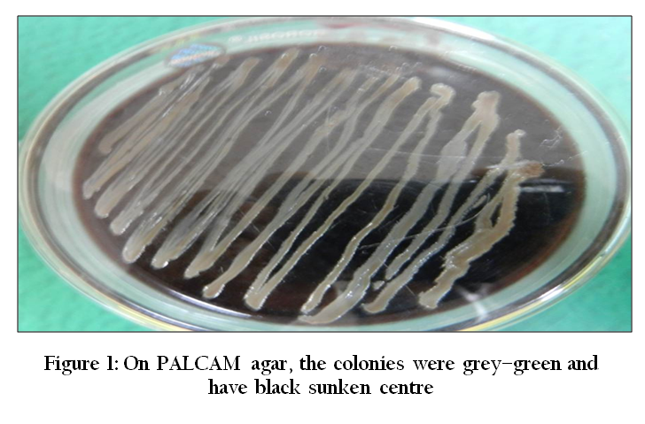
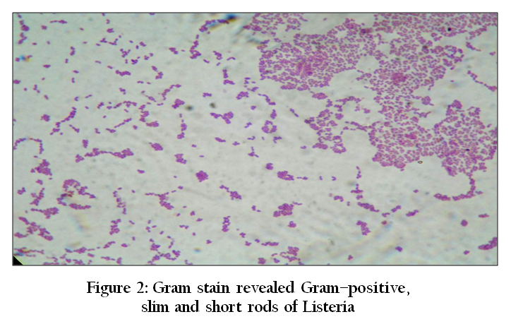
Twenty one brain samples were found positive for Listeria by cultural, biochemical, phenotypic and genotypic characteristics. Among these, 9 were found positive for Listeria monocytogenes. On PALCAM agar, the colonies were gray–green, measuring 1.5–2mm in diameter and had black sunken center (Figure 1). Esculin, ferric iron, D–mannitol and phenol red contribute to this colour formation which is characteristics of Listeria. The cold enrichment has the disadvantage of being a lengthy procedure which can continue for several months and is prone to environmental contamination (Gronstol et al., 1986). Hence, in our opinion, the organism can be readily isolated from brain tissues by enrichment in the selective media UVM I and UVM II and plating on PALCAM agar which is very simple and use full method in isolating Listeria from morbid samples such as brain. Simplified genus identification has been done such as Gram stain (revealed Gram–positive, slim, short rods) (Figure 2), positive catalase (3% hydrogen peroxide solution) methyl red and Voges–Proskauer (V–P) reaction and observation of motility. These isolates were negative for oxidase and nitrate reduction tests. The sugar fermentation tests of Listeria spp. showed negative for Xylose and Mannitol and positive for Ramnose. Antibiogram of these isolates revealed sensitivity to certain antibiotics such as oxytetracycline, ampicillin, gentamycin, amoxycillin, enrofloxacin, ceftriaxone and ciprofloxacin and resistant against cloxacillin. The actA virulence gene (839 bp) of L. monocytogenes was detected by PCR from the nine brain samples. PCR is the only test utilized for rapid detection of L. monocytogenes morbid specimens. Multiplex PCR assay has been used for the detection of Listeria virulence genes (Suarez and Vazquez–Boland, 2001; Rawool et al., 2007). Various test protocols were evaluated for cerebrospinal fluid samples and tissue samples (fresh or in paraffin blocks) (Jaton, et al., 1992)
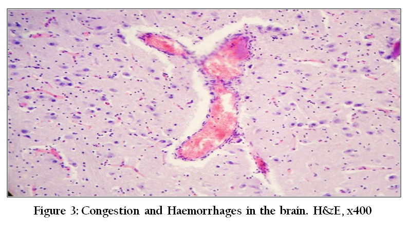
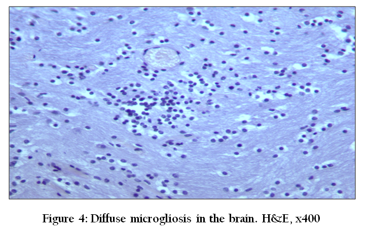
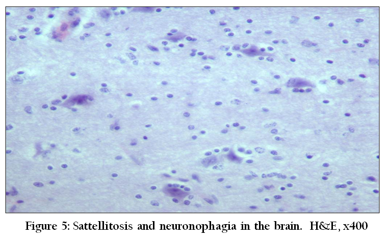
Out of 21 brain samples processed, the prominent change was vascular congestion in the grey matter, followed by degenerative changes in the neurons at different anatomical sites. The less frequent lesions noticed were leptomeningitis, vascular wall thickening, and multifocal congestion and haemorrhages in the grey matter (Figure 3), focal to diffuse gliosis (Figure 4), perivascular infiltration of mononuclear cells, sattellitosis and neuronophagia at many sites (Figure 5). But no characteristic lesions of Listeriosis have been found. This indicates the possibility of septicemic listeriosis and presence of listeria in brain even in the septicemic form. The animals with injured mucosa are most susceptible for entry of pathogen through the nerve endings (particularly trigeminal nerve), considered as the potential route of entry in encephalitic listeriosis (Otter and Blakmore, 1989a, 1989b). Therefore, the source of infection might be the faecal contaminated grazing areas and pastures. In India, Listeria monocytogenes has been isolated from blood samples of slaughtered goats (Banu Rekha, 1997) and goat milk and meat (Barbuddhe et al., 2000). Antibodies against LLO have also been detected in apparently healthy goats (Barbuddhe et al., 2000). The outbreaks of listerial encephalitis were reported in the migratory flocks of sheep in Punjab (Kumar et al., 2007).
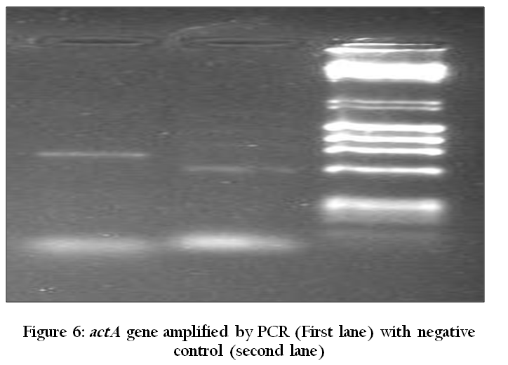
Vishwanathan and Uppal (1981) isolated L. grayi from the brain of a sheep that died after exhibiting signs of the cerebral form of listeriosis. On the basis of clinico–histopathological findings, an outbreak of listeric encephalitis with a morbidity rate of 3.8% in sheep and 1.2% in goats, primarily affecting adult animals, has been recorded in an organized farm at Bikaner, Rajasthan (Chattopadhyay et al., 1985). There are few studies in which Listeria has been isolated from brain. In the present study, we were able to isolate, identify and characterize the most pathogenic, public health significant Listeria monocytogenes from goats. The actA virulence gene (839bp) of Listeria was detected by PCR (Figure 6). The bacterial surface protein actA is a major virulence factor of L. monocytogenes that enables bacterial propulsion in the cytosol leading to the invasion of yet uninfected neighbouring cells by a process called cell–to–cell spreading. actA is also implicated in initial cellular invasion (Alvarez–Dominguez, C. 1997). The precise mechanism involved in actA–mediated invasion remains to be elucidated. However, for comprehensive characterization of Listeria, more studies are required to be undertaken (Nishibori et al., 1995), considering that there are five genes (plcA, prfA, hlyA, actA, and iap) which are associated with virulence of Listeria spp.
ACKNOWLEDGEMENTS
The authors are thankful to the Director, Central Institute for Research on Goats (CIRG), Makhdoom, for providing necessary facilities to carry out the research work.
CONFLICT OF INTEREST
There is no conflict of interests among authors.
REFERENCES
Alvarez–Dominguez C (1997). Host cell heparan sulfate proteoglycans mediate attachment and entry of Listeria monocytogenes, and the listerial surface protein ActA is involved in heparan sulfate receptor recognition. Infect. Immun. 65: 78–88.
PMid:8975895 PMCid:PMC174559
Banu Rekha G (1997). Virulence factor based diagnosis of Listeria monocytogenes infection in goats. M.V.Sc. Thesis. Indian Vet. Res I, Izatnagar, India.
Barbuddhe SB, Malik SVS, Bhilegaonkar KN, Kumar P and Gupta LK (2000). Isolation of Listeria monocytogenes and antilisteriolysin–O detection in sheep and goats. Small Ruminant Res. 38: 151–155.
http://dx.doi.org/10.1016/S0921-4488(00)00155-3
Barlow RM and McGorum B (1985). Ovine listerial encephalitis: analysis, hypothesis and synthesis. Vet. Rec.116: 233–236.
http://dx.doi.org/10.1136/vr.116.9.233
PMid:4002594
Chattopadhyay SK, Harbola PC and Bhagwan PSK (1985). Infectious encephalitis in sheep and goats in a farm at Arid Zone in India. Indian J Clin. Microbiol. Immunol. Infect. Diseases 6: 127—132.
Gronstol H, Rocourt J and Catimel B (1986). Study of 214 Listeria strains isolated from animals and silage in Norway. In Listeria – listeriosis 1985–1986 (A.L. Courtieu, E.P. Espaze and A.E. Reynaud, eds). Université de Nantes, Nantes, France, 325–329.
Jaton K, Sahli R and Bille J (1992). Development of polymerase chain reaction assays for detection of Listeria monocytogenes in clinical cerebrospinal fluid samples. J Clin. Microbiol. 30: 1931–1936.
PMid:1500495 PMCid:PMC265418
Kumar H Singh, BB, Bal, MS, Kamalpreet Kaur, Randhir Singh, Sidhu PK and Sandhu KS (2007). Pathological and epidemiological investigations into listerial encephalitis in sheep. Small Ruminant Res71: 293–297.
Low JC and Donachie W (1997). A review of Listeria monocytogenes and listeriosis. Vet. J 153: 9–29.
http://dx.doi.org/10.1016/S1090-0233(97)80005-6
Luna LG (1968). Manual of Histologic Staining Methods of the Armed Forces Inst Pathol, 3rd ed. McGraw–Hill Book Company, New York.
Maxie GM and Youssef S (2007). Nervous system. In: Maxie, G.M. (Ed.), Jubb, Kennedy and Palmer's Pathology of Domestic Animals. 5th ed. Saunders Elsevier, Edinburgh, pp. 281–457.
McClain D and Lee WH (1988). Development of USDA–FSIS method for isolation of Listeria monocytogenes from raw meat and poultry. J AssocOff Anal Chem 71: 660–663.
PMid:3134339
Nishibori T, Cooray K, Xiong H, Kawamuru I, Fujita M and Mitsuyama M (1995). Correlation between the presences of virulence associated genes as determined by PCR and actual virulence to mice in various strains of Listeria spp. Microbiol. Immun. 39: 343–349.
http://dx.doi.org/10.1111/j.1348-0421.1995.tb02211.x
PMid:7565175
Otter A and Blakmore WF (1989a). Observation on the presence of Listeria monocytogenes in axons. Acta Microbiologica et Immunologica Hungarica 36: 125–131.
Otter A and Blakmore WF (1989b). Observation on the neural transport of Listeria monocytogenes in a mouse model. Neuropathol. App. Neurol. 15: 590.
Radostits OM, Blood DC and Gay CC (1997). Vet Med, 9th Ed. W.B. Saunders, London, 660–666.
Rawool DB, Malik SVS, Shakuntala I, Sahare AM and Barbuddhe SB (2007). Detection of multiple virulence–associated genes in Listeria monocytogenes isolated from bovine mastitis cases. Int. J Food Microbio.113: 201–207.
http://dx.doi.org/10.1016/j.ijfoodmicro.2006.06.029
PMid:16979771
Sherman DM (2011). The spread of pathogens through trade in small ruminants and their products. Revue scientifique et technique (International Office of Epizootics) 30: 207–217.
PMid:21809765
Suarez M and Vazquez–Boland JA (2001). The bacterial actin nucleter protein ActA is involved in epithelial cell invasion by Listeria monocytogenes. Vet. Rec 133:165–166.
Summers BA, Cummings JF and Lahunta A (1995). Vet Neuropathol. Mosby–Year Book, St. Louis, pp. 53.
Zachary JF (2006). Nervous system. In: McGavin, M.D., Zachary, J.F. (Eds.), Pathol Basis Vet Disease. Mosby Elsevier, St. Louis, pp. 833–971.


