Advances in Animal and Veterinary Sciences
Review Article
Advances in Animal and Veterinary Sciences 1 (1): 1 – 4Vertebral Scale System to Measure Heart Size in Dogs in Thoracic Radiographs
Mudasir Bashir Gugjoo*, Mozammel Hoque, Abhishek Chander Saxena, Malik Mohammed Shamsuz Zama, Amarpal
-
Indian Veterinary Research Institute-Izatnagar, India.
*Corresponding author:mbgugjoo@gmail.com
ARTICLE CITATION:
Gugjoo MB, Hoque M, Saxena AC, Zama MMS and Amarpal (2013). Vertebral scale system to measure heart size in dogs in thoracic radiographs. Adv. Anim. Vet. Sci. 1(1): 1 – 4.
Received: 2013-03-16, Revised: 2013-03-22, Accepted: 2013-03-26
The electronic version of this article is the complete one and can be found online at
(
http://www.nexusacademicpublishers.com/table_contents_detail/4/21/html
)
which permits unrestricted use, distribution, and reproduction in any medium, provided the original work is properly cited
ABSTRACT
Radiographs of thoracic cavity are useful as a diagnostic modality to detect heart diseases and have a potential to provide information equivalent to other cardiac diagnostic modalities. Radiographic interpretation can be done by number of ways viz., by gross examination or by using different measurements like cardiothoracic ratio, relationship with inter-costal spaces or vertebral heart score. Cardiothoracic ratio and a specification of 2.5 to 3.5 inter-costal spaces had been introduced in veterinary medicine as an indicator of normal heart size in lateral radiographic views for dogs, but due to some limitations, use of these techniques has been supplanted by another cardiac measurement technique called the vertebral heart score, in which the heart length and width on the thoracic radiograph is compared with the vertebral length. The vertebral heart score may be useful in assessing the change in size of the heart in a patient over time as there is good correlation between the growth of different visceral organs and vertebral body length. Knowledge of inter-breed variation in the thoracic conformation and selection of proper reference value may further enhance the value of vertebral heart score technique in diagnosis of cardiac enlargement in dogs.
INTRODUCTION
Recent advances in veterinary cardiology, most notably in the areas of diagnostic imaging and cardiovascular therapeutics, have considerably added to our understanding on cardiac diseases in small animals. Cardiac diseases during recent years are considered as an important health problem in dogs and are being diagnosed with an increased frequency (Devi et al., 2009). Despite the availability of other diagnostic techniques like echocardiography (Root and Bahr, 2002), the thoracic radiography is one of the most commonly performed radiographic examinations in small animal practice. Important information about major medical problems, such as heart disease and cancer, is often obtained from thoracic radiographs (Dark et al., 1996). Follow up radiographs are used to evaluate clinical response to therapy or progression of disease (Thrall, 2007). A general guideline of 2.5-3.5 intercostal spaces for dogs with a deep and wide thorax, respectively, has been used as an indicator of normal heart size in lateral radiographic views (Owens, 1985; Kealy, 1987). Nevertheless, limitations in terms of, variations in the axis of the heart and its silhoutte, thoracic conformation, respiratory phase, rib superimposition and imprecise measurement points (Buchanan and Bucheler, 1995; Gulanber et al., 2005), have declined the use of this method. Cardiothoracic ratios have been used in the past on the basis of change in dimensions of thoracic cavity. In case of progressive cardiomegaly in dogs, chest cavity also expands. Thus, the cardiac dimensions relative to thoracic depth and width may remain same. In other words, if reduction in heart size occurs, a reduction in chest size may also be observed, thus there may be little or no change in the cardiothoracic ratio.
VERTEBRAL HEART SCORE
Vertebral heart score (VHS), first described by Buchanan and Bucheler (1995), employs measuring the cardiac silhouette by involving its long axis (taken from left main stem bronchus ventral border to the cardiac apex with a measuring scale) and short axis (taken from central third region of heart perpendicular to the longitudinal axis with a measuring scale) on a lateral radiograph (Figure).

Figure: Long axis (LA) and short axis (SA) measurements of heart in lateral recumbency for calculation of VHS starting from 4th thoracic vertebra (T4)
The sum of these measurements is then compared to the mid-thoracic vertebral bodies starting from the anterior edge of the 4th thoracic vertebra (T4). Long axis of heart includes left atrium and left ventricle in lateral radiograph and right atrium and left ventricle in dorso-ventral radiograph. The short axis of the canine heart includes right atrium and left heart chambers in lateral radiograph and left and right heart structures in dorso-ventral radiograph. Precise measurements for statistical analysis are taken to the margin of 0.1 vertebrae. Summation of number of vertebrae in relation to long and short axis of heart indicates vertebral heart score.
The mean VHS reported by Buchanan and Bucheler (1995) was 9.7±0.5 and 10.2±0.83 vertebrae in lateral and ventrodorsal radiographs, respectively, of different dog breeds. Also the VHS values in lateral radiographs were unaffected by the depth or broadness of the chest of dogs which is in contrast to intercostal space method where such variation does occur, as reported by Jepsen-Grant et al. (2013). VHS method has been reported to be unaffected by the experience of observer but does depend on the selection of the reference points of longitudinal and transverse axis of the heart and their conversion into VHS units (Hansson et al., 2005). Variations in the normal canine heart are more than in any other organ, and the heart is inherently variable in size because of its contractility during the cardiac cycle. Additionally, there is considerable breed variation with regards to normal heart size and shape. So, it is desirable to consider the breed specific value whenever the heart is evaluated (Root and Bahr, 2002; Owens and Biery, 1999; Toal et al., 1985; Silverman and Suter, 1975; Toombs and Ogburn, 1985).
Vertebral heart score in puppies calculated by Sleeper and Buchanan reported that vertebral heart size in puppies does not change with the growth and lies within adult dog’s reference range (9.7±0.5). Thus, similar standards exist to determine cardiac enlargement in puppies and adult dogs (Sleeper and Buchanan, 2001).
Effect of radiographic positioning on VHS in dogs was first studied by Fox (2003). Orientation of the heart is approximately at 45 degree angle in the lateral view, lies between 3rd to 8th thoracic vertebrae, covers about 3 intercostal spaces, and has VHS of 8.5-10.6 (av. 9.7). Roughly, it has an elliptical shape with a curved right ventricular and relatively straight left ventricular border in the ventro-dorsal (DV) or dorso-ventral (DV) view. Lateral radiographic positioning in clinical practice may be preferred over ventro-dorsal as it is less stressful for cardiac patients (Fox, 2003). Furthermore, image magnification occurs in VD views as the distance between heart and the X-ray cassette increases. In addition to magnification, higher VHS may occur as the VD/DV long axis includes the right atrium and left ventricle, whereas in lateral projections only the left atrium and left ventricle is included (Gulanber et al., 2005). Reports on comparison between dorso-ventral (DV) and ventro-dorsal (VD) views have revealed that VD heart sizes are wider (7%) and longer (5%) than DV heart sizes (Buchanan and Bucheler, 1995). However, correlation between DV/VD and lateral heart sizes appears to be poor. Determination of VHS in DV or VD projection in deep chested dogs appears to be of little value as there is relatively vertical long axis of heart in such dogs. Mean VHS in ventro-dorsal or dorso-ventral views has been reported to be significantly larger than that in lateral view. Training also has significant effect on VHS and there is higher VHS in trained dogs compared to non-trained (Bavegems et al., 2005). Possible reason could be more workload on heart in racing than non-racing animals which leads to compensatory increase in cardiac size. There are conflicting reports regarding the difference in VHS between left and right lateral recumbency in dogs. In one report, Greco et al. (2008) compared VHS calculated in right and left lateral recumbency and reported significantly higher VHS value in right (9.8±0.6) compared to left lateral recumbency (9.5±0.8). Similar findings were also reported in Whippets (Bavegems et al., 2005), Beagles (Kraetschmer et al., 2008), Doberman and German shepherd dogs (Ghadiri et al., 2010). In our study on Labrador retriever, also statistically non-significant differences were found between VHS taken in right and left lateral recumbency; however, slightly higher values were observed in right lateral recumbency (Gugjoo et al., 2013). The higher VHS in right recumbency could be explained by the fact that divergence of X-ray beam and more distance of the heart from the cassette occurs in right lateral recumbency which leads to image magnification. In contrast to above reports, findings on mixed breed and native dogs of Iran showed non-significant difference in VHS when taken in right or left lateral recumbency (Ghadiri et al., 2010). Gender, body size of the animal (Buchanan and Bucheler, 1995; Bavegems et al., 2005; Ghadiri et al., 2010; Gugjoo et al., 2013) and respiratory diseases (Lamb et al., 2001) did not significantly influence VHS values in dogs However, Lamb et al. (2001) reported significant effect of gender on VHS in which males had a higher mean VHS than females. In general, the vertebrae and internal organ size show a comparable development and can be attributed to non-significant effect of body weight on VHS.
BREED VARIATIONS
VHS of different dog breeds is given in Table. Boxer dog breed has significantly higher mean VHS measurements compared to the dogs of other breed including Yorkshire terrier and German shepherd while Labrador retrievers have significantly higher mean VHS measurements than other breeds except the boxer and the Cavalier King Charles spaniel (Lamb et al., 2001). However, Hansson et al. (2005) reported the mean VHS value of 10.8±0.5 Vertebrae in the normal cavalier King Charles spaniels. VHS in normal poodle dogs has been reported to be smaller or equal to 10.5 in 80% of dogs (Fonsecapinto and Iwasaki, 2004).
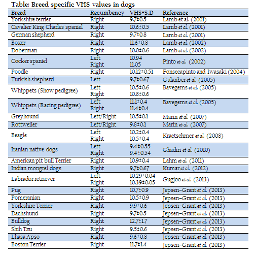
VHS values in Turkish shepherd have been reported to be comparable to other dog breeds. Also no significant difference with respect to age and sex of the dogs has been reported (Gulanber et al., 2005). In case of Whippets, two different values have been established. Bavegems et al. (2005) reported mean VHS on lateral radiographs of Whippets to be significantly larger than the VHS reported earlier. It has also been reported that there is significant difference between show and racing pedigree lines of the Whippet breed.
VHS in retired racing Greyhound has been reported to be significantly higher when compared with that of Rottweiler and to other dog breeds using both analog and digital radiography. The mean VHS established on lateral radiographs was 10.5±0.1 for Greyhounds, 9.8±0.1 for Rottweiler and 10.1±0.2 for mixed breed dogs. Also, Non-significant effect of radiographic positioning (Right or left recumbency) on VHS was observed in all the studied breeds (Marin et al., 2007). A slight modification of the original technique was done by Spasojevic Kosic et al. (2007) for calculating VHS in German shepherd. However, non-significant differences in VHS were reported when compared to those measured by standard technique (Spasojevic Kosic et al., 2007).
The mean VHS in the beagle dogs has been reported to be 10.3 which are significantly different from the mean value of 9.7 shown in Buchanan and Bucheler (1995) study. It has also been reported that inspiration has no significant effect on the vertebral heart scale of Beagle dogs (Kraetschmer et al., 2008).
The influence of breed on VHS in dogs in Iran has been reported by Ghadiri et al. (2010). VHS reported was similar in right lateral radiograph (9.4) as that of left lateral radiograph (9.4) in Iranian native dog breeds. Native dogs were found to have the lower VHS than all other breeds, both in left lateral and right lateral radiographs. The mean VHS values have been determined as 9.6 and 9.8 for German shepherd dogs and 9.6 and 9.7 for mixed breeds in left lateral and right lateral radiographs, respectively. The mean VHS values in left lateral views were significantly smaller than in the right lateral view in Doberman and German shepherd dogs. However, the VHS of left lateral and right lateral radiographs did not differ significantly in mixed breed and native dogs.
Mean value of VHS in American pit bull Terrier has been reported as 10.9±0.4 and found to be significantly different from the values of general dog population given earlier in Buchanan and Bucheler (1995) study (Lahm et al., 2011).
VHS in eight dog breeds (Pug, Pomeranian, Yorkshire terrier, Dachshund, Bulldog, Shih Tzu, Lhasa Apso, and Boston terrier dog breeds) was reported by Jepsen-Grant et al. (2013). They reported that the VHS in Pug, Pomeranian, Bulldog, and Boston terrier was significantly greater than 9.7±0.5 given by Buchanan and Bucheler (1995) for general dog population. VHS of Lhasa Apso was significantly affected by body condition score (BCS). Anomalous vertebrae in the thoracic column were associated with a significant increase in VHS of the Bulldog and Boston terrier and thoracic depth to width ratio did not have a significant effect on VHS.
Unpublished data of VHS in Indian mongrel dogs (Kumar et al. 2011) showed that the VHS in clinically normal mongrel dogs was 9.7±0.67. VHS distribution range was 8.4 to 10.9 and no significant difference was found between male and female animals.
The authors have themselves studied effect of gender, body weight and radiographic positioning on VHS in Labrador retrievers. Vertebral heart score was 10.29±0.04 and 10.39±0.05 in left and right lateral recumbency and was not affected by gender, body weight and radiographic positioning. These values were comparatively higher to those reported in German shepherd and Doberman (Gugjoo et al., 2013). However, Lamb et al. (2001) reported VHS value to be 10.8±0.6 in right lateral recumbency in Labrador retrievers.
Higher VHS in Labrador retriever, Boxer and Cocker spaniels as compared to other breeds may be due to slightly shorter vertebrae in such breeds (Lamb et al., 2001).
CLINICAL UTILITY
VHS can be used for diagnosis of different heart problems with variable accuracy. It is reported that electrocardiographic and echocardiographic parameters provide findings comparable to VHS for evaluation of heart size (Nakayama et al., 2001). Accuracy of the VHS in the diagnosis of cardiac disease was first evaluated by Lamb et al. (2001). Thoracic radiographs of 126 dogs, including animals with proven cardiac disease (50 dogs), other thoracic diseases (26 dogs), and no clinical signs of cardiovascular or respiratory disease (50 dogs) were mixed and examined. VHS value above 10.7 was found to be moderately an accurate sign of cardiac disease in most of the cases. In a study, the effect of different drugs (pimobendan and ramipril) upon size of the heart affected by myxomatous mitral valve disease was evaluated by VHS method. Pimobendan was found more effective than remipril as there was more reduction in VHS in pemobendan group (Woolley et al., 2007). VHS has been reported to be fairly accurate for exclusion of a cough of cardiac origin in dogs with mitral valve disease (MVD) (Guglielmini et al., 2009). In another report on congestive heart failure (CHF) due to mitral valve regurgitation, VHS was found to be useful for detecting onset of CHF in Cavalier Kings Charles Spaniels with mitral regurgitation (Lord et al., 2011). VHS has been found as the most accurate radiographic index for identifying dogs with pericardial effusion (PE) and also to differentiate it from other cardiac diseases (Guglielmini et al., 2012). Authors have applied VHS along with the other cardiac diagnostic modalities viz., ECG and echocardiography in diagnosing dilatation cardiomyopathy (DCM). It was observed that VHS increases significantly in DCM (Gugjoo et al., 2012) and inference drawn was that VHS can be used to diagnose such conditions very effectively. However, Inter-observer variability in selection of reference points for calculation of VHS should be considered while evaluating as this might affect the results (Hansson et al., 2005).
CONCLUSION
VHS is one of the easily available, applicable and interpretable cardiac diagnostic techniques as it does not require any sophisticated equipment. However, the problems related to inter-observer variability in relation to reference point selection and also due to breed specific values, should be considered while interpreting the radiograph.
ACKNOWLEDGEMENT
The authors are highly thankful to the Director of the Institute for providing necessary facilities.
REFERENCES
Bavegems V, Caelenberg AV, Duchateau L, Sys SU, Bree HV and DeRick A (2005). Vertebral heart size ranges specific for Whippets. Vet. Radiol. Ultrasound, 46: 400-403.
http://dx.doi.org/10.1111/j.1740-8261.2005.00073.x
PMid:16250398
Buchanan JW and Bucheler J (1995). Vertebral scale system to measure canine heart size in radiographs. J. Am. Vet. Med. Assoc. 206: 194-199.
PMid:7751220
Dark P, Bonagura JD and Kelly DF (1996). Cardiac radiography, In: Color atlas of veterinary cardiology, Dark P, Bonagura JD, Kelly DF, editors, St Louis: Mosby, 32-37.
Devi S, Jani RG, Karlette Anne F and Sing RD (2009). Study on Clinical symptoms in canine cardiac diseases. Vet. World, 2(8): 307-309.
Fonsecapinto BC and Iwasaki M (2004). Radiographic evaluation of the cardiac silhouette in clinically normal Poodles through the vertebral heat size (VHS) method. Braz. J. Vet. Res. Anim. Sci. 41: 261-267.
Fox PR (2003). Thoracic radiography: The coughing/dyspneic dog and cat 28th world congress of the world small animal veterinary association, Bangkok, Thailand, 27: 23.
Ghadiri A, Avizeh R and Fazili G (2010). Vertebral heart scale of common large breeds of dogs in Iran. Int. J. Vet. Res. 2: 107-111.
Greco A, Meomartino L, Raiano V, Fatone G and Brunetti A (2008). Effect of left vs. right recumbency on the vertebral heart score in normal dogs. Vet. Radiol. Ultrasound, 49: 454-455.
http://dx.doi.org/10.1111/j.1740-8261.2008.00406.x
PMid:18833953
Gugjoo MB, Hoque M, Saxena AC and Zama MMS (2012). Dilatation cardiomyopathy in dogs. 36th annual congress of ISVS and international symposium, 1st - 3rd November, 2012, Kolkata, India, 62.
Gugjoo MB, Hoque M, Zama MMS, Saxena AC, Pawde AM, Ansari MM and Bhat SA (2013). Vertebral Scale System to measure heart size on thoracic radiographs of Labrador retriever dogs. Ind. Vet. J. 90(2): 71-73.
Guglielmini C, Diana A, Santarelli G, Torbidone A, Tommaso MD, Toaldo MB and Cipone M (2012). Accuracy of radiographic vertebral heart score and sphericity index in the detection of pericardial effusion in dogs. J. Am. Vet. Med. Assoc. 15 (8): 1048-1055.
http://dx.doi.org/10.2460/javma.241.8.1048
PMid:23039979
Guglielmini C, Diana A, Pietra M, Tommaso MD and Cipone M (2009). Use of the vertebral heart score in coughing dogs with chronic degenerative mitral valve disease. J. Vet. Med. Sci. 71 (1): 9-13.
http://dx.doi.org/10.1292/jvms.71.9
PMid:19194070
Gulanber EG, Gonenci G, Kaya U, Aksoy O and Birsik HS (2005). Vertebral scale system to measure heart size in thoracic radiographs of Turkish Shepherd (Kangal) Dogs. Turk. J. Vet. Anim. Sci. 29: 723-726.
Hansson K, Haggstrom J, Kvart C and Lord P (2005). Reader performance in radiographic diagnosis of signs of mitral regurgitation in cavalier King Charles spaniels. J. Small Anim. Pract. 50: 44-53.
http://dx.doi.org/10.1111/j.1748-5827.2009.00669.x
http://dx.doi.org/10.1111/j.1748-5827.2008.00669.x
Jepsen-Grant K, Pollard RE and Johnson LR (2013). Vertebral heart scores in eight dog breeds. Vet. Radiol. Ultrasound, 54 (1): 3-8.
http://dx.doi.org/10.1111/j.1740-8261.2012.01976.x
PMid:22994206
Kealy JK (1987). Diagnostic radiology of the dog and cat. 2nd edition. Philadelphia: Saunders WB, 258.
Kraetschmer S, Ludwig K, Menesses F, Nolte I and Simon D (2008). Vertebral heart scale in the Beagle dog. J. Small Anim. Pract. 49: 240-243.
http://dx.doi.org/10.1111/j.1748-5827.2007.00531.x
PMid:18422506
Kumar V, Hoque M, Sharma MC, Saxena AC, Zama MMS and Gugjoo MB (2011). Vertebral heart scale system to measure heart size in thoracic radiographs of Indian mongrel dog. Ind. J. Vet. Surg., (InPress).
Lahm JMC, Caludino JL and Melussi A (2011). Measurement of Heart size by VHS method in Healthy American pit bull terrier. Cience Rural, 41: 1.
Lamb CR, Wikeley H, Boswood A and Pfeiffer DU (2001). Use of breed-specific ranges for the vertebral heart scale as an aid to the radiographic diagnosis of cardiac disease in dogs. Vet. Rec. 148: 707-711.
http://dx.doi.org/10.1136/vr.148.23.707
PMid:11430680
Lord PF, Hansson K, Carnabuci C, Kvart C and Haggstrom J (2011). Radiographic heart size and its rate of increase as tests for onset of congestive heart failure in Cavalier King Charles Spaniels with mitral valve regurgitation. J. Vet. Int. Med. 25 (6): 1312-1319.
http://dx.doi.org/10.1111/j.1939-1676.2011.00792.x
PMid:22092622
Marin LM, Brown J, Mcbrien C, Baumwart R, Samii VF and Couto G (2007). Vertebral heart size in retired racing Greyhounds. Vet. Radiol. Ultrasound, 48: 332-334.
http://dx.doi.org/10.1111/j.1740-8261.2007.00252.x
PMid:17691632
Nakayama H, Nakayama T and Hamlin RL (2001). Correlation of cardiac enlargement as assessed by vertebral heart size and echocardiographic and electrocardiographic findings in dogs with evolving cardiomegaly due to rapid ventricular pacing. J. Vet. Int. Med. 15: 217-221.
http://dx.doi.org/10.1111/j.1939-1676.2001.tb02314.x
PMid:11380030
Owens JM (1985). Radiology of the heart. In: Tilley LP, Owens JM (ed). Manual of Small Animal Cardiology. Churchill Livingstone Inc., New York. 37.
Owens JM and Biery DN (1999). Radiographic interpretation for the small animal clinician. 2nd edition. Williams and Wilkins Company, 190.
Pinto AC (2002). Radiographic methods in the cardiac evaluation in dogs. Veterinaria Noticias Univ Fed Uberlandia Braz. 8: 67-75.
Root CR and Bahr RJ (2002). The heart and great vessels in Textbook of diagnostic veterinary radiology, Thrall DE (ed) 4th edition. Philadelphia: Saunders WB, 402-419.
Silverman S and Suter PF (1975). Influence of inspiration and expiration on canine thoracic radiographs. J. Am. Vet. Med. Assoc. 166: 502-510.
PMid:1112758
Sleeper MM and Buchanan JW (2001). Vertebral scale system to measure heart size in growing puppies. J. Am. Vet. Med. Assoc. 219: 57-59.
http://dx.doi.org/10.2460/javma.2001.219.57
PMid:11439770
Spasojevic Kosic LJ, Krstic N and Trailovic RD (2007). Comparison of three methods of measuring vertebral heart size in German shepherd dogs. Acta Veterinaria (beograd), 57: 133-141.
http://dx.doi.org/10.2298/AVB0703133S
Thrall DE (2007). Veterinary Radiology. 5th edition. Philadelphia: Saunders Elsevier, 479-485.
Toal RL, Losonsky JM, Coulter DB and Denovellis R (1985). Influence of cardiac cycle on the radiographic appearance of the feline heart. Vet. Radiol. 26: 63-69.
http://dx.doi.org/10.1111/j.1740-8261.1985.tb01118.x
Toombs JP and Ogburn PN (1985). Evaluating canine cardiovascular silhouettes: radiographic methods and normal radiographic anatomy. Compend. Contin. Edu. Pract. Vet. 7: 579-587.
Woolley R, Smith P, Munro E, Smith S, Swift S, Devine C, Corcoran B and French A (2007). Effect of treatment type on vertebral heart size in dogs with myxomatous mitral valve disease. Int. J. Appl. Res. Vet. Med. 5 (1): 43-48.


