Advances in Animal and Veterinary Sciences
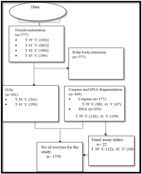
Flow of data collected in the study period.
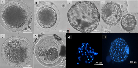
Assessment of heat stress on cattle oocytes at four temperatures: 39, 41, 42 and 43 ˚C. Produced blastocyst at day 7 and Hoechst (33342) staining of nuclei cells of produced blastocysts. Suitable maturation temperatures were selected according to the extrusion of polar bodies at a significant of (P < 0.05). Matured cattle oocytes from (A) 39˚C, (B) 41 ˚C, (C) 42 ˚C and (D) 43 ˚C. Day 7 Blastocyst from cattle oocytes matured at (E) 39 ˚C and (F) 41 ˚C, Hoechst 33342 (Blue) stained blastocyst from (G) 41 ˚C and (H) 39˚C matured cattle oocytes.
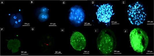
Representation of images following TUNEL Assay procedure. Hoechst 33342 (Blue) stained 2-4 cell embryos from (A) 39 ˚C, (B) 41 ˚C, (C) ≥ 8 cell embryos from both 39 and 41 ˚C, blastocysts from cattle oocytes matured at (D) 39 ˚C and (E) 41 ˚C. Alexa 488-stained (green) 2-4 cell embryos from 41 (F), 39 ˚C (G), ≥ 8cell embryos from 41 ˚C (H) and Alexa 488-stained (green) blastocysts from cattle oocytes matured at (I) 39 and (J) 41 ˚C maturation group.


