Advances in Animal and Veterinary Sciences
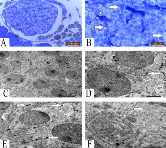
A: Photomicrograph showing semithin section of parathyroid gland of one year guinea pig. Methylene blue (100µm). B: Photomicrograph showing semithin section of parathyroid gland showing granulated (star), vacuolated (arrows) chief cells and blood capillaries (C). Methylene blue (20µm). C: TEM of mature parathyroid gland showing several chief cells with different organelles 5 μm. D: TEM of mature parathyroid gland showing tortuous plasma membrane (arrow) of chief cells 2 μm. E: TEM of chief cells contain RER (R), mitochondria (m) and secretory granules (arrows) 2 μm. F: TEM of chief cells contain concentric RER (R) and Golgi complex (arrow) 1 μm.
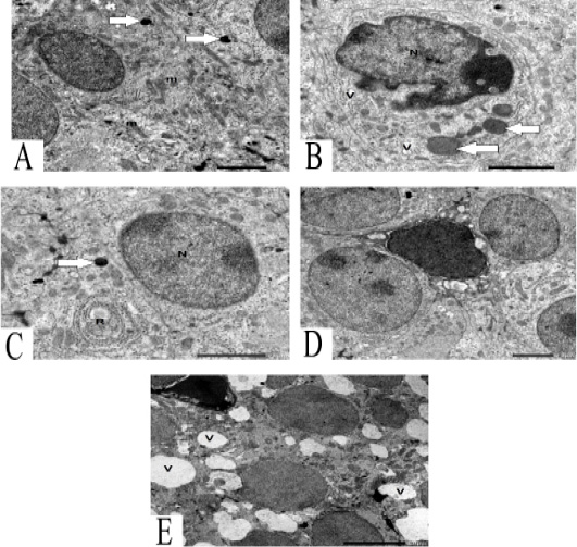
A: TEM of chief cells contains round and oval mitochondria (m) and lysosome (arrows) 2 μm. B: TEM of chief cells contain undulating nucleus (N) with condensation of chromatin, large secretory granules (arrows) and some vacuoles (V) 2 μm. C: TEM of chief cells contains nucleus (N) with chromatin islands, secretory granules (arrow) and characteristic RER (R) 2 μm. D: TEM of vacuolated chief cells between granulated chief cells. 2μm. E: TEM of vacuolated chief cells with dark nucleus and several vacuoles (V) .5 μm.
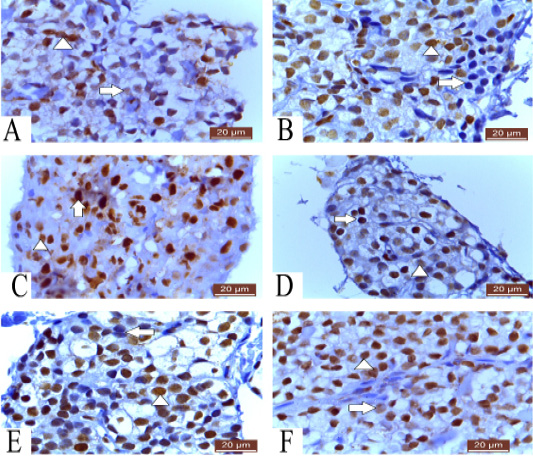
A: Photomicrograph of parathyroid gland of 60 ED showing negative nuclear reaction of few cells, strong nuclear reaction of some cells (arrowhead) and weak reaction of others (arrow) PCNA (20µm). B: Photomicrograph of parathyroid gland of 60 ED Guinea pig showing few cells give negative nuclear reaction (arrow) and most of cells show positive nuclear reaction (arrowhead) Ki-67 (20µm). C: Photomicrograph of parathyroid gland of 1 month showing strong nuclear reaction of some cells (arrow) and weak reaction of others (arrowhead) PCNA (20µm). D: Photomicrograph of parathyroid gland of 1 month Guinea pig showing strong (arrow) and weak (arrowhead) positive nuclear reaction Ki-67 (20µm). E: Photomicrograph of parathyroid gland of 12 months Guinea pig showing few negative (arrow) and most positive nuclear reaction (arrowhead) Ki-67 (20µm). F: Photomicrograph of parathyroid gland of 12 months showing strong nuclear reaction of most of cells (arrow head) and weak reaction of few cells (Arrow) PCNA (20µm).
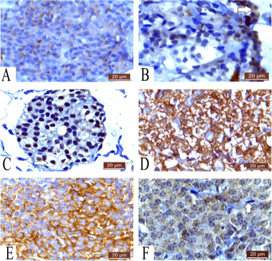
A: Photomicrograph of parathyroid gland of 60 ED showing very weak cytoplasmic reaction Caspase 3 (20µm). B: Photomicrograph of parathyroid gland of 60 ED Guinea pig showing negative cytoplasmic reaction of most of cells and positive cytoplasmic reaction of some cells (arrow) Bax (20µm). C: Photomicrograph of parathyroid gland of 1 month Guinea pig showing positive reaction and negative cytoplasmic reaction (arrow) Bax (20µm). D: Photomicrograph of parathyroid gland of 1 month showing positive cytoplasmic reaction of cells Caspase3 (20µm). E: Photomicrograph of parathyroid gland of 12 months showing strong cytoplasmic reaction of most of cells Caspase 3 (20µm). F: Photomicrograph of parathyroid gland of 12 months Guinea pig showing positive cytoplasmic reaction and negative fibroblast (arrow) Bax (20µm).


