Advances in Animal and Veterinary Sciences
Research Article
Adv. Anim. Vet. Sci. 10(1): 126-130
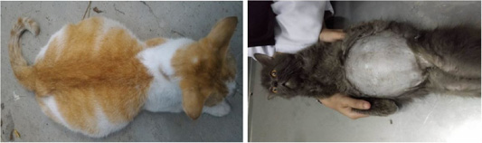
Figure 1
An enlarged abdomen due to ascites in a cat with effusive FIP.
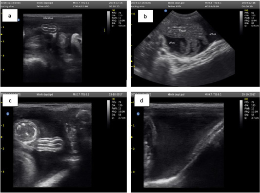
Figure 2
Ultrasound examination results showed an accumulation of anechoic fluid between the small intestine (a), liver (b), large intestine (c), and outside the bladder wall (d).


